Because occipital neuralgia typically involves specific nerve pathways rather than brain-based vascular changes, identifying the root cause helps determine the right treatment and long-term management strategy. The underlying causes of occipital neuralgia can include posture strain, arthritis, or a pinched nerve near the occipital region, and in some cases, underlying causes like infection or systemic inflammation. In this article, we’ll explore the anatomy of the three occipital nerves, the variety of causes behind the condition, how a doctor may confirm the diagnosis of occipital neuralgia, and the most effective treatment for occipital neuralgia—all through the lens of nervous system performance and adaptability.
Anatomy of the Occipital Nerves: Pathways Behind the Symptoms
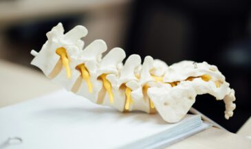
To understand what causes occipital neuralgia, it helps to look at the pathways that transmit pain messages to the brain. The three occipital nerves—the greater occipital nerve, the lesser occipital nerves, and the third occipital—emerge from the nerve root in the upper neck, traveling upward through the muscles and fascia of the occipital region to the scalp. When these nerves are compressed or stretched, their fibers can misfire, sending abnormal pain signals that feel like an electric jolt.
- Greater occipital nerve: Arises near the C2 spinal cord level and supplies most of the back of your head and central scalp.
- Lesser occipital nerves: Run along the side of the head and behind the ear, reaching into the upper scalp.
- Third occipital: Travels upward near C3, contributing to tenderness in the upper neck and lower occipital region.
Because these nerves pass through narrow channels of muscle and connective tissue, even minor irritation or compression of the occipital nerves can cause dramatic symptoms. Turning the head, pressing the skull base, or brushing the hair may activate the same pain in the back of the head. The affected skin near the occipital entry points often becomes tender and hypersensitive, showing just how reactive these small neural structures can be.
What Causes Occipital Neuralgia?
At its core, what causes occipital neuralgia is tension, inflammation, or compression of the nerve as it travels from the spinal cord to the scalp. Occipital neuralgia may arise from direct trauma, poor posture, repetitive strain, or deeper medical issues. Because the occipital nerves are embedded within multiple tissue layers, small distortions can easily irritate them.
Common underlying causes include:
- Mechanical factors: Chronic postural strain, tight neck and shoulder muscles, and prolonged computer work. These create local crowding near the occipital nerves and can set off sharp, shooting pain in the back of the head.
- Structural changes: Osteoarthritis or disc degeneration in the upper spine can pinch the nerve root, causing ongoing inflammation and neural tension.
- Head and neck injuries: Concussions, whiplash, or trauma caused by a head strike can disturb tissue alignment and irritate the greater occipital or lesser occipital nerves.
- Medical conditions: Infections, tumors, or vascular disorders such as arteriovenous malformations may compress the nerve. Autoimmune and metabolic diseases—diabetes, gout, rheumatoid arthritis, lupus—can also create neuropathic inflammation.
In some patients, no single cause of the pain is found. Occipital neuralgia can also occur spontaneously or from a mix of different causes that build over time. These overlapping factors explain why occipital neuralgia involves variable symptoms and why it is often misdiagnosed as migraine.
Learn more about INSiGHT scanning?
Fill this out and we’ll get in touch!
"*" indicates required fields
Recognizing the Symptoms of Occipital Neuralgia
The symptoms of occipital neuralgia reflect its nerve-based origin. The hallmark is shooting pain that radiates from the upper neck into the scalp. This may appear suddenly and last from seconds to minutes, recurring throughout the day.
Symptoms include:
- Sharp pain or electrical sensations traveling upward from the skull base.
- Pain behind the eye or radiating toward the forehead.
- Sensitivity of the scalp where even light touch can trigger pain.
- Pain localized to one or both sides of the head.
- Tenderness along the occipital nerve pathway and difficulty resting the head on a pillow.
- Occasional numbness or tingling in the skin near the occipital area.
Because occipital neuralgia is a headache, it’s frequently confused with migraine. However, neuralgia is a specific type of headache disorder that affects the occipital area through irritated nerve fibers rather than vascular changes in the brain. The pain of occipital neuralgia is typically localized, provoked by movement, and shorter in duration, while migraine tends to involve broader, throbbing pain and systemic symptoms such as nausea or light sensitivity.
Occipital Neuralgia vs. Migraine and Other Headache Types
Comparing occipital neuralgia with migraine and other headache types highlights key distinctions. Occipital neuralgia is a specific pattern of irritation along sensory nerves, whereas migraine is a disorder that causes widespread neurological responses.
| Feature | Occipital Neuralgia | Migraine |
| Source | Nerve compression or inflammation | Brain-based vascular changes |
| Pain quality | Sharp, shooting, electric | Throbbing or pulsating |
| Distribution | Back of the head, scalp, sometimes side of the head | Can move, often temples or periocular |
| Triggers | Touch, posture, neck movement | Hormones, light, stress, sleep changes |
| Duration | Seconds to minutes, can repeat | Hours to days |
| Relief test | Responds to an occipital nerve block | May not respond to a block |
The International Headache Society classifies neuralgia is a headache disorder, acknowledging that trigeminal neuralgia and occipital neuralgia share similar “electric” symptoms along different cranial nerves. Understanding these differences ensures patients receive the right treatment for their specific type of headache rather than relying on generic headache medication.
How Occipital Neuralgia Is Diagnosed
Accurate identification starts with diagnosing occipital neuralgia through a targeted physical exam and history. A doctor may locate tender points where the nerve exits near the skull base and check whether pressing these areas reproduces the familiar sharp sensations. Neck range of motion testing can reveal if movement intensifies symptoms.
Imaging may follow when underlying causes—arthritis, infection, or vascular lesions—are suspected. In complex cases, referral to a neurologist helps rule out migraines and other headache disorders. Together, these steps establish an evidence-informed baseline to plan the right treatment path forward.
Care and Treatment Options for Occipital Neuralgia
Occipital neuralgia treatment is most effective when approached progressively. Conservative strategies form the base of care, with interventional and surgical approaches reserved for chronic or severe pain.
Conservative Care
- Correct posture and ergonomics to reduce strain on the upper neck.
- Apply gentle heat to relax tight muscles near the occipital nerves.
- Chiropractic adjustments that restore motion and reduce neurological interference in the spinal region sEMG evidence.
- Stretching and targeted strengthening to support the head and neck.
- Occipital release surgery: A decompression procedure that frees the nerve from tight fascia. This surgical treatment for occipital neuralgia can be part of a larger plan to treat occipital neuralgia when other measures fail.
- Occipital nerve stimulation and spinal cord stimulation: Devices that send mild impulses to block pain messages for long-term management of chronic pain.
- Cutting or ablating the nerve is a surgical treatment of last resort due to potential numbness.
Each treatment option aims to restore normal signaling and reduce hypersensitivity. The best strategy combines postural correction, conservative care, and collaboration with medical specialists when needed.
The Chiropractor’s Role: Restoring Balance and Adaptability
From a neurologically focused chiropractic perspective, what causes occipital neuralgia is not only local irritation but also systemic overload. The nervous system can remain in a chronic sympathetic overdrive, keeping the tissues near the occipital nerves reactive. This sympathetic–immune relationship has been explored in peer-reviewed inflammation research.
Chiropractors assess:
- Postural alignment: How forward-head posture increases load on the upper neck.
- Muscle tone and nerve tension: Especially around C2–C3, where greater occipital nerve pathways emerge.
- Adaptive capacity: The system’s ability to recover from stress and avoid flare-ups, often tracked by HRV HRV review.
Adjustments and neuromuscular retraining help reduce strain, improving nervous system performance and coordination across the spine. Broader, interdisciplinary findings suggest chiropractic care may influence markers within the neuroendocrine-immune system, as reported in an open-access integrative review.
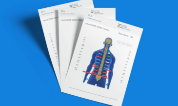
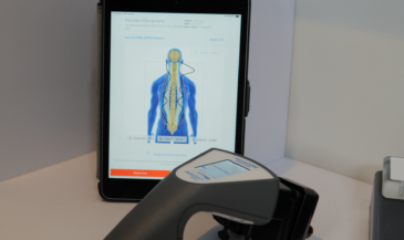
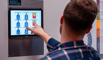
How INSiGHT Scanning Technology Supports Clinical Decisions
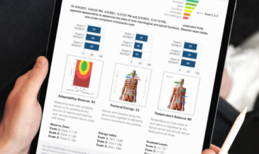
Objective data elevates clinical confidence. INSiGHT scanning technology—composed of the neuroCORE, neuroTHERMAL, and neuroPULSE instruments—translates complex neural behavior into simple, visual metrics that show how the body is adapting.
- neuroCORE (sEMG): Tracks muscular energy use and postural strain. Patterns of hyperactivity at the top of the spine often correlate with tension near the occipital nerves. Normative and clinical utility for paraspinal sEMG are reported in a pilot study.
- neuroTHERMAL: Detects asymmetrical temperature changes that reveal autonomic imbalance in the cervical segments, including those affecting the occipital region. Thermography’s reliability and clinical use in chiropractic are summarized in a technical review (see sEMG context; thermal reliability is covered in allied literature).
- neuroPULSE (HRV): Measures heart rate variability to identify sympathetic overdrive and depleted recovery reserves. Sustained HRV improvement during chiropractic care has been reported in a case series.
Autonomic regulation also relates to vagal pathways and the cholinergic anti-inflammatory response, described in vagal research. Together, these insights help clinicians communicate findings with clear scan views and show objective proof your care is making a difference over time.
When to Refer and Collaborate
Even with the best conservative efforts, some patients require multidisciplinary management. A doctor may recommend an occipital nerve block, advanced imaging, or evaluation for occipital release surgery when pain remains severe. Collaborative planning ensures each step supports long-term adaptability rather than chasing symptoms alone.
- Persistent, disabling symptoms after a thorough conservative trial.
- Signs of infection, tumor, or vascular compromise.
- Candidates for surgical treatment for occipital neuralgia, occipital nerve stimulation, or spinal cord stimulation.
Chiropractors can continue objective monitoring with INSiGHT scans before and after referrals, aligning findings with changes in HRV or upper cervical sEMG patterns as the plan progresses.
Bringing It All Together: From Cause to Clarity
Ultimately, understanding what causes occipital neuralgia means recognizing how mechanical, neurological, and systemic factors intersect. Occipital neuralgia can cause sharp, shooting pain across the back of the head, and even mild crowding around the greater occipital or lesser occipital nerves can trigger intense flare-ups.
When evaluating the causes and treatments of this headache disorder, focus on restoring balance through postural correction, functional adjustments, and objective analysis. The INSiGHT scanning technology provides a framework to measure nervous system performance, making invisible tension patterns visible and actionable.
By combining precise assessment, individualized care, and smart collaboration, clinicians can manage occipital neuralgia with confidence—helping patients reduce flare-ups and live with fewer limitations from occipital pain.