When you focus on the nervous system, muscle tone becomes the canary in the coal mine. It tells the truth long before a patient does. Muscle tone reflects how well the nervous system is adapting—something supported by chiropractic research.
But here’s the problem: you can’t feel tone through palpation. You can feel tension but you need to measure tone. And you sure can’t guess it.
That’s where surface EMG comes in. This widely used and non-invasive technology takes the mystery out of tone. It shows you, in real time, how the nervous system is managing energy. And once you see that, you shift from chasing symptoms to tracking adaptation.
So let’s talk about it—what is a surface EMG, what does it measure, and why should it be the backbone of every neurologically-focused chiropractic office?
What Is a Surface Electromyography (sEMG)?
Surface electromyography—commonly called surface EMG or sEMG—is a way to measure the electrical signals muscles produce when activated by the nervous system. Using adhesive electrodes on the skin surface, it picks up these subtle signals and displays them as visual data.
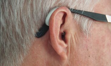
This isn’t like needle EMG or concentric needle studies used in hospitals. Surface EMG is entirely non-invasive. It’s safe for newborns and pregnant patients, and has been approved by regulatory bodies including Health Canada and the FDA.
More importantly, surface EMG gives you insight into function—not pathology. Unlike traditional diagnostics, it focuses on performance, not disease. In our world, that’s the more important question.
If you’ve ever wondered what is a surface EMG used for in chiropractic care—it’s for measuring the motor system’s output. Not in a gym, but in the adjusting area. And that changes everything.
How Surface EMG Works (Plain English Edition)
Let’s break this down. Every motor signal starts in the brain. That signal—called an action potential—travels down the spinal cord to activate a motor unit. That’s one nerve and all the muscle fibers it controls. Those muscle fibers contract and produce a tiny electrical current.
Now here’s where it gets interesting: that current travels up through the skin. And with surface electrodes placed along the spine, we can pick up that surface EMG signal. No poking or prodding. Just clean, measurable data.
But it’s not raw. The system filters out background noise, amplifies the true signal, and processes it using smart signal analysis techniques. It tracks things like:
- Amplitude: how strong the contraction is.
- Frequency: how often it’s firing.
- Symmetry: whether both sides are balanced.
Think of it like tuning a radio. The nervous system is always broadcasting. Surface EMG is the antenna that pulls in the station—without the static. And what you hear tells you a lot about how your patient is adapting to life.
Learn more about INSiGHT scanning?
Fill this out and we’ll get in touch!
"*" indicates required fields
Why Muscle Tone Tells the Truth
Muscle tone isn’t the same as tightness. It’s the constant low-level contraction happening even when we’re still. It’s regulated by the nervous system, and it reflects how much effort the body is putting out just to stay upright.
When tone is distorted—too high, too low, or asymmetrical—it means the nervous system is working inefficiently. That inefficiency has a name: dysponesis, a term used within chiropractic to describe the neurological mismanagement of energy.
Surface EMG helps you detect dysponesis long before it becomes a symptom. These subtle inefficiencies show up as red (overactive) or yellow (underactive) bars on the scan. Patients may feel “fine,” but research shows their system is running hot—and adapting poorly to stress.
Here’s the truth: you can’t palpate tone. You can’t see neurological fatigue. But with surface EMG, you can measure it—and that makes all the difference.
Understanding the Data – What You See in the Scan
The surface EMG scan gives you a full spine view of muscle tone, visualized in color-coded bar graphs. Each bar represents muscle activation at that level of the spine.
What do the colors mean?
- Red bars: Overactivation. The system is working too hard.
- Yellow bars: Underactivation. The system may be tired or inhibited.
- Blue/green bars: Less energy usage but still much more than optimal
These surface EMG recordings give you three core metrics:
- Pattern: Are certain areas consistently over- or underactive?
- Symmetry: Is the tone balanced left to right?
- Energy Index: How much energy is the system spending just to maintain posture?
All three are part of the neuroCORE scan, designed to track neurological efficiency.
This isn’t a test of strength. Studies confirm that surface EMG assesses postural tone—not voluntary muscular contraction. And that’s what makes it so powerful. You’re seeing what the nervous system is doing, not what the patient thinks they’re feeling.
Static vs. Dynamic EMG—Why INSiGHT Focuses on Static
There are different EMG techniques out there. Dynamic EMG is used in sports science to measure muscles during motion. But motion introduces variables—like signal noise and artifacts—that make it hard to track changes over time.
Chiropractic literature supports static surface EMG as the more reproducible, reliable option in clinical settings.
INSiGHT uses static surface EMG for a reason: it’s clean, consistent, and reproducible. It measures tone while the patient is standing still. That gives you a true baseline—and the ability to compare apples to apples from visit to visit.
What Surface EMG Tells Us About the Nervous System
Surface EMG gives you more than muscle data—it gives you a view into the motor division of the nervous system. Every bar on the scan tells you how that person’s system is adapting—or failing to adapt.
High-frequency signals may indicate the system is on high alert. Low signals may show neurological fatigue or inhibition. And when you look at the full pattern, researchers have shown you can begin to assess true adaptability.
This is the power of sEMG: it shows you how someone is functioning, not just whether they have a symptom. That’s the future of chiropractic care—and the cornerstone of performance-based care planning.
From Scan to Care Plan: How Chiropractors Use Surface EMG
Here’s how surface EMG fits into a neurologically-focused workflow:
- First Exam: Establish a baseline of tone and pattern.
- Care Planning: Identify which spinal regions are neurologically overburdened.
- Progress Exams: Use comparative scans to validate change and fine-tune care.
- Patient Education: Show how their nervous system is responding—visually, objectively.
In one study, sEMG was found to help guide adjustment focus and reinforce care decisions over time.
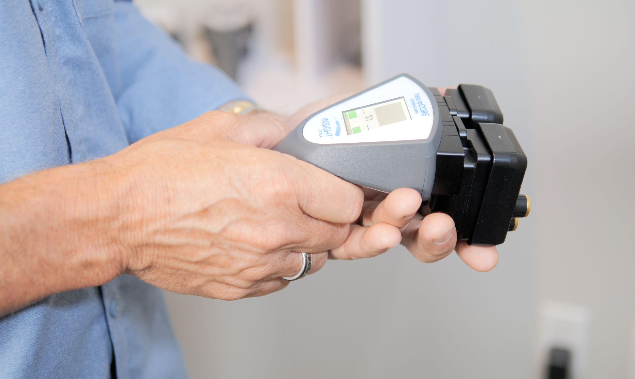
INSiGHT Technology and Surface EMG – Where It All Comes Together
INSiGHT CLA built its surface EMG instrument—neuroCORE—specifically for chiropractors. This isn’t lab equipment adapted for clinical use. It’s chiropractic scanning technology designed for daily, real-world practice.
The neuroCORE system is FDA-cleared, CE certified, and approved by Health Canada—making it one of the most trusted tools in vitalistic care.
It also contributes to the CORESCORE composite, which combines sEMG with HRV and neuroTHERMAL analysis for a full neurological picture.
Helping Patients Understand the Power of the Scan
Your patients don’t need to understand conduction velocity or electrode placement. They just need to see their scan—and understand what it means.
Here’s how to make that conversation stick:
- Show the scan: Red and yellow bars speak louder than words.
- Talk energy, not anatomy: “Your system is working too hard just to stay upright.”
- Celebrate wins: “Look how much less effort your nervous system is using now.”
Reading the Signals, Changing the Conversation
If you want to see how the nervous system is performing, you need a way to measure it. And that’s exactly what surface EMG does.
It shows how the nervous system is distributing energy. It highlights areas of inefficiency, exhaustion, and overcompensation. And it gives you objective proof that your care is making a difference.
With INSiGHT’s neuroCORE, you’re not just scanning muscles—you’re seeing adaptation. You’re turning tone into a measurable outcome. And that means you can build care plans with confidence, explain progress with clarity, and shift the conversation from pain relief to lifelong performance.
Because the nervous system doesn’t lie—and with surface EMG, neither do you.
Frequently Asked Questions About sEMG
What is a surface EMG?
A surface EMG, or surface electromyography, is a non-invasive technique used to measure the electrical activity of muscles through electrodes placed on the skin. It captures the EMG signal generated by motor unit action potentials during muscle contractions, allowing for an analysis of muscle function and neuromuscular performance.
How does the sEMG signal work?
The sEMG signal works by detecting the electrical impulses generated when a muscle contracts. Surface electrodes pick up the action potentials from muscle fibers, which are then processed to provide a representation of muscle activity. This signal can be influenced by factors like muscle fatigue, electrode placement, and the distance between electrodes.
What are the applications of surface electromyography?
Surface electromyography has various applications besides chiropractic including rehabilitation, sports science, and biomechanics. It is commonly used to assess muscle function, monitor neuromuscular disorders, and evaluate the effectiveness of therapeutic interventions. Additionally, it plays a role in ergonomics and human-computer interaction studies.
What is the difference between surface EMG and intramuscular EMG?
The primary difference between surface EMG and intramuscular EMG lies in the electrode placement. Surface EMG uses electrodes placed on the skin surface, while intramuscular EMG involves inserting needle electrodes directly into the muscle. Intramuscular EMG provides more localized readings and is less susceptible to noise, but it is also more invasive.
How does signal processing improve sEMG data analysis?
Signal processing techniques enhance the quality of sEMG data by filtering out noise and unwanted signals, enabling better interpretation of the electromyographic signal. Methods such as signal decomposition and median frequency analysis can help identify muscle fatigue and differentiate between muscle activation patterns, improving the reliability of the results.
What factors affect the quality of an EMG signal?
Several factors can affect the quality of an EMG signal, including electrode placement, the type of electrodes used, skin preparation, and the presence of motion artifacts. The spatial arrangement of closely spaced electrodes and the use of high-density surface electrodes can also enhance signal quality and provide more detailed information about muscle activity.