Here is the hard part. You can feel it with your hands, and you can see it in the way they move, but you still need a clear, objective way to show it. Symptoms can improve early, then fluctuate. A “better week” can create false certainty. That is exactly why a Surface EMG machine (more commonly referred to as an sEMG instrument) has become one of the most useful chiropractic tools for explaining what the body is doing now and verifying what is changing over time.
Surface EMG gives chiropractors reproducible data about muscle activity along the spine, especially the paraspinal muscles, without relying on guesswork. It is non-invasive, fast, and easy to repeat, which makes it ideal for baseline scanning, re-scans, and assessment of patient progress.
What a Surface EMG Instrument Measures
Electromyography is the measurement of electrical signals produced when motor nerves activate muscle fibers. Surface electromyography applies that concept with a clinical advantage: instead of inserting anything, the signal is captured from the skin above the muscle using surface electrodes.
That one difference changes how chiropractors use it. Because it is efficient and repeatable, sEMG is often used as a functional scan of neuromuscular output rather than a one-time diagnostic event. In plain language, it helps you estimate the amount of electrical activity being expressed by the motor system in the paraspinal region.
This is why Surface EMG fits chiropractic so well. When the body is adapting to neurological distress, muscle tone often shifts. Some regions begin firing too much. Other regions shut down and hand the workload to neighboring tissue. Those shifts may appear as muscle differentials around the spine, and the scan can highlight that distribution throughout the spine.
Why Chiropractors Use Surface EMG in Practice
Surface EMG is not about proving someone has a condition. It is about making function visible. When a patient’s symptoms fluctuate, your exam still needs a consistent anchor point. A Surface EMG instrument can provide that anchor, because it is focused on measurable output rather than subjective reporting.
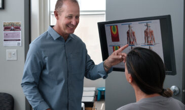
In many cases, the value is communication. The scan gives you a shared language for explaining why the body feels strained even when imaging looks “fine,” and why the patient may have health concerns even when they cannot clearly describe them.
In chiropractic, that becomes especially useful when you are discussing subluxation. Not as a simplistic “bone out of place” idea, but as a functional picture that can include altered segmental behavior, joint restriction, neurological interference, and changes in motor tone. When the conversation turns to vertebral subluxation, the goal is not to label every scan. The goal is to use objective scanning to support clinical interpretation.
This is also where patients commonly bring up x-ray. Imaging can show structure, but it does not show how the neuromuscular system is behaving today. Surface EMG can help bridge that gap, especially when postural compensation is obvious but hard to quantify.
Learn more about INSiGHT scanning?
Fill this out and we’ll get in touch!
"*" indicates required fields
The Anatomy of a Surface EMG Scan
A Surface EMG scan depends on consistency. If the setup is sloppy, the comparison over time becomes meaningless. If the setup is standardized, the scan becomes a strong objective measure of change.
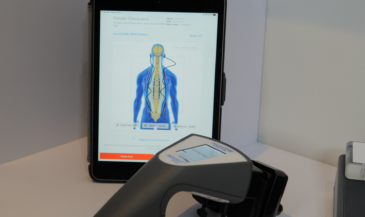
Skin preparation and placement
Start with skin preparation, because a clean surface improves signal quality. Then remember the key phrase patients understand easily: the sensors are placed on the skin. More specifically, an electrode is positioned at defined sites so the scan can compare left and right output along the spine.
This is where you will hear clinicians refer to electrodes placed in a consistent pattern. That matters because you are trying to measure the electrical activity in the muscle in a repeatable way. If placement shifts visit to visit, you are not tracking change. You are tracking inconsistency.
What the instrument captures
A Surface EMG instrument is capturing electrical activity of muscles as a reflection of motor unit recruitment. In simpler terms, it is capturing nerve firing in the muscles, expressed as electrical activity your muscles release. That is the entire point of sEMG in clinical practice: turning invisible effort into visible data.
For patients, it can help to say it this way: “We are looking at how the muscles around your spine are working to hold you up.” That wording keeps it accurate and understandable.
Interpreting sEMG in a Chiropractic Context
Let’s keep this grounded. Surface EMG provides detailed information about muscle activity, but it does not diagnose on its own. It is an electromyographic snapshot that must be integrated with the rest of your chiropractic exam.
When chiropractors interpret a Surface EMG scan, the most common clinical themes are symmetry, distribution, and relative output. You are looking for muscle differentials around the spine that suggest compensation or fatigue.
- High output: can reflect tight or contracted muscles, guarding, or workload concentration. In some cases, it aligns with spasm.
- Low output: may reflect inhibition, exhaustion, or poor recruitment strategy.
- Asymmetry: can suggest altered biomechanics, rotation, or imbalance in segmental control.
When the scan shows abnormal muscle firing, you are not obligated to “name a diagnosis.” What you can do is describe function: muscles are firing abnormally, abnormally distributed, or overloaded in specific regions. That makes your report more honest and more clinically useful.
In many chiropractic offices, this is where the subluxation discussion becomes more understandable for the patient. You can explain that subluxations often involve more than joints. They can involve how motor nerves are driving tone around specific segments. That is one reason sEMG is often used when chiropractors are evaluating vertebral subluxations and related functional stress.
Patients also search terms like spinal misalignments. You can acknowledge the search language, but keep your explanation clinical: the scan is showing motor control changes, not “bones out.” Still, when you connect those findings to what you see manually, the patient understands why your recommendations are not random.
How Surface EMG Supports Baselines, Re-Scans, and Progress Checks
The first scan is not the finish line. It is the starting point. The baseline scan becomes the reference you use for future comparisons, which is where sEMG becomes truly valuable in chiropractic.
When you re-scan, you are not just hoping the patient feels improved. You are checking whether the motor system is behaving more efficiently. That is why sEMG can be a powerful assessment of patient progress, even when symptoms change inconsistently.
In practical terms, you are using repeated Surface EMG scans to assess muscle function over time, verifying whether muscle activity is becoming more balanced and less exhausting. That is the kind of objective clarity that improves follow-through in chiropractic care.
It also makes the continuation conversation cleaner. Patients often feel better before the system is stable. With scanning, you can show whether the body is still firing too much in key regions, or whether distribution is improving.
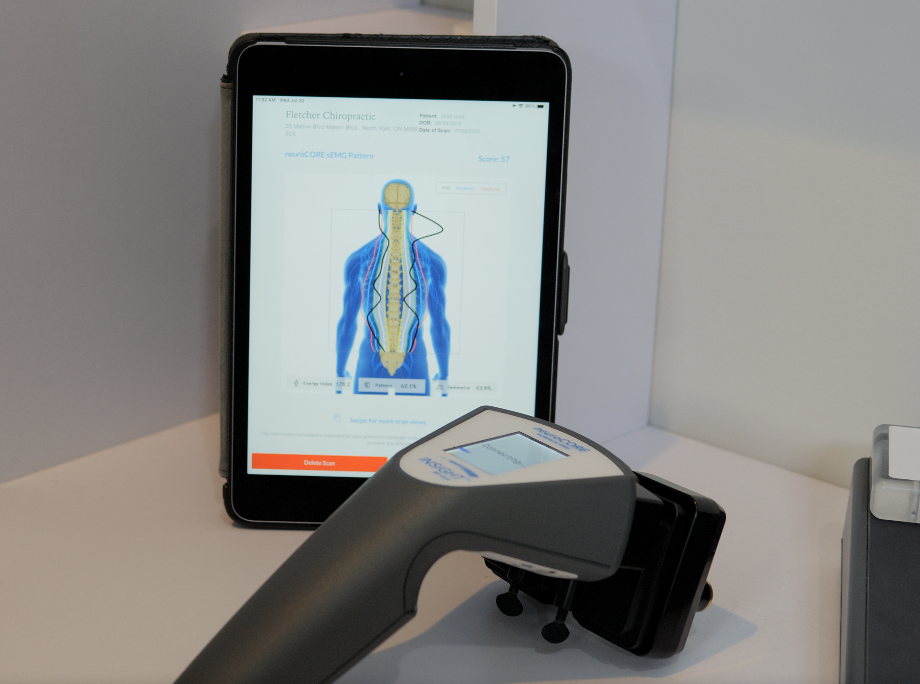
neuroCORE and the INSiGHT Approach to sEMG Scanning
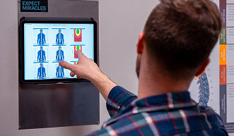
If Surface EMG is the motor lens, then neuroCORE is how that lens fits inside a bigger scan-led process. neuroCORE is our sEMG scanning technology within the INSiGHT ecosystem. It is designed to help chiropractors measure muscle differentials around the spine using objective Surface EMG data, then organize that information into reports that support clearer communication.
In neuroCORE workflows, the scan focuses on paraspinal output and the muscular component of function. It helps you see where motor output is concentrated, where it is asymmetrical, and where the system is spending excess energy to maintain posture.
This is where scanning technology becomes more than “a test.” It becomes a system for reporting, re-assessing, and building patient understanding without over-explaining. The INSiGHT provides objective exam data and scan reports. The chiropractor interprets that information, integrates it with the rest of the exam, and then designs the care plan.
When neuroCORE is combined with thermography and other objective scans in the same ecosystem, you can speak to function from multiple angles without drifting into speculation. That is how you keep your communication clear while staying clinically responsible.
Practical Patient Language That Keeps the Conversation Simple
Here are a few phrases that work well in a report of findings, especially when you want to explain Surface EMG without turning it into a lecture:
- “This scan shows the electrical activity in the muscle, which reflects how your motor nerves are driving tone.”
- “We are comparing the muscles around your spine side to side to see where your body is doing extra work.”
- “We will re-scan so we have an objective measure of change, not just a feeling-based guess.”
That is the heart of it. Surface EMG is most powerful when it stays in its lane: showing neuromuscular output, supporting interpretation, and reducing uncertainty.
Where This Lands for a Chiropractic Office
A Surface EMG instrument gives chiropractic teams a practical way to quantify what their hands are already detecting. It helps you show the amount of electrical activity across regions, identify workload concentration, and communicate why stability takes time. It also helps you explain why a patient may have symptoms one week and less the next, while the underlying strategy is still inefficient.
Used consistently, Surface EMG becomes one of the most useful chiropractic tools for building clarity. It supports communication about subluxation without oversimplifying it, and it strengthens your ability to track progress with objective data. That is the real promise of sEMG in a modern practice: less guessing, better explanation, and a clearer path forward.
When you integrate the neuroCORE into your practice, as part of the INSiGHT scanning technology, you are no longer relying on memory or subjective impressions alone. You are using repeatable scanning and clear reporting to guide decisions and verify what is changing throughout the spine.