That shift matters in today’s chiropractic because patients are overwhelmed with opinions and underwhelmed by vague answers. They want to know why you are recommending care, how you will track it, and what success actually looks like. Thermal scans give you an objective place to start. When you can show a temperature difference or asymmetry from one side of the spine to the other, you are no longer trying to “win” against symptoms that fluctuate. You are simply observing a measurable output and using it to guide your clinical thinking.
Used responsibly, thermal scans are a non-invasive, radiation-free way to evaluate paraspinal thermal trends, using infrared technology to assess skin temperature along the spine. They are often discussed under the umbrella of thermography, and they work best when they support a bigger neurological scanning workflow. The goal is not a dramatic reading. The goal is clarity, repeatability, and communication that leads to better follow-through.
What Thermal Scans Are and Why Chiropractors Use Them
In chiropractic, thermal scans are commonly used as a quick physiological check that compares temperature along the spine on the left and right sides. This is not structural imaging. It is functional insight. A thermal scan measures surface heat, then highlights temperature differences along comparable points so a chiropractor can observe whether regulation looks even or uneven in the paraspinal region.
This is where thermography is often explained in plain language. Every object emits infrared energy relative to its temperature, and a thermograph or thermogram can display that information as a simple visual map. In practice, many clinicians talk about thermal scans as a way to observe autonomic nervous system regulation in the tissues near spinal nerves, because changes in vascular tone can influence surface skin temperature. Chiropractors also use thermal scans to support conversations around subluxation concepts and neuro-spinal stress. When you hear phrases like nerve interference or neuro-toxicity, the intent in clinical practice is to describe altered neurological function rather than point to a pathology.
Thermal scans earn their place in chiropractic care for one reason: they are repeatable. A patient might report back pain one week and feel fine the next, then show up again describing “back problems” after travel, poor sleep, or a stressful stretch. Symptoms change. But thermal scans can be repeated with regular scans to observe whether the temperature along the spine is stabilizing over time and whether nerve interference is changing. That gives your conversations a steadier foundation and helps you talk about nervous system performance without turning every visit into a long discussion.
- A baseline you can re-check so changes are not guesswork
- A simple way to explain asymmetry without over-teaching neurology
- Objective scan measures that support a more inviting report conversation
- A fast, patient-friendly entry point into the spine and nervous system controls
How Chiropractic Thermal Scans Work in the Exam Room
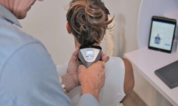
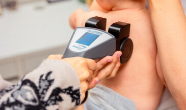
To use thermal scans well, it helps to keep the purpose simple. You are not trying to create a diagnosis from a picture. You are running an analysis that compares skin temperature side-to-side and segment-to-segment. That is why many chiropractors describe these scans as infrared thermal or digital infrared imaging. The technology uses infrared temperature detection to identify small differences that are not obvious to the naked eye. Most offices keep the process quick and consistent so the scan data is useful at follow-up.
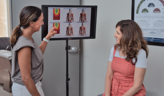
From a workflow standpoint, the scanning procedure is straightforward. The patient is positioned comfortably, the scanning environment is kept reasonably consistent, and the instrument is guided along the spine in a controlled way. Some offices describe this as a rolling scan, while others emphasize segmental regions, including the lumbar area. The goal is to capture a reliable left-right comparison and observe a temperature difference where the paraspinal thermal readings do not match. Many reports display results in color-coded bars or thermal images to highlight deviations and make the conversation easy.
This is also where patient communication gets easier. Thermal scans give you a shared visual reference point. You can show where the temperature difference appears, explain that you are observing regulation along the spine, and then move the conversation forward. Most patients do not need a lecture. They need a clear reason why you are recommending chiropractic adjustments and how you will measure change over time.
Learn more about INSiGHT scanning?
Fill this out and we’ll get in touch!
"*" indicates required fields
Interpreting Thermal Scan Findings What They Can Suggest and What They Cannot
This section is where thermal scans either become a powerful tool in your practice, or they become a confusing graphic nobody wants to explain. The key is to interpret what you actually measured. Thermal scans detect temperature differences that show up as side-to-side inconsistencies or segmental shifts. That is the core idea. A temperature difference is not a diagnosis. It is an observation. In chiropractic analysis, that observation is often discussed as a possible sign of altered autonomic regulation and vascular tone near the spine. It can also support a discussion of neuro-spinal stress without overstating what the scan can do.
In everyday chiropractic communication, you will hear two common explanations. A warmer region may be described as an area that can indicate inflammation or increased physiological activity. A cooler region may be described as reduced activity, altered circulation, or functional change that some clinicians associate with nerve interference. This is where wording matters. You can be clear, confident, and responsible at the same time. You can say, “This is showing uneven regulation,” and then connect it to function and the exam findings. You can also acknowledge the limits: thermography and infrared thermographic approaches can highlight differences, but they should be interpreted in context. That is how you stay aligned with scientific evidence and keep your reporting credible.
Thermal scans also fit into the profession’s longer conversation about subluxations and the body of evidence exploring functional change along the spine. You might reference vertebral subluxation research as part of chiropractic research history, but the scan itself is not declaring a condition. It is providing objective scan data that you, as the clinician, interpret alongside orthopedic, neurologic, and postural findings. That is why many offices find thermal scans helpful even when symptoms improve early. The scan can help a patient understand that feeling better is not always the same as stability, especially when the nervous system’s outputs are still inconsistent.
Used this way, thermal scans become less about “finding a problem” and more about tracking progress responsibly. You are observing temperature differences along the spine as a trend, not as a verdict. That is a healthier mindset for the doctor and the patient, and it tends to lead to better results in follow-through.
How Thermal Scans Fit Into a Modern Neurological Scanning Workflow
Thermal scans are strongest when they are not asked to carry the entire story. They are one window into regulation. A complete scan-led workflow gives you more than one window, which improves clarity for the patient and certainty for the clinician. This is one of the defining shifts in modern chiropractic: moving from symptom-chasing to objective neurological scans that reflect how the nervous system is functioning. Thermal scans contribute by showing a temperature difference along the spine, but they become more meaningful when you can correlate them with other objective findings.
This is where the three-scan approach has become so practical in clinical practice. When you pair thermal scans with HRV and surface EMG, you can tell a clearer story without drowning the patient in numbers. Thermal scanning reflects paraspinal thermal regulation. sEMG reflects neuromuscular output and symmetry, often helpful when postural tension is obvious. HRV helps you speak to adaptability and the autonomic nervous system balance across the whole system. When all three line up, the recommendation becomes easier to communicate and more defensible. It also helps you explain neck pain or recurring symptoms without turning the report into a guessing game.
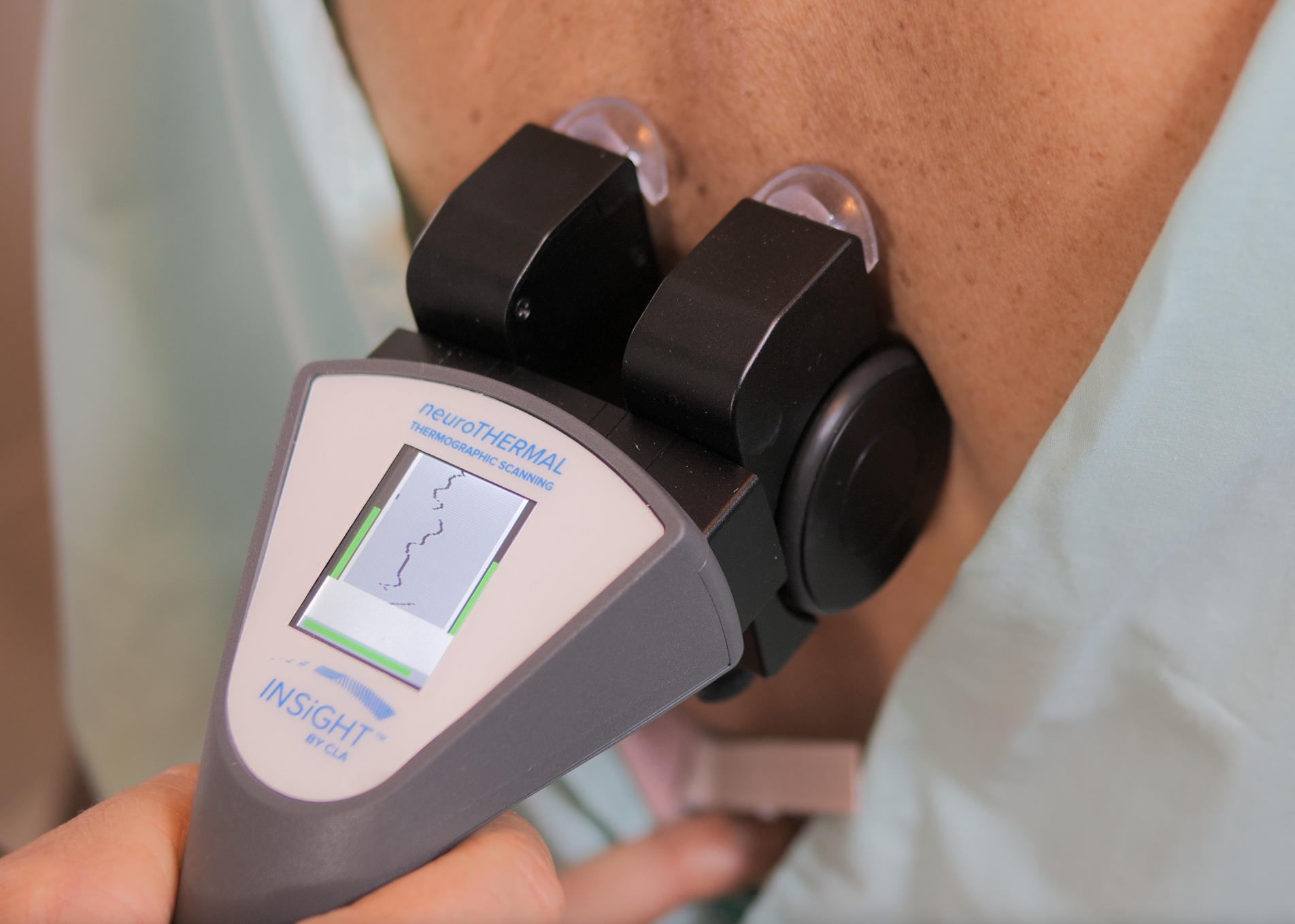
INSiGHT scanning and CLA’s products fit naturally into this workflow because the tools are designed to provide objective reports that are simple to communicate. The INSiGHT neuroTHERMAL focuses on thermal scan trends and left-right temperature comparison along the spine. The INSiGHT neuroCORE sEMG adds a view of neuromuscular function and symmetry. The INSiGHT neuroPULSE HRV adds adaptability and resilience, which helps you explain how well your nervous system is responding to neurological distress. Synapse software brings these scan views together so the patient can understand what they are seeing without a lecture in neurology. Just as important, the boundary stays clear: INSiGHT technology generates the scan measures and reports, and the chiropractor can use those findings, along with the full exam, to recommend a care plan.
Using Thermal Scans to Improve Patient Understanding, Retention, and Long-Term Follow-Through
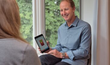
Most patients are silently asking the same three questions, even if they never say them out loud. What does this mean for me? How do we know it’s changing? What happens if I stop care? When you rely only on symptoms, those questions force you into long explanations because symptoms fluctuate. Thermal scans make those conversations simpler because they provide a shared reference point that is not based on opinion. You can show what the scan measures, explain that you are tracking regulation along the spine, and connect it to function in a calm, professional way. That alone can help your chiropractor sound more confident and reduce the pressure to over-explain.
Retention improves when the patient understands what you are stabilizing. Regular scans give you a repeatable way to show improvement, even when symptoms shift. That does not mean you chase perfect readings. It means you track trends. If temperature differences along the spine are reducing and symmetry is improving, you can show that. If the data remains inconsistent, you can explain that early improvement does not always mean the system has stabilized. This is a respectful way to answer the “what happens if I stop” question without fear tactics. It is also one reason many offices choose to use thermal scans consistently, because they support better care and clearer communication over time.
- Start with a baseline and explain what the scan is showing in one sentence
- Re-scan at meaningful milestones so the patient sees change, not just feels it
- Keep the conversation focused on stability and nervous system performance, not perfection
- Use thermography as part of a bigger exam story rather than a standalone claim
A Final Word on Using Thermal Scans with Confidence
Thermal scans are not about flashy technology. They are about clarity. They give you a fast, repeatable way to observe temperature along the spine, highlight temperature differences along the left and right sides, and bring the nervous system conversation into focus. They can aid in spinal decision-making when used responsibly, and they can help your chiropractor communicate why care continues past early symptom changes. They also help you stay grounded in what is objective, because the nervous system is functioning whether the patient can describe it well or not.
If you want to use thermal scans well, keep them in their proper role. Use thermography as physiological insight, not a diagnosis. Respect what the medical literature says about investigational uses and the limits of standalone thermographic imaging. Then do what chiropractors do best: interpret the findings in context, connect them to function, and track change over time with regular scans. When you bring thermal scans into the full INSiGHT scanning workflow, neuroTHERMAL plus neuroCORE sEMG plus neuroPULSE HRV, you give patients a clear, objective story about stability in the spine and nervous system. That is the kind of communication that helps patients commit, helps teams report with confidence, and helps practices lead with certainty.
And if you are wondering why this matters so much, it comes back to one simple idea. The best practices do not guess. They measure, they re-check, and they communicate what they see. Thermal scans help you do that, and when they are paired with INSiGHT scan reports and Synapse visuals, they become one of the simplest ways to show progress in a way patients can trust.