In chiropractic, that matters because many patients arrive thinking their situation is purely structural. They want to talk about joints, popping, and where it hurts. Infrared imaging is often the moment the conversation shifts toward regulation, adaptation, and nervous system function. It is a scan that helps you talk about what is happening at the surface, what may be changing underneath, and why consistency matters even when symptoms are inconsistent.
Used responsibly, infrared imaging becomes a practical way to establish a baseline, track subtle changes, and help patients visualize progress without turning every visit into a long explanation. That is why thermography continues to show up in practices that value clarity, consistency, and neurological thinking.
Why Infrared Imaging Belongs in a Neurologically Focused Chiropractic Conversation
In the early days of chiropractic, doctors relied on observation, palpation, and pattern recognition long before modern technologies were available. We still do. The difference now is that neurological scanning gives you a way to support those observations with objective information that is easy to explain. Infrared imaging fits beautifully in that world because it helps you focus the conversation on function, not just symptoms.
Here is the clinical mindset that keeps thermography useful. Thermography is not a diagnosis. It is a way to assess surface temperature and compare left-to-right symmetry, segment to segment, and visit to visit. The nervous system influences vascular tone and blood flow, so shifts in regulation can show up as changes in skin temperature. When you see a consistent temperature difference, it does not prove a condition, but it can point you toward areas of concern that deserve attention and a better exam.
This is where chiropractors use infrared imaging well. They use it as a steady, repeatable scan that supports chiropractic analysis and helps guide decisions around when care is needed, when the body looks stable, and when the care plan should be refined. You are not chasing a single reading. You are watching temperature patterns over time, looking for symmetrical regulation, and checking whether the system appears to be settling or still adapting.
- It helps you detect asymmetries that may indicate nerve tension and local physiological stress.
- It supports early detection of subtle changes before the patient can clearly describe them.
- It provides a simple way to pinpoint where you want to focus your exam along the spine.
- It gives the patient a visual anchor that makes follow through easier.
When you stay inside these boundaries, you protect your credibility. Thermography does not replace x-ray when structural imaging is clinically indicated. It does not diagnose inflammation, vascular disease, or nerve compression. What it can do is help you detect trends that may indicate nerve interference, then correlate those findings with history, exam, and your clinical reasoning.
What Infrared Imaging and Thermography Are
Thermography is a non-invasive way to analyze temperature readings on the surface of the body, most often along the spinal regions. In chiropractic language, the key word is symmetry. Skin temperature should be relatively symmetrical from left to right. When it is not, that difference can be meaningful, especially when it repeats and forms a consistent thermal pattern.
Infrared thermography is the method most commonly used in chiropractic. Using an infrared camera, the instrument detects infrared energy that the body can emit as heat. That information is displayed as thermal images, often called a thermogram. Modern systems use digital infrared capture, which simply means the scan is collected quickly, stored in software, and compared across visits. When you hear digital infrared imaging, think of it as imaging technology built for repeatability, documentation, and comparison, not a one-time snapshot.
From a practical chiropractic standpoint, the value of infrared imaging is not in the colors on the screen. The value is in the comparison. You are comparing left to right. You are comparing one spinal region to the next. You are comparing today’s scan to the baseline. That is how you turn information into clinical direction without overclaiming.
When asymmetry shows up, it may align with local inflammation, circulation issues, or a regulatory shift that involves the autonomic system. It can also align with clinical findings that suggest nerve dysfunction. Again, the scan does not diagnose the cause. It helps you identify patterns and decide what to do next with greater confidence.
Learn more about INSiGHT scanning?
Fill this out and we’ll get in touch!
"*" indicates required fields
How Thermography Scanning Is Performed in Chiropractic Settings
Thermography is simple when it is done consistently, but it is not casual. If you want infrared imaging to be meaningful, you have to respect what influences surface temperature. That is why many chiropractic offices coach patients through a few basic steps before a scan. Things like vigorous exercise, caffeine, and extreme heat or cold exposure can affect temperature measurement at the skin level. A controlled environment helps you reduce those variables so your scan is comparable over time.
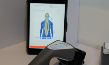
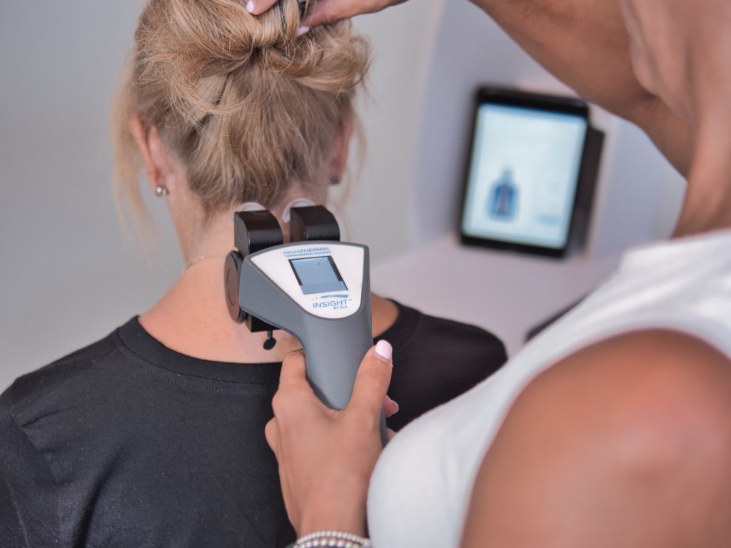
The scan process itself is straightforward. The patient is positioned, the room is kept stable, and a thermal camera captures the surface temperature along the spine or targeted regions. The result is a thermogram that displays temperature patterns, allowing you to compare temperature readings from side to side and segment to segment. The clinical goal is not to chase perfect numbers. The clinical goal is to look for symmetrical patterns and stable regulation.
Many chiropractors prefer to scan before an adjustment because care can temporarily change local circulation and surface temperature. Scanning first gives you cleaner baseline information, especially when you are using thermography to monitor stability. Some practices scan every visit, whether an adjustment is needed or not, because the scan becomes a consistent check on whether the body is holding or drifting. That repeated scan sequence is what makes subtle changes visible and supports better decisions across the care plan.
After the scan, interpretation should stay grounded. You are looking for asymmetries and anomalies that stand out from the patient’s baseline. Those findings can help you detect possible inflammation trends, identify areas of concern, and refine your exam. Used this way, thermography offers a practical bridge between what the patient feels and what you observe objectively.
Clinical Use Cases Chiropractors Actually Rely On
In real chiropractic practice, infrared imaging earns its place when it makes decisions and communication easier. The most common use case is simple: establish a baseline, then compare future scans to that baseline. When you establish a baseline, you stop guessing what “normal” should look like for that patient and start tracking their own trend line. That one step alone helps you keep the conversation steady when symptoms fluctuate.
Infrared imaging also supports early detection in a practical, chiropractic sense. Patients often cannot describe what is changing until it becomes obvious. A re-scan can help you detect subtle changes in temperature patterns that might otherwise be missed. When those patterns show up, they may indicate inflammation, may indicate nerve dysfunction, or may indicate a regulatory shift that deserves a closer look. The key is that you use the scan to guide assessment, not to replace it. That is what keeps your language honest and your clinical thinking sharp.
Another common use is helping the patient visualize progress. When a patient sees their scan views over time, the conversation becomes less emotional and more objective. Instead of debating how they feel on a single day, you can talk about whether the scan is becoming more symmetrical and stable. This is especially common in upper cervical models where stability and holding patterns are emphasized, but it applies across the spine in general practice as well.
Finally, infrared imaging can help you pinpoint where to focus when a patient cannot be specific. Some patients can only gesture at a broad area. A scan that highlights asymmetry in a paraspinal region can help you narrow your attention and refine your exam. It can also support decisions around whether additional imaging, including x-ray, is warranted. This keeps the scan in its proper role: a helpful guide that supports chiropractic analysis and better decision-making.
- Progress tracking that is easy to explain to patients
- Support for identifying patterns that repeat over time
- Objective comparison that makes care conversations calmer
- Practical guidance for where to assess more carefully
When the scan shows asymmetry, you can responsibly say it may indicate nerve interference. You can also note when a misalignment or spinal misalignments are part of your examination findings and the scan appears consistent with regulatory asymmetry. That does not prove a subluxation, but it can support a thoughtful discussion about subluxations, postural compensation, and musculoskeletal dysfunction without slipping into exaggerated claims. In many practices, this is exactly how thermography in chiropractic care is positioned: a stability and trend tool that helps you guide decisions and refine the care plan rather than guess.
How Infrared Imaging Fits Into the INSiGHT CLA Neurological Scanning Framework
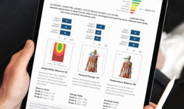
Infrared imaging works best when it is part of a bigger neurological picture. Thermography gives you insight into surface regulation and symmetry, but you still want objective information about how the nervous system is adapting and how the muscles are expressing stress. This is where INSiGHT scanning technology fits naturally into a scan-led workflow. The INSiGHT does not create a care plan. It provides objective exam data and reports that help the chiropractor interpret what is happening and make clearer decisions.
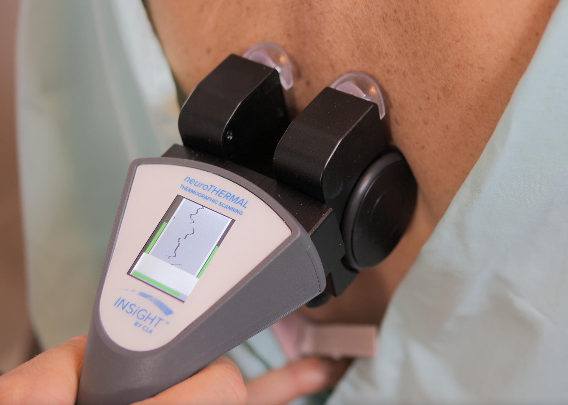
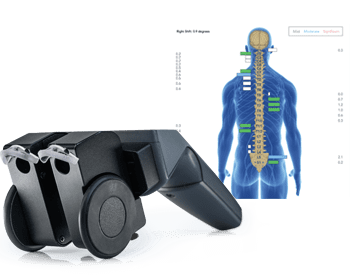
Within that framework, the neuroTHERMAL supports segmental thermography scanning so you can compare trends with consistency and clarity. It helps you detect changes in thermal patterns and track whether the scan is stabilizing over time. That thermography layer pairs well with neuroCORE sEMG, because muscle activity and motor tone often reveal how the body is compensating. Surface electromyography in chiropractic can help you map paraspinal electrical activity, observe imbalance, and monitor change as care progresses. Then neuroPULSE HRV supports the autonomic conversation, giving you an objective look at adaptability and regulation that complements what you observe through thermography.
When these tools are used together, the narrative becomes simple for both doctor and patient. Thermography shows you surface temperature regulation. sEMG helps you observe neuromuscular expression and postural stress. HRV helps you observe autonomic balance and adaptability. You are not relying on a single data point. You are building a clinical story supported by objective scanning and repeatable reporting. That makes your communication cleaner, your documentation stronger, and your care plan decisions more confident.
- Use thermal scanning to track whether regulation becomes more symmetrical over time
- Use sEMG to observe neuromuscular imbalance and compensation trends
- Use HRV to support the autonomic conversation and overall neurological regulation
- Re-scan consistently to make progress visible and objective
In a practical office flow, you do not need to overcomplicate this. Using an infrared scan as part of a broader neurological exam gives you a baseline, a way to compare changes, and a way to communicate without pressure. That is the INSiGHT CLA approach at its best: objective data that supports better conversations, better follow through, and better clinical certainty.
A Clear, Responsible Next Step for the Chiropractor
Infrared imaging is most valuable when you treat it as a trend tool, not a verdict. Thermography helps you observe surface temperature, identify patterns, and compare changes over time. It can help you detect asymmetry, detect inflammation trends, and decide where you want to assess more carefully. It can also help the patient visualize what you are tracking, which often improves understanding and consistency.
The professional win is not simply adding thermography to your office. The win is using infrared imaging to support clearer decisions, cleaner communication, and a more confident care plan. That happens when you respect what the scan can do, avoid overclaiming what it cannot, and integrate it into neurological thinking. When you anchor thermography inside INSiGHT scanning technology, the scan becomes part of a complete, objective story. You are no longer guessing. You are comparing. You are documenting. And you are guiding patients with data they can understand.
That is why infrared imaging continues to matter in chiropractic. It helps you stay grounded, stay consistent, and keep the focus on progress, stability, and nervous system function, one scan at a time.
Keyword placement notes for completeness: This article referenced digital infrared, digital infrared imaging, infrared thermography, thermography in chiropractic care, thermography scanning, infrared camera, thermal camera, thermal images, thermogram, temperature difference, temperature readings, temperature measurement, temperature patterns, thermal pattern, surface temperature, skin temperature, blood flow, vascular, physiological, autonomic, assess, detect, detect inflammation, indicate inflammation, pinpoint, paraspinal, spinal, spine, vertebral, postural, musculoskeletal, nerve, nerve dysfunction, nerve compression, dysfunction, misalignment, spinal misalignments, subluxation, subluxations, non-invasive, noninvasive, chiropractic care, chiropractor, neurological, care plan, treatment plan, use thermal, using an infrared, imaging technology, x-ray, overall health, subtle changes, areas of concern.