By Dr. Christopher Kent
Videofluoroscopy has a distinguished track record in clinical research and practice. Its judicious use in chiropractic practice may be valuable in detecting and characterizing spinal kinesiopathology associated with the vertebral subluxation complex.
The first known fluoroscopic image was produced by Roentgen in 1895. Roentgen placed his hand between an x-ray source and a fluorescent screen, and was astonished to see an image of the bones of his hand on the screen. One year later fluoroscopic screens became available, and the technique was employed for “real time” observation of human structures.[1]
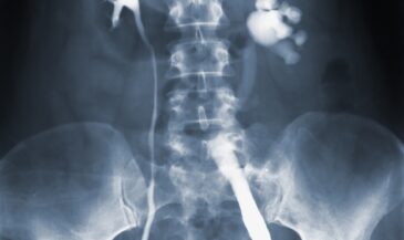
A videofluoroscopic system consists of an x-ray generator capable of operating at low (1/4 to 5) milliamperage settings, an x-ray tube assembly, an image intensifier tube, a television camera, a VCR, and a monitor. The heart of the system is the image intensifier tube. This tube permits imaging at very low radiation levels. It is used instead of intensifying screens and film as an image receptor.
Clinical applications
The role of videofluoroscopy in the evaluation of abnormalities of spinal motion has been discussed in textbooks, medical journals, and chiropractic publications. Observational and case cases have appeared in the literature comparing the diagnostic yield of fluoroscopic studies vs. plain films. In addition, studies have been published reporting abnormalities detected by fluoroscopy which could not be assessed using plain films.
Schaff described cases where instability of the upper cervical spine was appreciated on videofluoroscopic studies. It was observed that all cases of upper cervical instability are not revealed by static flexion-extension studies. The role of videofluoroscopy in assessing the upper cervical spine in Down’s syndrome patients competing in Special Olympics events was discussed.[2]
Wood and Wagner reviewed the use of radiographic methods for the analysis of cervical sagittal motion. They reported that videofluoroscopic studies may reveal kinematic irregularities not detectable by examining the extremes of range of motion alone.[3]
Wallace et al studied the reliability of certain methods of fluoroscopic measurements, reporting that independent examiners could replicate the measurements reliably.[4]
Van Mameren et al used fluoroscopy to determine the variability of instantaneous centers of rotation in the cervical spine. These investigators concluded that their procedure “shows variability of such low extent that it seems feasible to use it to diagnose abnormal mobility or in assessing therapy in the neck region.”[5]
Bland states, “Clearly, cineradiography is the best method for the study of biomechanics and dynamics of motion in the cervical spine…The determination of normal motion, sites of greatest and least motion, contribution by joints, discs, ligaments, tendons, and muscles to motion (and their limitations), and the biomechanics of normal motion of the occiput-atlas-axis complex all have been studied very successfully through cineradiography.”[6]
According to Ochs, “Cineradiography, using film or videotape, is shown in a study of 34 painful or injured necks to be a valuable diagnostic tool. It is useful in fracture management, diagnosis of instability and demonstration of solid healing. A video tape system featuring instant replay, clear image and low radiation exposure was found to be ideal for routine use.”[7]
Buonocare, Hartman, and Nelson examined the cervical spines of 107 patients using cineradiography, including 57 who sustained flexion-extension injuries. They concluded, “The ability to demonstrate localized abnormal motion in the cervical spine allows one to predict soft-tissue injuries and the quality of spinal fusions, spinal stability, and early subluxation of the cervical spine–conditions that may not be identified on static roentgenograms nor at physical examination.”[8]
Jones studied abnormalities of the upper cervical spine using cineradiography, and concluded, “Cineradiography has been used to detect instability not ascertainable by routine roentgenograms obtained in flexion and extension…”[9]
In a case study of abnormal atlanto-axial motion, Tasharski noted, “Interpretation by means of standard static radiographs failed to disclose the nature of the functional post-traumatic disorder. Cinefluorographic visualization of the articulation in motion demonstrated abnormal mobility.”[10]
Woesner and Mitts also concluded that fluoroscopic studies often revealed abnormalities undetected on plain films. They stated, “There were, however, a significant number of instances in which cineroentgenography demonstrated abnormal motion not detected on conventional roentgenograms.[11]
Cineroentgenography is, therefore, a valuable adjunctive technique and its continued utilization in the analysis of cervical spine motion is justified.”
Numerous applications for spinal fluoroscopy have been reported in the medical literature. These include recording the effects of cervical spine traction, evaluating cervical spine laminectomies, examining athletes presenting with pain, to assist in surgical planning, evaluating atlanto-axial rotatory fixation, examining the effects of cervical collars, characterizing joint disorders in the cervical spine, studying degenerative disease of the cervical spine, and determining the effects of occipitalization and odontoid hypoplasia on spinal motion.[12-20]
In addition to the studies cited, applications for fluoroscopy in chiropractic have been reported in chiropractic trade publications, indexed peer reviewed literature, and presented at chiropractic symposia.
Gillet, Henderson and Dorman, and Howe used fluoroscopy to study cervical spine kinetics.[21-24] Shippel and Robinson described a case where fluoroscopy and magnetic resonance imaging were used to evaluate cervical spine instability.[25] Leung used fluoroscopy to evaluate the cervical spine and concluded, “Cineradiography has been found to be the method of examination that conveys most functional abnormalities. The diagnostic value of cineradiography is substantiated…The effect of chiropractic adjustment in removal of cervical fixations was proven with cineradiography.”[26]
Chiropractors Foreman and Croft in their textbook, “Whiplash Injuries,” state, “This motion study of the spine may be quite useful in detecting abnormal biomechanics secondary to ligamentous damage that may be unappreciated with plain film radiography… Cineradiography or fluorovideo radiography plays an important role in the diagnosis of aberrant spinal biomechanics that may be secondary to chronic muscle contracture, scar tissue formation, or ligamentous instability.”[27]
Antos, Robinson, Keating and Jacobs presented the results of an interexaminer reliability study of cinefluoroscopic detection of fixation in the mid-cervical spine. Two examiners reviewed 50 videotapes of fluoroscopic examinations of the cervical spine. The examiners achieved 84% agreement for the presence of fixation, 96% agreement for the absence of fixation, and 93% total agreement. The Kappa value was .80 (p<.0001). Only the C4/C5 level was examined. The authors concluded, “The current data indicate that VF determination of fixation in the cervical spine is a reliable procedure.”[28]
Other chiropractic authors have described applications for fluoroscopy.
Taylor and Skippings used the procedure to study paradoxical motion of the atlas in flexion.[29] Betge described applications for fluoroscopy in the diagnosis of dysfunctions of the cervical spine.[30] Masters, and Mertz both used fluoroscopy to evaluate spinal motion.[31,32] Robinson, and Sweat have also published articles concerning chiropractic applications for fluoroscopy.[33,34]
In addition to diagnostic studies, fluoroscopy has been used to study normal motion in the spine.
Bronfort and Jochumsen used cineradiography to evaluate intermediate stages and extremes of intervertebral motion in the lumbar spine.[35] Fielding, and Howe described normal motion of the cervical spine based on cineradiographic examinations.[36,37]
Few technologies in chiropractic enjoy the literature support of videofluoroscopy. Unfortunately, it is currently under-utilized. Doctors of chiropractic should consider exploring the potential of this technology in the assessment of subluxation induced pathomechanics.
References
1. Glasser O: “Dr. W.C. Roentgen.” Springfield, IL, Charles C. Thomas, 1945.
2. Shaff AM: “Video fluoroscopy as a method of detecting occipitoatlantal instability in Down’s syndrome for Special Olympics.” Chiropractic Sports Medicine 8(4):144, 1994.
3. Wood J, Wagner N: “A review of methods for radiographic analysis of cervical sagittal motion.” Chiropractic Technique 4(3):83, 1992.
4. Wallace H, Wagnon R, Pierce W: “Inter-examiner reliability using videofluoroscope to measure cervical spine kinematics: a sagittal plane (lateral view).” Proceedings of the International Conference on Spinal Manipulation May 1992, pages 7-8.
5. Van Mameren H, Sanches H, Beursgens J, Drukker J: “Cervical spine motion in the sagittal plane II.” Spine 17(5):467, 1992.
6. Bland JH: “Disorders of the Cervical Spine.” Philadelphia, PA, W.B. Saunders Co. 1987. P. 144.
7. Ochs CW: “Radiographic examination of the cervical spine in motion.” US Navy Med 64:21, 1974.
8. Buonocare E, Hartman JT, Nelson CL: “Cineradiograms of cervical spine in diagnosis of soft-tissue injuries.” JAMA 198(1):143, 1966.
9. Jones MD: “Cineradiographic studies of abnormalities of high cervical spine.” AMA Arch Surg 94:206, 1967.
10. Tasharski CC: “Dynamic atlanto-axial aberration: a case study and cinefluorographic approach to diagnosis.” JMPT 4(2):65, 1981.
11. Woesner ME, Mitts MG: “The evaluation of cervical spine motion below C-2: a comparison of cineroentgenographic methods.” Am J Roent Rad Ther & Nuc Med 115(1):148, 1972.
12. Bard G, Jones MD: “Cineradiographic recording of traction of the cervical spine.” Arch Phys Med 45:403, 1964.
13. Bard G, Jones MD: “Cineradiographic analysis of laminectomy in cervical spine.” AMA Arch Surg 97:672, 1968.
14. Becker E Griffiths HJ: “Radiologic diagnosis of pain in the athlete.” Clin in Sports Med 6(4):699, 1987.
15. Brunton FJ, Wilkerson JA, Wise KS, Simonis RB: “Cine radiography in cervical spondylosis as a means of determining the level for anterior fusion.” J Bone and Joint Surg 64-B(4):399, 1982.
16. Fielding JW, Hawkins RJ: “Atlanto-axial rotatory fixation.” J Bone and Joint Surg 59-A(1):37, 1977.
17. Jones MD: “Cineradiographic studies of collar immobilized cervical spine.” J Neurosurg 17:633, 1960.
18. Jones MD: “Cineradiographic studies of various joint diseases in the cervical spine.” Arthritis & Rheumatism 4:422, 1961.
19. Jones MD: “Cineradiographic studies of degenerative disease of the cervical spine.” J Canad Assoc Radiol 12:52, 1961.
20. Jones MD, Stone BS, Bard G: “Occipitalization of atlas with hypoplastic odontoid process, a cineroentgenographic study.” Calif Med 104:309, 1966.
21. Gillet H: “A cineradiographic study of the kinetic relationship between the cervical vertebrae.” Bull Eur Chiro Union 28(3):44, 1980.
22. Henderson DJ, Dormon TM: “Functional roentgenometric evaluation spine in the saggital plane.” JMPT 8(4):219, 1985.
23. Henderson DJ: “Kinetic roentgenographic analysis of the cervical spine in the saggital plane: a preliminary study.” Int Review of Chiro 35:2, 1981.
24. Howe JW: “Observations from cineroentgenological studies of the spinal column.” ACA J of Chiro 7(10):65, 1970.
25. Shippel AH, Robinson GK: “Radiological and magnetic resonance imaging of cervical spine instability: A case report.” JMPT 10(6):316, 1987.
26. Leung ST: “The value of cineradiographic motion studies in diagnosis of dysfunctions of the cervical spine.” Bull Eur Chiro Union 25(2):28, 1977.
27. Foreman SM, Croft AC: “Whiplash Injuries: The Cervical Acceleration/Deceleration Syndrome.” Baltimore, MD, Williams and Wilkins, 1988. P. 114, 133.
28. Antos J, Robinson GK, Keating JC, Jacobs GE: “Interexaminer reliability of cinefluoroscopic detection of fixation in the mid-cervical spine.” Proceedings of the Scientific Symposium on Spinal Biomechanics, International Chiropractors Association, 1989. P. 41.
29. Taylor M, Skippings R: “Paradoxical motion of atlas in flexion: a fluoroscopic study of chiropractic patients.” Euro J Chiro 35:116, 1987.
30. Betge G: “The value of cineradiographic motion studies in the diagnosis of dysfunction of the cervical spine.” J Clin Chiro 2(6):40, 1979.
31. Masters B: “A cineradiographic study of the kinetic relationship between the cervical vertebrae.” Bull Eur Chiro Union 28(1):11, 1980.
32. Mertz JA: “Videofluoroscopy of the cervical and lumbar spine.” ACA J of Chiro 18(8):74, 1981.
33. Robinson GK: “Interpretation of videofluoroscopic joint motion studies in the cervical spine C-2 to C-7.” The Verdict February 1988.
34. Sweat RW: “C-Arm Cinefluorography.” Today’s Chiropractic 13(4):31, 1984.
35. Bronfort G, Jochumson OH: “The functional radiographic examination of patients with low back pain.” JMPT 7(2):89, 1984.
36. Fielding JW: “Normal and selected abnormal motion of cervical spine from second cervical vertebra based on cineroentgenography.” J Bone and Joint Surg 46-A:1779, 1964.
37. Howe JW: “Cineradiographic evaluation of normal and abnormal cervical spinal function.” J of Clinical Chiro 2:76, 1972.


