In a medical setting, a spine scan usually means structural imaging. In a chiropractic setting, especially a neurologically focused chiropractic office, a spine scan can mean something broader and more meaningful. It can include a scan of structure when imaging is used, but it can also include objective analysis of nerve function, postural tension, autonomic regulation, and the impact of stress on the body. Chiropractors know that the spine and nervous system work together, and many of the most important changes in nervous system function cannot be seen on x-ray.
That is why this topic matters. A patient may have a dramatic report from MRI, CT scans, or x-ray technology and still not understand why they do not feel resilient. Another may have very little structural change and yet show obvious stress on your nervous system, altered adaptability, and clear signs of nerve interference along the spine. A useful spine scan in chiropractic is not just about what the spine looks like. It is about what the spine and nervous system are doing.
What Most People Mean When They Ask for a Spine Scan
In conventional healthcare, the term spine scan usually refers to imaging of the cervical, thoracic, or lumbar spine. That imaging is used to look at anatomy. MRI is often chosen when the goal is to see soft tissue, discs, nerves, or the spinal cord. CT scan is often chosen when the goal is to evaluate bone detail, trauma, fractures, or other urgent structural findings. In some cases, x-ray is still used to review alignment, gross structure, and certain mechanical changes in the spinal vertebrae.
MRI and CT scans have an important place. MRI gives very clear images of soft tissue and does not use radiation. CT uses x-ray technology and computerize processing to build cross-sectional views of the body. These scans can help identify fractures, infections, tumors, disc problems, stenosis, or major structural compromise. That is why a spine scan can be highly valuable when the clinical question is structural.
Patients also associate a spine scan with the experience of the test itself. They think about the scanner, the table, the noise, the contrast dye, and the wait for an answer. MRI can take longer and may be uncomfortable for someone who feels confined. CT is faster and often described as painless. But even when the scan goes well, structural imaging still has limits. A picture can show anatomy clearly while saying very little about adaptation, regulation, or how the body is handling stress.
- MRI is often best for soft tissue, discs, nerves, and the spinal cord.
- CT scan is often best for bone detail, fractures, and rapid structural assessment.
- X-ray can help evaluate structure and alignment but gives a limited view of function.
- Imaging is valuable when the question is structural, but it does not fully explain why a patient is not adapting well.
When Imaging Matters and When It Does Not Tell the Whole Story
Every responsible chiropractor should say this clearly: there are times when imaging matters a great deal. If a patient presents with severe trauma, suspected fracture, infection, tumor, progressive weakness, or serious neurological red flags, imaging is used for a reason. MRI, x-ray, or CT scans may be the right next step. Recognizing those cases is part of sound chiropractic education and good clinical judgment.
At the same time, it is very common for patients to ask for a spine scan when a scan is not the first thing they need. In many straightforward spinal complaints, a careful history and examination are more valuable than rushing into imaging. That is not dismissing the patient. It is being accurate. A structural image does not always explain why a spine is symptomatic, why tension keeps returning, or why the body is showing the effects of stress and anxiety in daily life.
This is where mixed messages often start. A report may describe degeneration, bulging, or narrowing in language that sounds dramatic. Patients naturally worry. But findings on imaging do not always match the patient’s presentation. A disc bulge may be present without major symptoms. A visible change may sound serious and still not be the main reason a person is struggling. A healthcare provider who understands the spine should be able to put the scan in context and explain what matters and what does not.
That is especially important in chiropractic. A chiropractor to see is one who understands both when a spine scan is necessary and when a deeper functional evaluation is needed. MRI and CT scans show structure. They do not directly assess nerve function, the impact of stress on the autonomic nervous system, or how nerve interference disturbs adaptation. They do not show every piece of what stress and subluxations are doing to the person in front of you.
- Red flags may justify MRI or CT scans.
- Routine back or neck complaints do not always require immediate imaging.
- Structural findings do not always explain clinical signs.
- Clinical context matters more than dramatic wording on a report.
Learn more about INSiGHT scanning?
Fill this out and we’ll get in touch!
"*" indicates required fields
What a Chiropractic Spine Scan Should Really Assess
A chiropractic spine scan should do more than look for what is broken. It should help the doctor understand how the body is adapting, where stress is accumulating, and whether there is nerve interference along the spine. That is a different question from whether a disc is bulging or a joint looks worn. It is the question of function. And in a neurologically focused chiropractic practice, function matters every day.
The nervous system controls posture, balance, coordination, recovery, and organ function. It influences how well a person handles physical, chemical, and emotional load. That means a spine scan in chiropractic should help assess nerve function, not just anatomy. It should help the chiropractor accurately assess whether the body is stuck in sympathetic overdrive, whether spinal nerves are under strain, and whether the effects of stress and anxiety are showing up in measurable ways.
This is also where the conversation around subluxation becomes more useful. In modern chiropractic, subluxation is not just a bone-out-of-place idea. A spinal subluxation reflects altered motion, altered control, and altered adaptation. Subluxations can involve spinal misalignment, postural tension, and changes in how the body organizes itself under load. That is why a spine scan built around function can be so helpful. It looks for the patterns of nerve interference, autonomic imbalance, and muscular guarding that may accompany a subluxation or misalignment.
For many patients, this is the first time they understand that the issue is not simply where the symptoms are. The issue may be how the body is regulating. A person may feel tension on one side of your spine, restriction along your spine, or instability in the upper cervical region without realizing how closely that connects to stress on your autonomic nerves, motor nerves, and overall adaptability. A meaningful spine scan should help reveal that bigger picture.
- Structure tells you what the body looks like.
- Function tells you how the body is adapting right now.
- Nerve interference may affect performance even when imaging looks unimpressive.
- Spinal health is about more than anatomy alone.
The Technologies Behind a Functional Spine Scan in Chiropractic
If chiropractors are going to talk about a spine scan in a way that is truly helpful, they need to explain the tools that assess function. This is where scan technology changes the conversation. Rather than relying only on structure, chiropractors can use non-invasive technology to evaluate thermal patterns, muscular response, and autonomic adaptability. These tools are used by chiropractors because they bring objectivity to an area that patients often feel but cannot see.
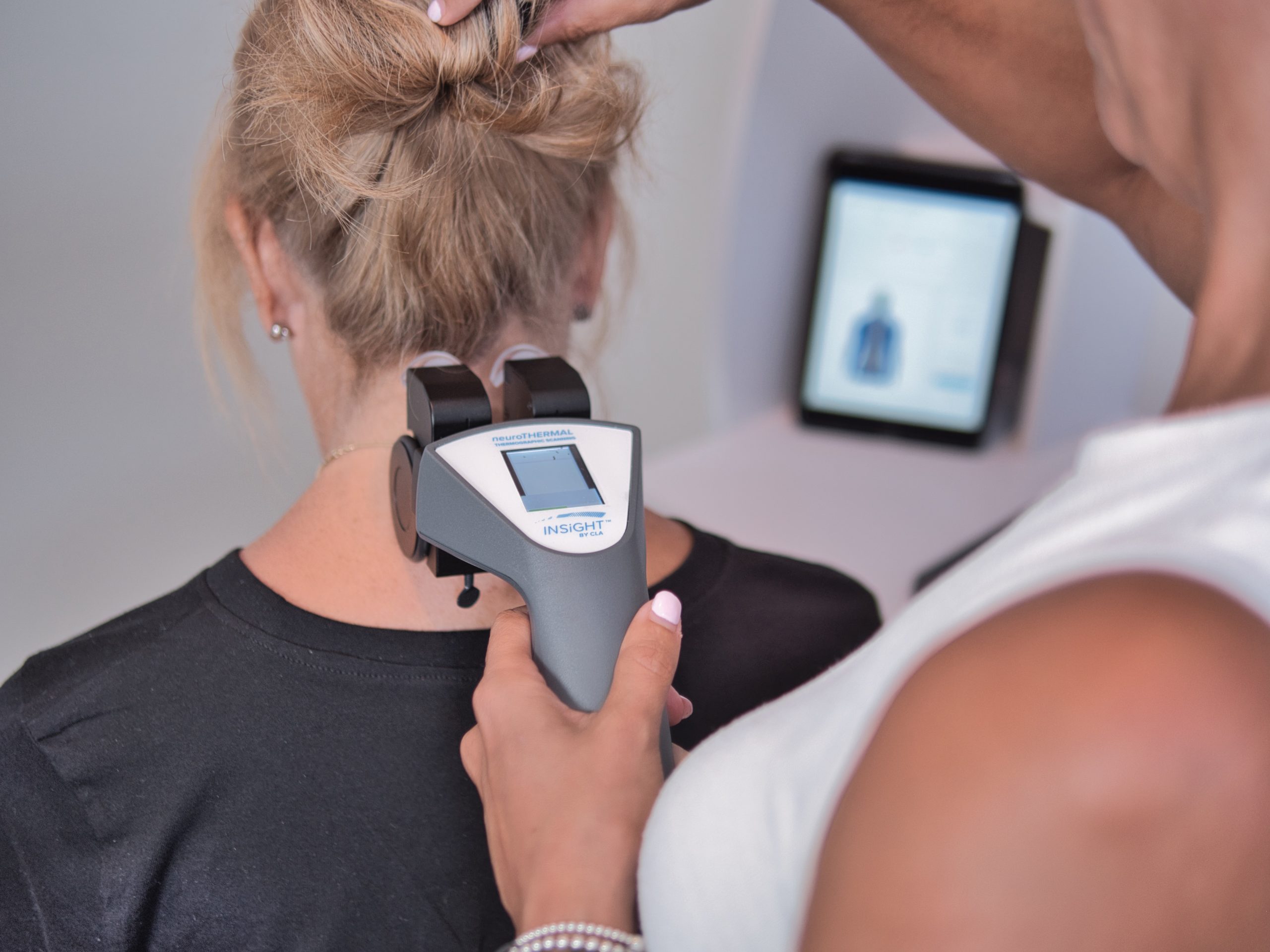
One part of a functional spine scan is thermography. A thermal scan uses infrared analysis to look for asymmetries in temperature along the spine. Because the autonomic nervous system influences circulation and gland regulation, temperature differences may reflect altered autonomic control. In practice, a thermal scan glides along your spine quickly, is painless, and can reveal patterns that matter clinically. It helps the doctor look at stress on your nervous system and how that stress may be showing up segment by segment.
Another part is surface emg. Surface EMG, also called sEMG, is a form of electromyography that evaluates the electrical activity of the muscles, especially the paraspinal muscles. In simple terms, it measures the electrical output of those muscles to show whether the body is overworking, guarding, or compensating. Surface electromyology can help identify muscular imbalance, postural tension, and spasms or weakness. It gives the chiropractor more information about how the motor system is reacting to load and how nerve interference disturbs normal muscular control.
The third piece is heart rate variability. Heart rate variability, or hrv, is not a scan of tissue along the spine in the same way, but it belongs in the conversation because it measures adaptability and reserve. HRV gives insight into autonomic balance and the broader stress response. It helps a chiropractor assess nerve function from the standpoint of recovery, resilience, and the body’s ability to shift between activation and restoration. In that sense, heart rate variability strengthens the value of a spine scan by showing the bigger autonomic picture.
- Thermography helps evaluate autonomic patterns with infrared analysis.
- Surface EMG measures the electrical activity of the paraspinal muscles.
- HRV shows variability, adaptability, and autonomic reserve.
- These scans help a chiropractor assess nerve function, postural response, and functional change over time.
Why INSiGHT Changes the Meaning of a Spine Scan
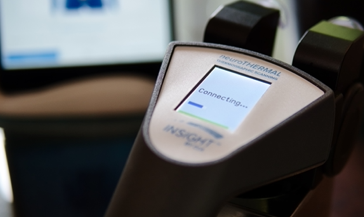
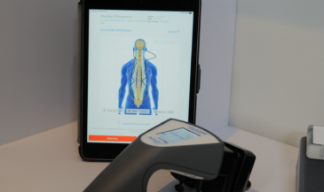
This is where INSiGHT scanning technology becomes so important. It takes the broad idea of a spine scan and gives it a practical, measurable, and neurologically focused framework. Instead of relying on vague impressions, the chiropractor can use advanced technology to gather objective examination findings and communicate them clearly. That is one reason an insight scan has become such a valuable part of the modern chiropractic office.
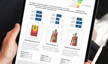
INSiGHT scanning technology brings together neuroTHERMAL, neuroCORE, and neuroPULSE, supported by Synapse software. neuroTHERMAL provides a thermal scan that evaluates autonomic patterns along the spine. neuroCORE uses surface EMG to analyze postural tension and muscular activity. neuroPULSE evaluates heart rate variability and helps the doctor understand autonomic adaptability. Together, these tools create a fuller view of nervous system performance and give the chiropractor technology to help measure subluxations in a more objective way. This is the role of INSiGHT technology in the chiropractic profession.
What makes this especially useful is that these findings address what often cannot be seen on x-ray. A structural image may reveal anatomy, but an insight scan can show trends in nerve function, postural stress, asymmetry, and autonomic response. It can help identify nerve interference along the spine, stress on your nervous system, and the impact of stress on regulation. This is the kind of state-of-the-art, non-invasive scan technology that helps a chiropractor accurately assess whether care is making a difference over time.
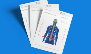
It also improves communication. When patients see their analysis in a visual format, they understand the conversation differently. The focus shifts from bones and joints to the nervous system controls that influence adaptation, balance, and overall health. That does not mean INSiGHT replaces clinical reasoning. It does not produce the care plan on its own. The chiropractor interprets the findings, combines them with the examination, and builds the care plan. But this non-invasive technology can support increased accuracy in diagnosis, stronger communication, and better patient outcomes in a chiropractic practice.
- neuroTHERMAL evaluates thermal and autonomic findings along the spine.
- neuroCORE uses sEMG to assess paraspinal muscular response.
- neuroPULSE evaluates heart rate variability and autonomic adaptability.
- Synapse software helps organize the data into reports that are easier to explain and track.
A Better Way to Think About a Spine Scan
When someone asks for a spine scan, the best answer is not always a quick referral for imaging. Sometimes MRI, x-ray, or CT scan is exactly the right next step. But many times, the better question is this: what are we really trying to learn? Are we trying to find a fracture or major structural issue, or are we trying to understand how the body is functioning under stress?
That distinction matters. The spine and nervous system are dynamic. Stress can disturb adaptation, alter muscle tone, influence autonomic balance, and change how a person functions day to day. Those changes matter in chiropractic care, and they often do not appear clearly on structural imaging alone. A good spine scan in chiropractic respects structure while also looking at nervous system function, spinal nerves, motor nerves, autonomic control, and the real-world effects of stress on the body.
That is why the future of the spine scan conversation in chiropractic is not structure versus function. It is structure and function, each in the right place. When chiropractors understand both, they communicate with more clarity, build a more informed care plan, and serve patients with more certainty. And when that process is supported by insight scanning technology, the scan becomes more than an image. It becomes a meaningful part of clinical decision-making, patient understanding, and resilient spinal health.