Ask any chiropractor what they’d most like to know about a patient on Day One, and the answer is usually the same: “Where is the stress showing up—and how is it affecting the nervous system?” That’s where surface EMG steps in.

Surface electromyography (sEMG) is more than just a reading of muscle activity. It’s a real-time analysis of how efficiently the motor nerves are directing energy to postural muscles along the spine. And when surface EMG is abnormal, it tells us something vital: the nervous system is working harder than it should—or not working enough—to maintain upright posture and spinal control.

So, what happens if surface EMG is abnormal?

It means the nervous system isn’t coordinating muscle tone properly. That can show up as postural fatigue, protective tension, poor adaptation, or early signs of neurological interference—often before symptoms even appear. In this article, we’ll explore what those abnormal EMG patterns really mean, why they appear, and how they shape care plans in practices that use INSiGHT’s advanced scanning technology.

If you’re ready to shift the conversation from posture to performance, let’s start with what EMG actually measures—and why it matters so much.

What Surface EMG Measures—and Why It Matters

Surface EMG (electromyography) is a non-invasive diagnostic test that evaluates how motor nerves interact with spinal postural muscles. Rather than analyzing symptoms of nerve or muscle dysfunction through physical exams alone, surface EMG measures the electrical activity that underlies muscle tone and coordination.

It’s important to clarify the difference between surface EMG and needle EMG. A traditional EMG test may involve a needle electrode inserted into the muscle to diagnose muscle disorder or nerve damage, often in hospital or medical center settings. This is typically done by a neurologist to identify nerve compression syndromes like carpal tunnel or neuropathy. These emg and nerve conduction studies can be uncomfortable and are diagnostic in nature.

By contrast, surface EMG uses surface electrodes placed on the surface of your skin, with no penetration. The electrical signals detected come from underlying nerve and muscle function, offering a view of postural efficiency and neurological distress.

Within the INSiGHT CLA suite, this technology is called neuroCORE. It records the electrical impulse through spinal motor nerves and translates this data into easy-to-understand scan views using the following core metrics:

  • Total Energy Index: Measures overall energy use to maintain posture. Reference value = 100 for adults, 125 for children.
  • Symmetry Score: Evaluates left/right neuromuscular balance.
  • Pattern Score: Measures the top-to-bottom organization of postural muscle tone.

When EMG results are abnormal, chiropractors gain insight into inefficiency, overexertion, and compensation—all of which help guide neurologically focused care.

What Abnormal Surface EMG Results Actually Mean

When EMG results show abnormal patterns, it’s not a red flag for disease—it’s a signal of nerve dysfunction. These patterns reveal how the nervous system is coordinating (or failing to coordinate) muscle tone under gravitational load.

Abnormal EMG scans typically present as:

  • Hypertonicity: Overactive firing due to spinal guarding or neurological interference.
  • Hypotonicity: Low tone areas where the nervous system isn’t recruiting muscle properly.
  • Asymmetry: One side working harder, indicating nerve or muscle imbalance and compensation.

These aren’t isolated “muscle problems.” They’re signs of abnormal electrical activity along the nerve roots, often caused by vertebral subluxation, nerve compression, or prolonged sympathetic overdrive. A patient may appear symptom-free, but still show significant postural inefficiencies on their surface electromyography test.

Rather than waiting for muscle weakness, muscle pain, or loss of function, chiropractors using EMG scans can diagnose stress patterns early. This makes surface EMG an essential tool for catching changes in nerve and muscle function—even before the patient feels a thing.

Why Do These Abnormal Patterns Appear?

The root cause of abnormal EMG patterns is rarely muscular—it’s neurological. The nerves that communicate with postural muscles are under constant influence from the brain and spinal cord. When this communication is altered by subluxation, environmental toxins, or nerve compression, the muscles respond accordingly.

For example:

  • A nerve is compressed in the cervical region → upper trapezius overfires (hypertonicity).
  • Early developmental interference from antibiotics → lumbar region underfires (hypotonicity).
  • Chronic desk posture → full-spine asymmetry and increased energy output.

The RED model helps decode this. The “E” stands for Energy, which is exactly what surface EMG reflects. When energy usage is high, symmetry is low, and patterns are disorganized, the nervous system is expending more than it’s conserving. Over time, this depletes reserve and leads to dysfunction—even without pain.

That’s what happens if surface EMG is abnormal: the nervous system is waving a flag that it’s adapting inefficiently, and that it needs help before symptoms of nerve disorder or muscle disease surface.

How Chiropractors Use This Information to Shape Care Plans

Abnormal EMG patterns guide chiropractic care in a way no physical exam can match. With neuroCORE’s surface EMG technology, chiropractors build care plans based on real-time feedback from the nervous system—not just subjective symptoms.

  • Hypertonic zones indicate overcompensation. Adjustments reduce neurological tension and restore tone balance.
  • Hypotonic zones reveal disconnection. These areas need neural stimulation and stabilization.
  • Asymmetrical patterns show spinal rotation, torsion, or energy waste. Adjustments re-establish coordinated nerve and muscle interaction.

Progress scans show reduced energy output, improved symmetry, and smoother tone patterns—signs that the nervous system is recalibrating. That’s how surface EMG provides proof your care is making a difference.

This isn’t about chasing dysfunction—it’s about restoring function. Chiropractors trained in neurological scanning aren’t reacting to symptoms—they’re responding to signals.

From Baseline to Breakthrough: The Power of Re-Scanning

When patients undergo EMG at multiple intervals, they provide the chiropractor with a window into neuroplastic change. INSiGHT’s scanning protocol includes:

  1. Initial Scan – Establishes the baseline.
  2. Progress Scan – Measures early change.
  3. Comparative Scan – Confirms neuroadaptive trends.
  4. Continuation Scan – Guides long-term wellness care.

Sometimes, a scan may look “worse” before it looks better—especially when patients undergoing EMG begin unwinding long-held patterns. This is not damage to nerves and muscles, but part of the adjustment process.

With this system, chiropractors can assess whether to increase or decrease frequency, modify adjustments, or address underlying nerve disorder traits more directly. The scan becomes the map—and the nervous system becomes the guide.

INSiGHT neuroCORE and the CORESCORE: Making the Invisible Visible

INSiGHT’s neuroCORE scanning system is a cornerstone of neurological analysis. With surface EMG, chiropractors gain a clear, reproducible view of spinal motor activity in less than 30 seconds.

  • FDA-cleared Class II device
  • Safe for children, pregnant patients, and the elderly
  • Built for reproducibility with high inter-examiner reliability

It also integrates with the CORESCORE, which combines:

  • neuroCORE (sEMG)
  • neuroTHERMAL (thermal differentials)
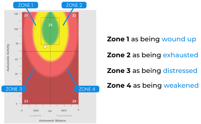
  • neuroPULSE (HRV and autonomic tone)

This creates a three-dimensional analysis of neurological status, capturing both the electrical activity and adaptability of the nervous system. EMG is often used to diagnose inefficiencies in postural tone and reserve capacity—and when it’s part of CORESCORE, the data becomes more than visual. It becomes undeniable.

When Patterns Speak Louder Than Symptoms

In chiropractic, the most meaningful care doesn’t chase symptoms—it tracks performance. That’s why understanding what happens if surface EMG is abnormal is so essential. It shifts the focus from short-term relief to long-term neurological change.

Surface EMG shows you where energy is being spent, where coordination is breaking down, and where the nervous system is asking for attention. It does what no subjective exam can do: it records the electrical activity that powers posture and movement.

Whether you’re working with kids who’ve never had symptoms or adults stuck in years of sympathetic overdrive, abnormal EMG findings give you a place to start—and a path to follow.

Because when you can see the nervous system in motion, you stop adjusting in the dark. You adjust with purpose, precision, and proof.

Have you ever wondered, what should heart rate variability be—and why it matters at all? You’re not alone. As health trackers and wearables spit out numbers and scores, it’s easy to assume that HRV is just another performance stat. But buried inside that rhythm between heartbeats is one of the most powerful indicators of how well your body is really functioning.

Heart rate variability (HRV) isn’t just about your heart—it’s about your nervous system. It tells us whether you’re stuck in fight-or-flight, or if your body has the flexibility to adapt, recover, and respond to life’s demands. In fact, HRV may be the most sensitive and accessible measure of your health and resilience.

But here’s the catch: there’s no single “normal” HRV value. Instead, the right question to ask is: how adaptable is your nervous system—and how can I track that over time?

Let’s explore why HRV is so much more than just data, how it reflects the story of your stress and recovery, and how chiropractic care brings that story into sharper focus.

What Is Heart Rate Variability and Why It Matters

Heart rate variability (HRV) is the measurement of the time gaps between each of your heartbeats. While your heart may beat at an average heart rate of 70 beats per minute, those beats don’t land perfectly on the second. One interval might be 0.86 seconds, the next 0.94—and that fluctuation is exactly what we want. HRV is the signal that your body is flexible and responsive.

To be clear, HRV is not the same as resting heart rate. Heart rate tells us how fast your heart is beating. HRV tells us how intelligently it’s responding to what’s going on around you. It’s a measure of your heart rhythm variability, not just beat count.

What makes HRV so powerful is its connection to the autonomic nervous system—the part of your brain and spinal cord that controls involuntary functions like breathing, digestion, immunity, and stress response. This system has two main branches:

  • The sympathetic nervous system (fight-or-flight), which helps you respond to threats.
  • The parasympathetic nervous system (rest-and-digest), which helps you recover, repair, and regenerate.

When your parasympathetic nervous system is engaged and working well—primarily via the vagus nerve—HRV goes up. When stress hijacks the system and you get stuck in sympathetic overdrive, HRV goes down.

HRV reflects nervous system and heart adaptability. It helps us see whether the body is shifting gears appropriately. When HRV is balanced and active, it’s a sign of strong neurological coordination—a healthy heart supported by a healthy brain-body connection.

What Should Heart Rate Variability Be? Understanding the Numbers

It’s the most common question people ask when they first see an HRV report: “What should my heart rate variability be?” And it’s a fair question—especially when you’re trying to make sense of numbers, trends, and zones on a heart rate variability chart.

Here’s the truth: There is no universal answer to what should heart rate variability be. HRV is like a fingerprint—unique to you. It’s shaped by factors like age, fitness, stress, and even your breathing rate. That’s why trying to compare your HRV score to someone else’s can feel like comparing apples to oranges.

Still, we can speak in general reference ranges. For RMSSD, a healthy adult might land between 30 and 100 ms. For SDNN, normal values range from 20 ms to over 200 ms.

Let’s look at some of the factors affecting HRV:

  • HRV naturally decreases with age. A 25-year-old will typically have a higher HRV than a 65-year-old.
  • Men often have slightly higher HRV than women, though this difference narrows with age.
  • Fitness level: Athletes and physically active individuals tend to show better HRV.
  • Time of day: HRV is generally higher during resting HRV periods and lower during activity.
  • Chronic heart conditions can reduce HRV.

So, what should heart rate variability be? It should be a reflection of adaptability, not perfection. As long as your HRV over time is stable or improving, you’re on the right track toward good heart rate variability and overall health.

Factors Affecting Heart Rate Variability

Heart rate variability isn’t static. It’s one of the most dynamic metrics your body produces, responding in real time to life’s inputs. And because HRV is a highly sensitive signal, it picks up on the smallest disruptions in your internal environment.

  • Sleep quality: Most HRV recovery happens during deep sleep.
  • Spinal tension and vertebral subluxation affect your nervous system and HRV.
  • Diet and hydration influence variability and vagal tone.

When these stressors accumulate, low heart rate variability can become the new normal. But it’s not permanent. Chiropractic care and lifestyle strategies can help improve HRV over time.

How Your Nervous System Influences HRV

Your nervous system is the master switchboard behind heart function. It determines whether your body speeds up, slows down, or shifts into healing. That’s why HRV reflects more than stress—it reflects function.

The autonomic nervous system consists of:

  • The sympathetic system: accelerator mode.
  • The parasympathetic system: brake mode.

Vagus nerve tone is essential to maintaining high heart rate variability. It keeps heart beats rhythmic and modulated.

When the system is dysregulated, HRV falls due to imbalance. Over time, this wears down resilience and increases dysfunction.

Using the HRV Rainbow Graph to Measure Adaptive Reserve

At INSiGHT CLA, the neuroPULSE scan and Rainbow Graph bring clarity to adaptability. Plotted on a color-coded chart, it visualizes your nervous system status using AAI and ABI scores.

Here’s how the zones break down:

  • Zone 1: High reserve, sympathetic dominant.
  • Zone 2: Parasympathetic dominant, fatigued.
  • Zone 3: Low reserve, sympathetic overload.
  • Zone 4: Critically low reserve.
  • Zone 5: The Green Zone—optimal adaptability.

This model helps chiropractors check heart rate variability and track patient progression toward a normal heart rate variability pattern.

High HRV vs Low HRV: What the Numbers Really Indicate

Consistently low HRV may reflect chronic stress, poor sleep, or neurological tension. But a single low reading isn’t a red flag—it’s the trend that matters.

In contrast, high HRV may signal parasympathetic dominance—and not always in a good way. In cases of adrenal fatigue, HRV appears high, but the system is underperforming.

Always interpret high or low HRV in context—and over time.

Improve Your Heart Rate Variability with Nervous System-Centered Care

Here’s how to improve your HRV and support your nervous system:

  • Chiropractic adjustments restore neurological balance.
  • Breathwork stimulates the vagus nerve and boosts HRV.
  • Sleep, nutrition, posture, and hydration all support heart rate and blood pressure regulation.
  • Athletes use HRV to plan their workouts and optimize recovery.

HRV Is the Language of Adaptability

So, what should heart rate variability be? It should be adaptive, responsive, and improving. HRV tells the story of your body’s flexibility—not just its performance.

Chiropractors use heart rate monitoring to measure adaptability, and chiropractic care to improve it. In a stressed-out world, HRV is how we make resilience visible.

When it comes to optimizing nervous system function, chiropractors have historically relied on structural markers—palpation, posture, and radiographs. But what if you could see the stress on your patient’s nervous system? What if you had objective insight into how their body is actually adapting? That’s exactly what heart rate variability (HRV) measurement brings to the table. It’s the tool neurologically-focused chiropractors have been waiting for—especially when backed by research-grade technologies like INSiGHT’s neuroPULSE.

HRV isn’t just a buzzword in biohacking or elite athletic circles anymore. In a chiropractic setting, it’s a performance metric for the autonomic nervous system (ANS)—a measure of balance, adaptability, and resilience. As we’ll explore in this article, HRV measurement helps us quantify how well the nervous system responds to stress, and how chiropractic care can create long-term improvements in neurophysiological health.

What Is HRV and Why Does It Matter?

At its simplest, HRV refers to the variation in time between heartbeats, and it’s controlled by the dynamic interplay between the sympathetic (fight or flight) and parasympathetic (rest and recover) nervous systems. A healthy heart isn’t perfectly rhythmic—it adapts from moment to moment, constantly adjusting based on internal and external demands.

That moment-to-moment flexibility is what HRV measures. A high HRV is associated with a greater ability to recover, respond, and regulate. A low HRV—often seen in patients under chronic stress—is associated with decreased adaptability and increased risk of cardiovascular, emotional, and immune dysfunctions.

Studies show that sustained chiropractic care can significantly improve HRV over time, reinforcing its value as a functional health marker.

The Role of HRV Measurement in Chiropractic

In chiropractic, we’ve always aimed to reduce interference in the nervous system—what we call vertebral subluxation. But what if you could track your progress beyond posture and pain? That’s where HRV measurement becomes indispensable.

Using INSiGHT’s neuroPULSE system, chiropractors can:

  • Objectively assess autonomic balance
  • Monitor changes in adaptive reserve throughout care
  • Establish measurable baselines and progress milestones
  • Communicate care outcomes in ways that patients can understand

Research shows that chiropractic adjustments affect sympathetic and parasympathetic tone, validating the clinical impact HRV measurement can document.

HRV Metrics and Measurement Standards

There’s more to HRV than just the average beat-to-beat interval. True HRV assessment includes both time domain and frequency domain measures:

  • SDNN (Standard Deviation of NN Intervals): Indicates total variability and resilience.
  • RMSSD (Root Mean Square of Successive Differences): Reflects short-term vagal (parasympathetic) tone.
  • LF (Low Frequency): Represents both sympathetic and parasympathetic influence.
  • HF (High Frequency): Primarily vagal tone.
  • LF/HF Ratio: Shows the balance or dominance of either nervous system branch.

The neuroPULSE, for example, distills these into two key indices: the Autonomic Balance Index (ABI) and the Autonomic Activity Index (AAI), allowing chiropractors to communicate findings with clarity and precision.

These assessments are validated against ECG standards. One clinical study analyzing chiropractic HRV outcomes demonstrated statistically significant increases in total power, SDNN, and both frequency bands after adjustments.

Introducing the HRV Rainbow Graph

Let’s talk visuals. One of the most effective tools in a neurologically-focused chiropractic practice is the Rainbow Graph, a proprietary display used in INSiGHT CLA’s scanning suite.

This XY graph plots:

  • Autonomic Balance on the X-axis (sympathetic to parasympathetic)
  • Autonomic Activity on the Y-axis (low to high reserve)

The graph divides into 5 zones:

  • Zone 1 (Upper Left): Sympathetic dominant with high reserve—often athletes, yet stressed.
  • Zone 2 (Upper Right): Parasympathetic dominant, but insufficient—often a sign of fatigue or burnout.
  • Zone 3 (Lower Left): Sympathetic dominant with low reserve—common in new patients.
  • Zone 4 (Lower Right): Severely weakened nervous system—low tone, low reserve.
  • Zone 5 (Green Center): Ideal! Balanced and resilient.

With each scan, a white dot shows the patient’s current state—making invisible stress visible, trackable, and actionable. Read more about the Rainbow Graph model.

Measuring Change: HRV in Progress Exams

With HRV tracking, chiropractors can quantify progress, even in the absence of pain. This makes it a vital tool for communicating the true value of care beyond symptom relief.

Studies indicate that consistent HRV changes are observable within a 12-week window of consistent care. This timeline supports care planning focused on adaptive retraining rather than symptom chasing.

HRV Measurement and Patient Engagement

Patients feel better when they see results. HRV scanning creates moments of clarity during re-evaluations. They see that their body is less stuck in survival mode. They recognize that their nervous system is healing, not just their back pain fading.

In fact, the visual feedback loop that HRV provides boosts compliance and deepens trust. Patients begin to understand their health from the inside-out.

This isn’t about creating dependency—it’s about building autonomy. By visually tracking how care improves heart rate variability, they’re empowered to make choices for resilience, not just relief.

HRV as a Chiropractic Compass

Heart rate variability measurement has become more than a research tool—it’s a clinical compass for the modern chiropractor. It tells us where the patient’s nervous system is today, how well it’s adapting, and what’s changing over time.

From Zone 3 distress to Zone 5 coherence, HRV scanning makes progress real, visible, and measurable. In the hands of a neurologically-focused chiropractor, this technology does more than validate care. It transforms it.

If you’re still relying solely on symptoms or posture to guide your care plans, HRV is the missing link. It’s time to shift from pain to potential, from posture to performance, from structure to function.

Heart rate variability (HRV) is one of those concepts that sounds complicated at first, but once you see how it works, it becomes one of the most practical ways to understand your body’s resilience. At CLA, we’ve spent years showing chiropractors how HRV can reveal a patient’s nervous system status—how well they’re adapting to everyday demands, and where they may need help restoring balance.

So, what is normal heart rate variability? That’s a great question, and the truth is: there’s no one-size-fits-all answer. A “normal” HRV is unique to you—shaped by your age and gender, fitness level, lifestyle habits, and even how you slept last night. Rather than chasing an exact number, the real value comes from knowing your baseline , at rest HRV and watching how it changes over time. That’s where chiropractic’s nerve-first approach, supported by HRV analysis, becomes such a powerful tool.

Understanding Heart Rate Variability (HRV)

At its simplest, heart rate variability is a measure of how much the time between your heart beats changes from one beat to the next. If your heart is beating 60 times per minute, that doesn’t mean it’s firing every exact second like a metronome. In fact, a resilient autonomic nervous system will cause small, healthy variations—one beat might come at 1.02 seconds after the last, the next at 0.98 seconds. These tiny changes, measured in milliseconds, are what we call HRV.

This variability tells us far more than the average heart rate ever could. It reflects how well your parasympathetic nervous system (the “rest and recovery” side) and your sympathetic nervous system (the “fight-or-flight” side) are working together. A good heart rate variability score means these two branches are in healthy balance, shifting seamlessly depending on your needs.

When you’re calm, your parasympathetic nervous system slows the heart, promoting recovery. When you face a challenge, your sympathetic nervous system raises your heart rate to respond. HRV is a highly sensitive indicator of how quickly and effectively you can make these shifts—and it’s routinely used to evaluate the autonomic nervous system in chiropractic and clinical contexts via heart rate variability to assess autonomic function.

Chiropractic care looks at HRV not just as a cardiovascular measure, but as a direct window into nervous system performance. That’s because your heart rhythm is under the influence of the brain and spinal cord every second of your life. A change in HRV may be the first clue that the nervous system is stuck in sympathetic overdrive or is lacking parasympathetic responsiveness—long before symptoms ever appear.

Why HRV Matters for Health and Performance

Your heart rate variability is more than just an interesting statistic—it’s a reflection of how adaptable you are to the demands of life. Think of it as your body’s “readiness score,” showing how prepared your nervous system and heart are to shift gears between effort and recovery.

A higher HRV is generally a sign that your body can respond well to challenges and bounce back quickly. Sports science relies on HRV to assess response to training and manage recovery. But you don’t have to be an athlete to benefit—anyone with a better HRV trend over time is building long-term resilience.

On the other hand, a low HRV can signal that your system is working harder than it should just to keep up. Low HRV is often associated with regulating inflammation via sympathetic pathways: prolonged inflammatory load and poor recovery can keep adaptability low. While a single low reading isn’t cause for panic, lower heart rate variability over weeks or months may be a clue that your system needs attention.

In chiropractic, HRV matters because it’s tied directly to the autonomic nervous system—the same system influenced by neurological interference along the spine. When the brain and body aren’t communicating efficiently, adaptability suffers, and that’s exactly what shows up in your HRV analysis. By improving nervous system balance and reducing interference, we aim to see changes reflected in HRV that point to better recovery and overall performance.

What Is a Normal Heart Rate Variability Range?

One of the first things people ask when they see their HRV score is, “Is that good?” The answer is—it depends. Normal heart rate variability doesn’t look the same for everyone, and comparing your number to someone else’s can be misleading.

For most healthy adults, short-term HRV values often fall somewhere between 19 and 75 milliseconds when measured under resting conditions. And calculated using SDNN methods. But these numbers are only part of the story. Normal HRV for adults is shaped by many factors:

  • Age and gender – HRV naturally decreases as we get older, and younger people tend to have higher HRV. Women may have slightly higher values before menopause.
  • Fitness level – Well-conditioned individuals, especially endurance athletes, often see higher heart rate variability—sometimes well over 100 milliseconds.
  • Lifestyle and recovery – Sleep quality, nutrition, hydration, and daily stress loads all influence HRV.
  • Overall health – Cardiometabolic risk states such as high blood pressure often accompany lower HRV and autonomic imbalance.

That’s why your normal HRV range is best determined by tracking your own baseline HRV over time. For example, if your average is 55 ms and suddenly drops to 35 ms for several days, that’s a sign worth paying attention to—even if 35 ms falls within a published “average” range.

In the chiropractic setting, we look less at whether your HRV fits a textbook definition and more at whether it’s improving under care. A trend toward better HRV is one of the clearest signs your autonomic nervous system is finding its balance and building resilience.

High HRV vs. Low HRV – What It Means

When it comes to heart rate variability, context is everything. A number by itself doesn’t tell the whole story—you have to consider your baseline, your HRV trends over time, and what’s happening in your life when you take the reading.

High HRV is generally a good sign. It suggests your parasympathetic nervous system is doing its job, keeping you adaptable and ready to recover. People with high heart rate variability can usually handle both physical and emotional challenges without staying in a stressed state for long. Athletes in peak condition, or individuals with consistent healthy routines, tend to have higher HRV.

Low HRV, on the other hand, can mean your system is stuck in sympathetic overdrive or lacking parasympathetic responsiveness. Low HRV can signal overtraining, lack of sleep, poor nutrition, ongoing illness, or unresolved neurological interference. In some cases, it shows up alongside cardiometabolic challenges like high blood pressure or insulin resistance.

Here’s the important part: a single lower heart rate variability reading isn’t an emergency—it might simply reflect a tough workout yesterday, a poor night’s sleep, or temporary illness. But when HRV decreases and stays down, it’s a clue that your nervous system and heart are working harder than they should just to keep up. That’s where regular monitoring becomes invaluable.

By combining HRV data with a full neurological evaluation, chiropractors can spot these patterns early. Over time, improvements in HRV become visible proof that your care plan is making a difference—not just in how you feel, but in how your body performs and adapts.

How to Measure Heart Rate Variability

You can only manage what you can measure—and with heart rate variability, accuracy matters. HRV is usually expressed in milliseconds, and those tiny differences between times your heart beats are easy to misread without the right tools.

There are several ways to measure heart rate variability:

  • Electrocardiogram (ECG) – The gold standard in research, measuring the exact electrical signals of the heart. Used to detect pathologies.
  • Mobile Photoplethysmography (PPG) – Common in consumer wearables like smartwatches and rings; uses light sensors to detect pulse waves.
  • Clinical-grade HRV monitors – Designed for precise readings in a controlled environment.

While consumer devices are useful for tracking trends, they can vary in accuracy. A heart rate variability monitor used in a chiropractic setting is designed to remove those variables—controlling posture, movement and environment to ensure reproducible results. Within chiropractic, HRV is often paired with other objective assessments to evaluate autonomic patterns and direction of change.

That’s where INSiGHT neuroPULSE comes in. This instrument collects HRV data in just three minutes, using millisecond-level precision and built-in quality controls. The results are instantly plotted on the Rainbow Graph, showing:

  • Autonomic Balance Index (ABI) – Where you fall between sympathetic and parasympathetic influence.
  • Autonomic Activity Index (AAI) – How much adaptive reserve your system has in the tank.

By pairing this analysis with the patient’s baseline HRV and ongoing HRV trends, chiropractors can clearly see how care is impacting adaptability. And when patients see these scan views in living color, the conversation shifts from “How many visits will I need?” to “How far can I take my resilience?”

Inside the neuroPULSE Technology

While there are many ways to collect HRV data, few tools are built specifically for the chiropractic setting. The INSiGHT neuroPULSE was designed with one goal: to make measuring heart rate variability accurate, repeatable, and meaningful for both the chiropractor and the patient.

Here’s what sets neuroPULSE apart:

  1. Millisecond precision – Captures each heartbeat interval with research-grade timing accuracy.
  2. Standardized 3-minute protocol – Ensures consistent comparisons over time.
  3. Immediate scan views – Results are instantly plotted on the Rainbow Graph.
  4. ABI & AAI metrics – Provide a complete picture of balance and activity.
  5. Integrated CORESCORE reporting – Combines HRV data with other neurological scans.
  6. Trend tracking for long-term care – Shows true progress over time.

With neuroPULSE, HRV stops being an abstract concept and becomes an everyday part of your clinical conversation. Instead of a patient leaving with just a number, they leave with a clear visual map of where they are, where they’re headed, and how your care is helping them get there.

Factors That Influence HRV

Your heart rate variability isn’t set in stone—it shifts constantly based on what’s happening in and around your body. While this is normal, knowing what can raise or lower HRV helps you understand the patterns you see in your HRV trends over time.

Lifestyle and recovery – Sleep quality, hydration, nutrition, and exercise load can improve or lower HRV; sport and rehab settings use HRV to assess response to training and manage recovery. Health status – Chronic inflammation and sympathetic dysregulation are linked through neuroimmune signaling, with the SNS playing a central role in regulating inflammation. Mental and emotional state – Ongoing stress can suppress parasympathetic responsiveness; vagal activation helps restore balance via the cholinergic anti-inflammatory pathway. Environmental factors – Climate, altitude, and early exposure to antibiotics and other environmental toxins play a role. Age and gender – HRV naturally decreases with age, and hormonal changes can affect averages.

Recognizing these influences is key to interpreting your baseline HRV correctly. This is why a single low or high reading is less important than the patterns you see over weeks and months.

Improving Heart Rate Variability

The good news about heart rate variability is that it’s not fixed. Whether your HRV score is high, low, or somewhere in between, there are practical, drug-free ways to help your nervous system become more adaptable.

Chiropractic adjustments remove neurological interference and restore balance, often resulting in HRV increases that reflect improved adaptability—documented in retrospective analyses of chiropractic care’s impact to improve HRV. Lifestyle support – Sleep consistency, balanced training, mindfulness, and quality nutrition all contribute to better HRV; slow, controlled breathing engages vagal mechanisms described in the cholinergic anti-inflammatory pathway. Trend monitoring – Regular scans with the neuroPULSE confirm when interventions are working and guide future decisions as part of chiropractic objective assessments.

Improving HRV is not about chasing a single “perfect” number—it’s about increasing your nervous system’s ability to adapt to whatever life throws at it.

HRV in Different Life Stages

Pediatric HRV – Naturally higher and valuable for tracking growth and development. Adult HRV – Stable with healthy habits, but vulnerable to lifestyle overload and sympathetic overdrive. Senior HRVHRV naturally decreases with age, but even small gains can improve daily function and resilience.

At every stage, HRV is a sensitive marker of adaptability. The INSiGHT neuroPULSE helps chiropractors track these changes and keep patients’ resilience as high as possible.

Are You Using HRV In Your Practice?

Heart rate variability is more than a number—it’s a real-time reflection of how your nervous system is adapting to the world around you. Because normal heart rate variability is unique to each person, its true value comes from consistent tracking and understanding the story your numbers are telling. A healthy HRV doesn’t just indicate a strong heart; it shows a nervous system that can switch smoothly between effort and recovery, stress and restoration. That adaptability is the foundation for long-term health, vitality, and resilience.

In chiropractic, HRV bridges the gap between what we feel and how we function. With the INSiGHT neuroPULSE and Rainbow Graph, chiropractors can take a complex neurological process and make it visual, simple, and motivating for patients. As adaptability improves under care, the upward shift in HRV becomes undeniable proof that the body is regaining its balance. And when we can measure, understand, and improve that adaptability, we give patients more than relief—we give them the confidence that their nervous system is equipped for whatever life sends their way.

If you’ve ever stared at an MRI report and wondered, “Is a herniated disc the same as a bulging disc?”—you’re not alone. These two terms are often thrown around in the same breath, leaving patients (and sometimes even providers) unsure about what’s actually happening inside the spine.

Let’s get this straight from the start: a bulging disc and a herniated disc are not the same thing. While both involve changes to the spinal disc’s structure, they differ in how—and how much—the disc has changed. But here’s what matters more than the disc itself: how your nervous system is responding to the pressure, the tension, and the stress.

The truth is, disc findings on their own don’t always mean you’re headed for back pain or surgery. What really matters is your body’s adaptability. Is your nervous system resilient enough to handle the shift?

In this article, we’ll walk through the difference between a bulging disc and a herniated disc, what causes them, what symptoms may show up, and how chiropractors—especially those using INSiGHT scanning technology—are uniquely positioned to track nerve system performance and help patients build lasting resilience.

What Are Spinal Discs, Really?

Before we dive into the difference between a herniated disc and a bulging disc, it helps to understand what a spinal disc actually is. Think of each disc as a small but mighty cushion tucked between the vertebra of your spine. These spinal discs aren’t just shock absorbers—they’re critical parts of your spinal structure that allow you to move, bend, and twist without grinding bone against bone, as explained in this anatomy review.

Each disc is made up of two distinct parts:

  • The annulus fibrosus – the tough, fibrous outer layer of the disc that holds everything together
  • The nucleus pulposus – the soft, gel-like center that helps distribute pressure and maintain flexibility

Picture it like a jelly donut: the outer dough (annulus) protects the inner filling (nucleus). When everything’s intact and hydrated, your discs help your spine move smoothly while safeguarding the delicate nerve tissue nearby.

And that’s the key point—your discs aren’t just structural. They’re closely intertwined with your nervous system. Just millimeters away from these discs are spinal nerves that pass signals from your brain to your body. When disc material shifts—whether from a bulge or a rupture—it’s the nervous system that often ends up carrying the burden.

The Difference Between a Bulging Disc and a Herniated Disc

Let’s break down the key difference between a bulging disc and a herniated disc, because while they’re both disc conditions, they’re not interchangeable.

A bulging disc occurs when the disc’s outer layer—the annulus fibrosus—remains intact but begins to stretch and protrude outward. Think of it like pressing on a water balloon without popping it.

A herniated disc means the inner disc material—called the nucleus pulposus—has broken through a tear in the outer layer. When this disc material escapes, it can leak into the spinal canal and press directly on the nerve root. 

Feature Bulging Disc Herniated Disc
Outer Layer (Annulus) Intact Torn or ruptured
Disc Material Stays contained Escapes into the spinal canal
Onset Often gradual Sudden or gradual
Symptoms May be mild or absent More likely to cause nerve irritation
Common Locations Lumbar and cervical regions Lumbar and cervical regions

What Causes Disc Changes?

It’s easy to hear terms like “disc degeneration,” “slipped disc,” or “bulging or herniated disc” and assume something has gone wrong. But in many cases, disc changes are a natural part of life.

In fact, research confirms that degeneration is common with age and not always problematic. The disc may dry out, flatten, or lose its shape, especially in the lower back or neck.

Common contributors to disc degeneration and disc problems include:

  • Prolonged sitting, especially in slouched posture
  • Heavy lifting or repetitive motion jobs
  • Past spinal injuries or trauma
  • Lack of motion, which limits disc hydration
  • The natural aging process

What determines whether a bulging disc or herniated disc becomes a problem is often whether the nervous system can keep up. If nerve tension builds or adaptive reserve drops, symptoms can emerge.

Herniated Disc Symptoms vs. Bulging Disc Traits

When the nervous system starts to struggle, symptoms of a herniated or bulging disc may show up. These symptoms vary depending on the location and severity of the disc issue—but also on the adaptability of the nervous system.

Common herniated disc symptoms include:

  • Back pain or neck discomfort
  • Radiating pain into the arm or leg
  • Numbness, tingling, or “pins and needles”
  • Muscle weakness or instability
  • Symptoms that worsen with sitting, coughing, or sneezing

Even small disc changes can cause pain if the system is overwhelmed. In many cases, what we call a “disc problem” is really a nervous system communication problem.

The Role of Imaging—And Its Limits

MRIs and CT scans can confirm a bulging or herniated disc, but they don’t tell us the whole story. Imaging captures structure—not function. It doesn’t measure nerve tension, sympathetic overdrive, or adaptability.

That’s why Neurologically-Focused Chiropractors rely on functional assessments in addition to imaging. Nerve interference, not just disc shape, is the true driver of dysfunction.

Why the Nervous System’s Response Matters Most

The disc and a herniated disc both apply pressure—but the real difference lies in how the nervous system handles that pressure. If the system is stuck in stress mode, even a mild bulge can send things off track.

When a patient is in a state of sympathetic overdrive, their body is less capable of adapting to mechanical tension. This is what leads to persistent symptoms, even in the absence of major damage.

Chiropractors look at these issues through the lens of neurological performance. When interference is reduced and adaptability restored, symptoms often improve—regardless of whether the disc has changed shape.

The Chiropractic Approach: Restoring Resilience

Chiropractic care doesn’t directly treat the disc. Instead, we work to restore the nervous system’s ability to adapt to stress, injury, and inflammation.

Specific adjustments aim to reduce neurological interference, relieve postural tension, and support the body’s healing response. The approach is drug-free and adaptability-focused.

When a care plan is supported by neurological data, chiropractors can:

  • Improve spinal motion near the affected disc
  • Relieve muscle tension and guarding
  • Reduce nerve root irritation
  • Build adaptive reserve
  • Prevent disc flare-ups from recurring

Using INSiGHT Scanning Technology to Track Progress

To make care measurable, chiropractors use INSiGHT scanning technology, which tracks how disc interference may be affecting the nervous system in real time.

  • neuroTHERMAL scans reveal autonomic imbalances along the spine.
  • neuroCORE sEMG scans track energy imbalance and postural fatigue.
  • neuroPULSE HRV scans reveal adaptability under stress.
  • The CORESCORE compiles all metrics into one score, offering an easy-to-understand view of the nervous system.

These scans shift the conversation from structural fear to functional clarity.

From Disc Diagnosis to Nervous System Direction

So, is a herniated disc the same as a bulging disc? No—and yes. They differ in structure, severity, and onset. But what matters more than the difference between a bulging disc and a herniated disc… is how the nervous system is handling it.

Bulging and herniated discs may show up on scans long before symptoms do. But that doesn’t mean you’re broken. It means your body is doing its best to adapt.

And chiropractors using tools like INSiGHT scanning are uniquely trained to read that adaptation, support it with personalized care, and build lifelong resilience—without drugs, without fear, and without guessing.

Chiropractors talk a lot about adaptability—but how do you measure it?

Not symptoms. Not just how someone feels after their last adjustment. What we’re really looking for is a nervous system that can respond and recover.

That rhythm is exactly what heart rate variability (HRV) reveals.

Now, don’t let the word “heart” fool you. Healthy heart rate variability isn’t just about cardiac performance. It’s a trackable expression of how well the autonomic nervous system is working. HRV tells us whether a patient is stuck in fight-or-flight, or if their nervous system is adaptable, calm, and coordinated. It’s a measurement of resilience.

In this article, we’re going to unpack the science and clinical meaning behind HRV, explore the factors affecting HRV, and walk through how this one number can transform your care plans. If you’re ready to make adaptability visible—HRV is where it starts.

What Is Heart Rate Variability, Really?

Heart rate variability (HRV) is the variation in time between each heartbeat. Not the number of beats per minute—but the milliseconds between one beat and the next. These fluctuations in heart rate tell us how the nervous system is managing the constant shift between demand and recovery.

This rhythm is regulated by the autonomic nervous system, which controls everything from breathing to digestion to heart function. It has two branches:

  • The Sympathetic Nervous System (fight-or-flight)
  • The Parasympathetic Nervous System (rest-digest-repair)

A higher heart rate variability generally indicates better adaptability. A low HRV, especially when persistent, may suggest the nervous system is stuck in overdrive.

HRV isn’t just about the number of times your heart beats—it’s about how that rhythm fluctuates, and how quickly your system can adapt. In chiropractic, that adaptability is the key to long-term outcomes.

A Good HRV Is a Responsive HRV—Not Just a High One

Wearables and apps often say a “high HRV” is always good. But what matters most is responsiveness.

HRV tends to decrease with age, but that doesn’t mean an older patient is unhealthy. A good heart rate variability is one that reflects the person’s baseline, environment, and adaptability.

This is why tracking HRV over time is far more useful than comparing a patient to global averages. If the patient’s HRV is low but improving, that’s real progress.

So instead of fixating on “high or low HRV,” we look at whether HRV is becoming more responsive. That’s the mark of adaptability—and the true definition of healthy heart rate variability.

What Influences Heart Rate Variability?

HRV is influenced by both short-term and long-term inputs. Chiropractors should be watching for the following:

  • Vertebral subluxations: Incoherence in the nervous system limits adaptability
  • Sleep quality: Poor sleep leads to reduced HRV.
  • Physical activity: Regular movement can increase HRV, while overtraining can lower it.
  • Heart rate and blood pressure: Patients with high blood pressure often show lower HRV.
  • Healthy diet and hydration: These support vagal tone; poor diet undermines it.
  • Emotional distress: Drives sympathetic overactivity.
  • Structural tension: Postural distortion influences HRV via autonomic nervous system changes.

Remember, HRV naturally decreases with age. So when reviewing results, consider factors like age, mean heart rate, and individual trends.

How HRV Tells the Story of Your Nervous System’s Adaptability

In a chiropractic setting, HRV helps answer one critical question: How well is this person adapting to life?

The Parasympathetic Nervous System is responsible for recovery. If vagal tone is weak, even small stressors can disrupt regulation. That shows up as lower HRV.

Look for:

  • High resting heart rate paired with low variability
  • Average heart rate variability stuck in a narrow range
  • HRV changes that correlate with lifestyle or care updates

Variability reflects resilience. A flexible nervous system can shift gears smoothly. That’s what we’re trying to restore.

The Rainbow Graph: Turning Data into a Nervous System Story

The Rainbow Graph from INSiGHT’s neuroPULSE scan maps HRV scores on an easy-to-understand grid.

  • Zone 1: Revving Engine — Sympathetic dominant, high reserve
  • Zone 2: Parking Brake On — Parasympathetic dominant, low response
  • Zone 3: Distressed — Sympathetic dominance, low reserve
  • Zone 4: Weakened — Depleted adaptability, little responsiveness
  • Zone 5: Green Zone — Balanced, responsive, and thriving

With this graph, patients can check heart rate variability visually and understand where they are in their journey.

Using HRV to Build Better Chiropractic Care Plans

When you use heart rate variability to plan, you’re no longer relying on symptoms alone.

HRV helps identify:

  • Consistently low HRV before traits appear
  • When to shift frequency of care
  • How a patient is responding neurologically over time

When HRV improves over time, you know their system is regulating better—evidence of sustained improvement in HRV. That’s powerful clinical data.

Why the neuroPULSE Belongs in Every Chiropractor’s Scan Suite

The neuroPULSE is your instrument for measuring HRV with clarity. It works alongside the full INSiGHT suite:

  • neuroTHERMAL: Detects thermal asymmetries
  • neuroCORE: Assesses postural tension
  • neuroPULSE: Tracks adaptability reserve

These three scans come together in Synapse to generate a CORESCORE. With it, patients get the complete picture—and you gain confidence in how you adjust and plan care.

This is where HRV analysis becomes reality. Clinical research documents the effect of chiropractic care on HRV. You’re helping patients see their adaptability improve, visit by visit.

Your Practice Is Wired for This

Healthy heart rate variability is about potential. When you track HRV, you’re giving your patients more than a number—you’re giving them proof. You’re helping them see how their system is bouncing back, recalibrating, and handling more of life with less friction.

The chiropractic profession has always been centered around helping people adapt better. HRV gives us the tools to track that mission, guide it, and communicate it.

It’s not about adding more to your plate. It’s about turning what you already know—that the nervous system runs the show—into something visible, practical, and transformative.

Let HRV show you what the nervous system is saying. And let it show your patients what’s possible.

If you’ve ever had a patient ask, “What is syncope?”. Here’s the simple answer: syncope is the medical term for fainting—a short-lived loss of consciousness caused by a temporary dip in blood flow to your brain. It can happen quickly, and though many cases are benign, others may reflect a more serious concern, like an underlying heart condition or nervous system imbalance.

In chiropractic practice, especially in vitalistic care models, these episodes are more than just a fall—they’re signals. They tell us the body wasn’t able to adapt at that moment. Whether it’s a drop in blood pressure, a misfire in heart rhythm, or an overwhelmed autonomic circuit, syncope is a symptom that deserves clarity, not guesswork.

In this article, we’ll walk through the most common types of syncope, the warning signs to look for, how these events are evaluated, and how INSiGHT scanning technology gives chiropractors a window into the nervous system’s adaptability—often long before symptoms appear.

Types of Syncope (and What They Tell You)

Every chiropractor needs to know how to identify the type of syncope they’re seeing—or referring. All syncopal episodes boil down to one shared feature: blood flow to your brain dropped just long enough to cause a brief loss of consciousness.

Let’s break them down:

1. Reflex Syncope (Vasovagal and Situational)

This is the most common type of syncope, also known as neurally mediated syncope or neurocardiogenic syncope. It includes:

  • Vasovagal syncope – Triggered by heat, emotional stress, needles, or standing too long. It reflects an overreaction of the vagus nerve, causing a coordinated drop in heart rate and blood pressure.
  • Situational syncope – Happens during activities like coughing, laughing, swallowing, or urinating. The trigger causes a vagal reflex response.

Vasovagal syncope is usually harmless, especially when it includes a brief warning phase—dizziness, nausea, tunnel vision, warmth, or lightheadedness—and quick recovery.

2. Postural Syncope (Orthostatic Hypotension)

This occurs when standing up too quickly causes blood to pool in the legs, creating a drop in blood pressure and not enough blood flow reaching the brain. Common in dehydrated individuals or those on certain medications, postural syncope often includes lightheadedness just before the episode.

3. Cardiac Syncope

Here’s where we pump the brakes. Cardiac syncope reflects an underlying serious heart issue like a valve disorder, arrhythmia, or obstruction between the heart and the aorta. These episodes often occur without warning and may indicate elevated risk of sudden cardiac death.

Red flags include:

  • Fainting during exertion or while lying flat
  • A family history of sudden cardiac death
  • Known heart failure or prior arrhythmia

In these cases, it’s important to get treatment right away and co-manage with cardiology.

4. Neurologic Syncope

Though less common, neurologic syncope can be confused with seizure or stroke. These events may involve longer recovery, confusion, or focal neurological signs. A neurologic evaluation is warranted when symptoms point that way.

5. Syncope of Unknown Cause

Not every episode fits neatly into a category. Some require careful tracking, lifestyle analysis, and sometimes testing to get clear on the cause of syncope.

Recognizing the Symptoms and Triggers

Syncope often gives us warning—if we know how to listen.

Common symptoms of syncope:

  • Tunnel vision or “seeing spots”
  • Lightheadedness or dizziness
  • Clammy skin or nausea
  • Feeling off-balance or weak
  • Headache or warm flush
  • Sudden fatigue or confusion after

Possible causes include:

  • Dehydration
  • Standing up too quickly
  • Emotional stress or fear
  • Hot environments
  • Large meals or alcohol
  • Medications affecting blood pressure or heart rate
  • Underlying cardiac or neurological conditions

Many of these trigger syncope through autonomic overload—too much input, not enough adaptability. The warning signs of syncope are your patient’s early alert system. Helping them tune in is just as vital as evaluating the fall itself.

Evaluation: History, Testing, and the Right Questions

Every syncope episode tells a story. The key is asking the right questions and observing the right signals.

Start with:

  • A detailed history: what happened, what was felt, and what came before the spell
  • A full physical exam, including blood pressure and heart rate in various positions
  • A medication and hydration review

Tests commonly used to explore syncope symptoms:

  • Electrocardiogram (ECG): Helps record your heart rhythm
  • Ambulatory monitor: Detects intermittent abnormal heart rhythm
  • Echocardiogram: Assesses valve and flow problems
  • Tilt-table test: Assesses how heart rate and blood pressure respond to positional shifts
  • Autonomic reflex testing: Evaluates reflex imbalance
  • Labs or brain imaging if symptoms suggest seizure or vascular events

According to the American Heart Association, anyone with syncope should receive an initial evaluation to rule out several serious heart conditions. While many potential causes are benign, it’s critical to identify those that aren’t.

Most people don’t need follow-up treatment for simple reflex faints. But you do need treatment for certain causes, especially if the cause of your syncope stems from underlying cardiovascular risk.

INSiGHT Technology: Revealing What’s Beneath the Fall

In chiropractic, we don’t guess. We scan. And when it comes to syncope—especially recurring or unexplained episodes—objective scanning helps move the conversation from uncertainty to clarity.

The INSiGHT neuroTECH and Synapse software gives chiropractors a visual, multi-dimensional lens on nervous system adaptability:

  • neuroPULSE HRV scan shows us autonomic reserve—how well the system handles challenge and recovery.
  • neuroCORE surface EMG maps muscular tension and balance.
  • neuroTHERMAL thermography highlights patterns of sympathetic overdrive or interference.

Together, this technology reveals the hidden terrain beneath a fainting spell—stress patterns, adaptation fatigue, and autonomic instability. With each full spine nerve system scan, chiropractors can map progress, design better care plans, and build trust through results.

Looking Beyond the Fall

Syncope often feels like an isolated event. A quick scare. But more often than not, it’s a reflection of long-term imbalance—a system stuck in fight-or-flight with no reserve left to stand tall.

So, what is syncope? It’s a signal. One that tells you something in the background isn’t working as it should.

That’s why chiropractors using INSiGHT technology don’t just stabilize—they elevate. They identify the cause, analyze stress responses, and support patients in building resilience from the inside out.

Whether the trigger was emotional, positional, or rooted in a deeper heart condition, the goal of treatment is the same: restore adaptability, protect recovery, and prevent the next collapse—not by treating the symptom, but by supporting the system.

Because in a nerve-first practice, syncope is also a starting point—not an endpoint.

It’s a question that comes up in every practice: what’s the real difference between a bulging disc vs herniated disc? They sound similar, they show up on the same scans, and they both make patients nervous. But as chiropractors focused on performance and adaptability, we know the difference isn’t just about structure—it’s about the nervous system story beneath the surface.

A bulging disc means the disc’s outer wall has thinned or deformed, but the jelly-like core stays contained. A herniated disc means part of that inner material has escaped, usually through a small tear. That distinction matters because it often determines whether a nearby spinal nerve is simply nudged—or chemically irritated and inflamed.

But let’s go deeper. Discs aren’t the problem; they’re the symptom of a system under strain. At INSiGHT CLA, we’ve seen how the nervous system status—how well a person adapts to stress—determines whether a disc bulge turns into a breakdown, or quietly reintegrates under care. That’s the message patients need to hear.

The Key Difference Between a Bulging Disc and a Herniated Disc

Let’s get visual. If a disc is a jelly doughnut, then:

  • A bulging disc is when the doughnut flattens and bulges outward—still intact.
  • A herniated disc is when the jelly pushes through a crack in the dough—no longer contained.

Bulging discs involve a general widening of the disc, often due to aging, poor mechanics, or disc degeneration. The outer layer of the disc stretches but doesn’t rupture. It’s usually broader, affecting a quarter or more of the disc’s circumference.

Herniated discs, on the other hand, happen when that stretched wall gives way, and the inner core breaks through. This is more focal—and more likely to irritate a nearby spinal nerve.

Where They Show Up—and What They Feel Like

Lower back pain is the classic sign for both conditions—but not the only one.

  • A lumbar disc herniation may cause leg pain, numbness, or weakness that radiates into the foot.
  • A cervical disc issue can produce neck pain, arm tingling, or grip fatigue.
  • A bulging disc may feel like tightness, stiffness, or a sense of strain after long drives or poor posture.

Here’s the kicker: many people walk around with a bulging or herniated disc and have no clue—until a moment of overload sends their system into distress. Studies show that a surprising number of people have disc abnormalities on MRI but no symptoms.

Symptoms of a Herniated Disc vs Bulging Disc

Herniated disc symptoms tend to be more dramatic:

  • Radiating pain into the arm or leg
  • Sharp, electric sensations
  • Numbness or tingling in a nerve distribution
  • Muscle weakness (can’t lift the foot, poor grip strength)
  • Worsens with coughing or sitting

Bulging discs, especially in the cervical or lower back, tend to present with:

  • Dull, achy back or neck pain
  • Reduced flexibility or endurance
  • Symptoms that come and go with posture or position

Diagnosis: Going Beyond the Scan

You don’t need a radiology degree to know when a disc is talking. You need ears, hands, and the right instruments. A great evaluation includes:

  • Straight leg raise or Spurling’s to provoke symptoms
  • Reflex testing and strength checks
  • Sensory mapping
  • And—when it’s warranted—an MRI to confirm structural change

Why Neurological Scanning Changes the Game

This is where INSiGHT’s technology shines.

Using the INSiGHT neuroTECH suite, chiropractors can scan the spine in minutes and see what the body is hiding.

  • neuroTHERMAL identifies patterns of painless neurological distress
  • neuroCORE (sEMG) maps out protective muscle bracing
  • neuroPULSE HRV measures adaptability and recovery

Together, these scans give you the truth behind the complaint. They show whether the nervous system is adapting, inflamed locally, or fatigued. Best of all? They give you visual, undeniable proof your care is making a difference.

Treatment Options: What Helps and What Doesn’t

Most bulging discs and herniated discs don’t need surgery. They need space, motion, and a nervous system that isn’t in full-blown defense mode.

Studies confirm that conservative care is often effective—especially when customized to the nervous system’s needs.

Treatment for a bulging disc often includes:

  • Chiropractic adjustments
  • Postural re‑education
  • Progress tracking through objective scans

Occasionally, when symptoms escalate, an interventional referral is warranted.

Red Flags to Watch For

Refer out when:

  • Bladder or bowel control changes
  • Saddle anesthesia develops
  • Sudden weakness emerges
  • Fever, trauma, or cancer history is present

Why Objective Scanning Makes the Difference

You can’t improve what you can’t measure. With INSiGHT, chiropractors get reproducible, patient-friendly ways to evaluate disc-related patterns. Over 12,000 offices rely on:

  • neuroTHERMAL for autonomic asymmetries
  • neuroCORE for postural distortion
  • neuroPULSE for recovery potential

What This Means for You—and Your Patients

Bulging vs herniated disc isn’t just a technical distinction. It’s the fork in the road where you choose between chasing symptoms—or building a care plan that restores performance.

When we look beyond the disc and assess the nervous system’s adaptability, we’re no longer treating just back pain. We’re helping the body reorganize. Reconnect. Reclaim.

With the right scans, the right strategy, and a clear message, patients stop counting visits—and start valuing results.

If you’ve ever wondered what is electromyography and why chiropractors like me get so excited about it, let’s make it simple: EMG is a way of listening to the electrical chatter between your spine, muscles, and the nerves that control them. It turns invisible patterns into something you can see, understand, and use to make smarter care decisions.

I’ve found that when patients see their own EMG scan, the conversation shifts. Suddenly, they’re not just talking about symptoms—they’re looking at how well their body’s motor system is performing. And when we can track those changes over time, we’re no longer guessing. We’re showing proof your care is making a difference.

How an EMG Works: From Brain to Muscle

The story starts in your brain and spinal cord. From there, motor neurons send electrical impulses through a nerve toward a muscle. Those signals tell the muscle fibers to contract. Every one of those signals has a measurable action potential, and EMG is the technique for measuring it.

There are two main ways to do an EMG:

  • Needle EMG – Used in hospitals by a neurologist to diagnose specific nerve or muscle disorders, like amyotrophic lateral sclerosis and focal nerve damage.
  • Surface EMG (sEMG) – What we use in chiropractic. Small sensors (electrodes) on the skin pick up the electrical activity of multiple postural muscles at once. It’s quick, safe for all ages, and ideal for tracking changes visit by visit.

In both cases, the electrical activity picked up by the electrodes is amplified and shown as a form of waves. At rest, healthy muscle show little activity. When you contract your muscle, the waveforms change shape and size, showing us how the nerves that control that muscle are firing.

What EMG Reveals in Practice

In my years with surface EMG, I’ve learned it tells you three main things:

  • How much energy is being used – Are muscles working too hard just to keep you upright?
  • How balanced the system is – Are both sides of the spine sharing the load evenly?
  • How well the load is distributed – Are a few areas overcompensating for weaker spots?

When you see high amplitude during a light task, it can mean the system is burning extra fuel because of nerve dysfunction. Low readings may signal fatigue or disuse. Uneven patterns often reveal chronic compensation.

And here’s the part patients love: you can show them their own data. That’s powerful. It’s not about scaring them with “bad” results—it’s about helping them understand how their nervous system is performing and how it can improve.

The Limits of EMG

It’s just as important to know what EMG doesn’t do:

  • It’s not a scoliosis diagnosis tool, though it can show muscle imbalance around a curve.
  • It doesn’t “measure pain” or discomfort. EMG is purely motor.
  • Without a solid normative database, EMG numbers are just that—numbers.

Bottom line—EMG findings should always be part of a bigger picture, alongside your HRV and thermal exams, health history, and palpatory findings.

From Measurement to Mastery: INSiGHT’s neuroCORE

Here’s where things get exciting. In chiropractic, the EMG test really shines when you pair it with tools built for our profession. The INSiGHT neuroCORE instrument uses high-grade sensors and chiropractic-specific protocols to measure paraspinal muscle function with precision.

With neuroCORE, you can compare your patient’s results to a trusted normative database, track changes in motor control patterns, and show side-by-side scan views over time. That’s huge for both clinical certainty and patient engagement.

We often combine neuroCORE with our other INSiGHT scans—neuroTHERMAL and neuroPULSE HRV—so the patient gets a complete picture of their neurological performance. When you roll these into the CORESCORE, you’re not just showing them a number—you’re showing them their progress in living color.

Bringing the Nervous System into Focus

So, what is electromyography? It’s more than a diagnostic test. It’s a way of making the invisible visible. It’s how we can help detect neuromuscular abnormalities, track adaptation, and show the small but important changes that lead to bigger breakthroughs in resilience.

For me, EMG is one of the most valuable conversations I have with patients. It keeps them focused on function, not just symptoms. It helps them see that chiropractic care is about unlocking performance, not just relieving today’s stiffness.

Every scan is a chapter in their story. And with the right tools, we can read that story together—and guide it toward a healthier, more adaptable future.

+