By Dr. Christopher Kent and Dr. Patrick Gentempo, Jr. Paraspinal EMG scanning is becoming a popular technique in chiropractic. It is a useful tool for measuring changes in the electrical activity of paraspinal muscles. It is important for the chiropractor to understand the clinical significance of these changes, and to integrate the EMG findings with other examination findings. The limitations of EMG must also be understood. Unsubstantiated claims must be avoided if EMG scanning is to assume its proper place in chiropractic practice. The authors suggest that users familiarize themselves with the literature, and exercise caution when explaining the results of EMG scans to thirdparty payers and attorneys. Statement: Surface electrode EMG is a reliable procedure. Fact: The statement is true. Testretest reliability studies by Spector [1], Cram [2], Komi and Buskirk [3], and Thompson [4] have demonstrated verygoodtoexcellent reliability for surface techniques. Statement: Soft tissue injuries can be documented by paraspinal EMG studies. Fact: True. Soft tissue injuries may result in muscle involvement, which may cause EMG changes. [5,6] Statement: EMG scans detect vertebral subluxations. Fact: EMG scans measure changes in the electrical activity of paraspinal muscles. Changes in paraspinal muscle function are seen in vertebral sub
By Dr. Christopher Kent and Dr. Patrick Gentempo, Jr.
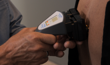
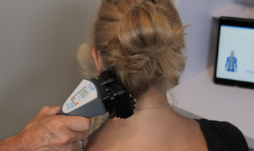
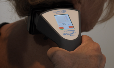
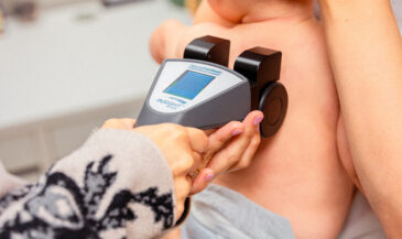
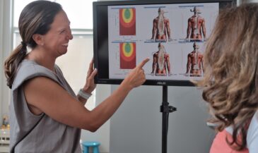
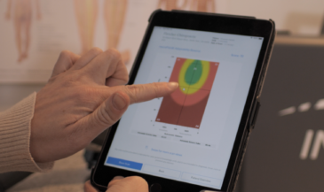
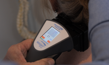
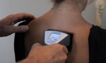
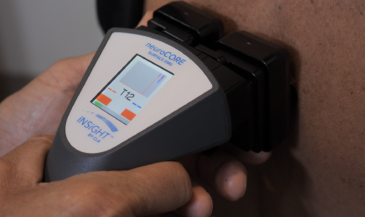
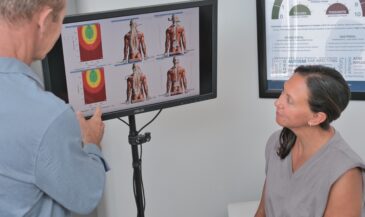
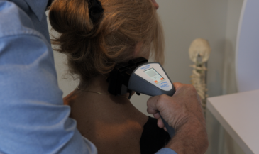
Paraspinal EMG scanning is becoming a popular technique in chiropractic. It is a useful tool for measuring changes in the electrical activity of paraspinal muscles. It is important for the chiropractor to understand the clinical significance of these changes, and to integrate the EMG findings with other examination findings.
The limitations of EMG must also be understood. Unsubstantiated claims must be avoided if EMG scanning is to assume its proper place in chiropractic practice. The authors suggest that users familiarize themselves with the literature, and exercise caution when explaining the results of EMG scans to thirdparty payers and attorneys.
Statement: Surface electrode EMG is a reliable procedure.
Fact: The statement is true. Testretest reliability studies by Spector [1], Cram [2], Komi and Buskirk [3], and Thompson [4] have demonstrated verygoodtoexcellent reliability for surface techniques.
Statement: Soft tissue injuries can be documented by paraspinal EMG studies.
Fact: True. Soft tissue injuries may result in muscle involvement, which may cause EMG changes. [5,6]
Statement: EMG scans detect vertebral subluxations.
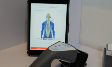
Fact: EMG scans measure changes in the electrical activity of paraspinal muscles. Changes in paraspinal muscle function are seen in vertebral subluxation. Taken in concert with other examination findings, EMG scans may be useful in detecting and characterizing the muscular dysfunction seen in subluxation. Because EMG changes may be due to conditions other than subluxation, EMG findings must be considered a part of the subluxation exam, not the sole criteria. [7,8]
Statement: EMG scans detect “pain” or measure “discomfort.”
Fact: This claim is false. EMG activity is purely motor. The EMG scan is not a sensory test. It is possible to have increased muscular activity without pain or discomfort. [9]
Statement: EMGs have been accepted as evidence in courts of law.
Fact: This is true. There have been 29 recorded cases where EMG evidence was accepted at the appellate and state supreme court level. EMG evidence should be admitted if a proper foundation is established.10,11
Statement: Paraspinal EMG scans detect and measure scoliosis.
Fact: Scoliosis cannot be detected by paraspinal EMG scans. A scoliosis may be associated with paraspinal muscle dysfunction. However, there are scoliotic curves that exist in the absence of paraspinal muscle spasms, and there are cases of paraspinal muscle spasm in the absence of scoliosis. The authors were unable to find any justification for the claim that EMG scans are diagnostic for scoliosis.
Statement: Paraspinal EMG scans can objectively document facet joint dysfunction.
Fact: This claim is not supported in the literature. Facet joint dysfunction may or may not be associated with abnormal paraspinal muscle activity. Conversely, paraspinal muscle dysfunction can be due to a variety of conditions other than facet joint dysfunction.
Statement: EMG scans can differentiate nerve root pressure from cord pressure.
Fact: This claim is not supported in the literature or by the clinical experience of the authors.
Statement: Normals are not necessary. Any reading over seven microvolts is abnormal.
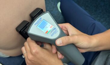
Fact: False. Measuring EMG potentials without a normative data base is as ridiculous as taking a temperature using an unmarked thermometer. A normative data base compiled by authors indicates that the EMG potentials in a painfree population vary from segment to segment.12
Statement: The patient’s skin should be sprayed with a conductive ECG solution prior to scanning.
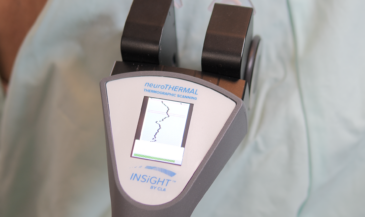
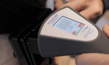
Fact: Using such a procedure produces electrode “bridging” which may invalidate readings. If a conductive gel or liquid is used, it should be applied to the electrodes, not the patient directly. High impedance scanners minimize this problem. Most patients can be scanned without skin preparation or coupling agents if equipment with an input impedance of one million megohms or greater is used.
Statement: All EMG systems are basically the same.
Fact: False. Be certain that your EMG system has an input impedance of at least one million megohms and has a normative data base collected at an accredited chiropractic college. Support is also important. Does the manufacturer provide you with access to chiropractic and legal EMG experts who are published authors? Is a tollfree number provided for computer and software support? Finally, be certain that your EMG system is FDA registered.
Paraspinal EMG scanning is one of the most exciting technologies in contemporary chiropractic practice. This procedure is a valuable tool in detecting and measuring changes in paraspinal muscular activity. Chiropractors using this equipment should understand the strengths and weaknesses of the technique.
References
1. Spector, B: “Surface electromyography as a model for the development of standardized procedures and reliability testing.” JMPT 2(4); 214, 1979.
2. Cram, J: “Clinical EMG: muscle scanning for surface recordings.” Seattle, WA. Biofeedback Institute of Seattle, 1986.
3. Komi, P and Buskirk, E: “Reproducibility of electromyographic measurements with inserted wire electrodes and surface electrodes.” Electromyography 10:357, 1970.
4. Thomson, J; Erickson, R and Offord, K: “EMG muscle scanning: stability of handheld electrodes.” Biofeedback Self Regulation 14(1):55, 1989.
5. Cailliet, R: “Soft Tissue Pain and Disability.” Philadelphia, PA. F.A. Davis Co. 1982.
6. Gentempo, P: “Evaluating soft tissue injuries with electromyography.” Today’s Chiropractic, May/June 1988, p.83.
7. Kent, C: “Documenting the vertebral subluxation complex with electromyography.” The Chiropractic Journal, April 1988. p. 20.
8. Gentempo, P and Kent, C: “The role of paraspinal EMG scanning in managing the vertebral subluxation complex.” The American Chiropractor, 12(3):7, 1990.
9. Yanof, H: “Biomedical Electronics.” Philadelphia, PA. F.A. Davis Co. 1972.
10. Kent, C and Fitzsimons, W: “Admissibility of electromyographic findings in personal injury cases.” Digest of Chiropractic Economics, March/April 1988. p.42.
11. Clements, H and Kent, C: “Going to court: using paraspinal EMG scans as evidence in personal injury cases.” The American Chiropractor, 11(8):28.
12. Kent, C and Gentempo, P: “Improving your practice with stateoftheart EMG.” Today’s Chiropractic, May/June 1989. p.56.