An important issue when selecting clinical examination procedures is reliability. Reliability is a measure of the ability to reproduce a measurement, which is expressed as a coefficient ranging from 0.00 to 1.00. Perfect reliability results in a coefficient of 1.00, while chance agreement would be 0.0.
As an example, Hass and Panzer (1) noted that the inter-examiner reliability of palpation for muscle tension is poor, with coefficients ranging 0.07 to 0.20. As presented below, research data indicates that the reliability of SEMG is clearly superior to palpation for muscle tension.
Decades of research by independent investigators show that surface electrode electromyography exhibits very good to excellent test-retest reliability. Spector (2) conducted a study at New York Chiropractic College which yielded correlation coefficients ranging from 0.73 and 0.97. Komi and Buskirk (3) compared the test-retest reliability of surface electrodes vs. needle electrodes in the deltoid muscle. The average test-retest reliability for surface electrodes was 0.88 compared to 0.62 for inserted electrodes.
Giroux and Lamontagne (4) compared the reliability of surface vs. intramuscular wire EMG of the trapezius and deltoid muscles during isometric and dynamic contractions. The statistical analysis on the integrated EMG was a factorial analysis model with repeated measures. They found that surface EMG was more reliable than inserted wire EMG on day-to-day investigations.
Andersson et al (5) compared the electrical activity in lumbar erector spinae muscles using inserted electrodes and surface electrodes. They found that the standard deviations and coefficients of variation for wire electrodes were greater than those for surface electrodes. They concluded, “Wire electrodes are more sensitive to electrode location and give estimates with less precision than surface electrodes.”
Other investigators have evaluated the reliability of surface electrode techniques using hand-held electrodes. This method is referred to as surface EMG scanning. Thompson et al (6) of the Mayo clinic found that the scanning electrode technique correlated well with the “gold standard” of attached electrode technique. Cram et al (7) evaluated the reliability of surface EMG scanning in 102 subjects in the sitting and standing positions. SEMG scans were performed on three occasions approximately one hour apart on the same day. The median correlation was 0.64. The authors concluded, “With adequate attention given to skin preparation, EMG sensors held in place by hand with a light pressure provide reliable results.”
In a review of surface EMG, Lofland et al (8) state that “Recent methodologically sound research has shown modern multichannel surface EMG to be reliable and valid.”
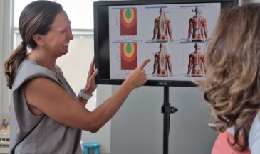
The most exciting recent study examining surface EMG reliability (9) was conducted at the NZCA School of Chiropractic in New Zealand. The study involved chiropractic care provided by 19 chiropractic interns in a teaching clinic. The equipment used was an Insight 7000 Subluxation Station.
Each of the 30 patients involved in the study received chiropractic examinations including static and motion palpation, joint play and end feel, Derefield-Thompson leg length assessment, and muscle challenge testing. Full spine x-rays were taken and analyzed when evidence of spinal dysfunction was determined.
One or more adjusting procedures, including Palmer Upper Cervical, Diversified, Gonstead, and Thompson Terminal Point Technic were used. Baseline SEMG scans were performed prior to initiation of chiropractic care. Follow up SEMG scans were performed one week after the first adjustment, and four weeks after the first adjustment.
To evaluate intra-examiner reliability, a two-tailed paired t-test was used to compare means of the intra-examiner trial population samples. This approach was used because correlation coefficients could reveal a high level of self-consistency, but mask examiner error. The objective was to determine if there was so much variability in the samples that they could be distinguished as different. The article reported that in 99.7% of the paired trials, variation was not sufficient to distinguish the samples as significantly different. This suggests an acceptable level of examiner consistency.
The investigators further concluded, “Under the conditions of this study, it is concluded that SEMG is an objective measure of change which can be used as an assessment of patient progress.”
References
1. Haas M, Panzer DM: “Palpatory diagnosis of subluxation.” In: Gatterman M (ed): “Foundations of Chiropractic Subluxation.” St. Louis, MO, Mosby, 1995.
2. Spector B: “Surface electromyography as a model for the development of standardized procedures and reliability testing.” JMPT 1979;2(4):214.
3. Komi P, Buskirk E: “Reproducibility of electromyographic measurements with inserted wire electrodes and surface electrodes.” Electromyography 1970;10:357.
4. Giroux B, Lamontagne M: “Comparisons between surface electrodes and intramuscular wire electrodes in isometric and dynamic conditions.” Electromyogr Clin Neurophysiol 1990;30:397.
5. Andersson G, Jonsson B, Ortengren R: “Myoelectric activity in individual lumbar erector spinae muscles in sitting. A study with surface and wire electrodes.” Scand J Rehab Med 1974 Suppl;3:91.
6. Thompson J, Erickson R, Offord K: “EMG muscle scanning: stability of hand-held electrodes.” Biofeedback Self Regul 1989;14(1):55.
7. Cram JR, Lloyd J, Cahn TS: “The reliability of EMG muscle scanning.” Int J Psychosomatics 1994;41:41.
8. Lofland KR, Mumby PB, Cassisi JE, et al: “Assessment of lumbar EMG during static and dynamic activity in pain-free normals: implications for muscle scanning protocols.” Biofeedback and Self-Regulation 1995;20(1):3.
9. Kelly S, Boone WR: “The clinical application of surface electromyography as an objective measure of change in the chiropractic assessment of patient progress: a pilot study.” Journal of Vertebral Subluxation Research 1998;2(4):175.