In traveling throughout the chiropractic world, I frequently encounter D.C.s who mindlessly regurgitate misinformation with the demeanor of one who has spoken directly with God. Examples are, “X-ray is useful only for pathology; line drawing is unreliable” or “14 x 36 full spine films expose the patient to more radiation than sectional studies” and the ever popular “there is no such thing as subluxation.”
Tracking down the primary source of this garbage poses a formidable challenge. But in many cases, it is easy to identify the messenger — a lazy chiropractic college faculty member who hasn’t bothered to check the facts, or, worse, is promoting a political agenda. Thankfully, these individuals are rare, but every college seems to have at least one. Students say these folks speak with such authority that they are rarely questioned. Some mention “recent studies” for which citations are rarely offered.
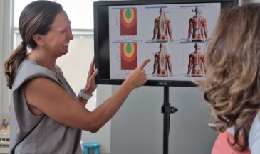
Let’s begin by examining the accuracy of lumbar spine film interpretation for pathology. A study was conducted where medical and chiropractic radiologists, medical and chiropractic clinicians, as well as medical and chiropractic students took a test on radiographic interpretation consisting of nineteen cases of clinically important findings. (1)
Here’s how they did (percent correct):
- Chiropractic radiologists — 71%
- Skeletal radiologists and fellows (medical) — 70.18%
- General medical radiologists — 51.64%
- Medical clinicians — 31.26%
- Chiropractic clinicians — 28.8%
- Chiropractic students — 20.45%
- Medical students — 5.74%
While it is gratifying that the D.C.s in the study outperformed their medical counterparts, where I went to school 71% was failing — at best a low “D.” With general medical radiologists (board certified) the results are barely above 50%. It’s a good thing these folks aren’t taking chiropractic licensing exams.
The other results speak for themselves. Please realize I am not suggesting that radiography be abandoned, or that all films must be read by a radiologist. I am simply trying to demonstrate that radiographic assessment of pathology is a very imperfect art.
So much for pathology. What about line drawing? Is it reliable? Reliability studies investigating several systems of biomechanical analysis have been published. A sampling follows.
Plaugher and Hendricks evaluated the inter-examiner reliability of the Gonstead pelvic marking system. Agreement for categorizing listings was impressive. Inter-examiner concordance for listings of the ilia, sacrum, symphysis pubis, and femur head height was evaluated by calculating the kappa values for each. The resulting kappa values ranged from .4849 (moderate) to .8161 (excellent). (2)
In addition to Gonstead pelvic analysis, proponents of 14 x 36 full spine radiography also use the procedure to evaluate vertebral body rotation and lateral flexion malposition. Zengel and Davis investigated how projectional distortion affects such determinations. They concluded, “as long as a given osseous segment is compared to its adjacent segment (as in analysis for subluxation), the apparent vertebral rotation may be regarded as a sufficiently accurate representation of the actual rotation of the vertebra.” (3,4)
Studies have yielded results supporting the reliability of cervical spinographic techniques. The preponderance of evidence supports the reliability of these procedures when properly performed.
Grostic and DeBoer did a retrospective study of 523 patients evaluating roentgenographic measurements of atlas laterality and rotation pre and post adjustment. Statistically significant changes in the postulated direction of atlas positioning were reported. (5)
Jackson et al. studied the inter-and intra-examiner reliability of upper cervical x-ray marking. Six practitioners evaluated 30 radiographs. The study revealed very good intra- and inter-examiner reliability for the procedure employed. (6) Leach investigated the effect of chiropractic care on hypolordosis of the cervical spine. A significant improvement in the cervical curve was noted in patients receiving chiropractic care. (7)
The inter- and intra-examiner reliability of certain mensuration procedures in the lumbar spine ranged from .66 to .98 in a study by Troyanovich et al. (8) The inter- and intra-examiner reliability of cervical geometric line drawing procedures used in the Chiropractic Technique ranged from .72 to .98. (9)
One negative study is often cited by critics of upper cervical spinographic analysis. Sigler and Howe conducted an inter- and intra- examiner reliability study of a method for measuring atlas laterality. Twenty x-rays were marked by three different doctors. This study concluded that because of the ranges of error, differences produced using this system will be just as likely due to marking error as from actual atlas position change. (10) This study has several significant shortcomings. These include small sample size and a conclusion which cannot properly be drawn from the data presented.
What about the radiation levels of full spine vs. sectional radiography? Dosimetry studies using supplemental filtration and single-speed screens revealed that the 14 x 36 AP spinograph actually resulted in lower radiation levels than sectional AP films of like-sized subjects. As Hildebrandt observed, “It has been shown that it is possible to produce reasonably good diagnostic quality full-spine roentgenographs with less radiation exposure to the patient than when the same full-spine areas are exposed by smaller sectional views.” (11)
Phillips stated, “Anteroposterior views of the spine on a 14 x 36 inch exposure can be produced with acceptable quality.”(Ref #) Hildebrandt cites a comparative study conducted by the Bureau of Radiological Health stating, “In this study, it was shown that it was in fact possible to obtain diagnostic full-spine films with a skin dose exposure as low as 128.8 mR, while separate lumbar and thoracic films taken according to standard exposure practices delivered 166.1 and 184.5 mR respectively.” (12)
Buehler and Hrejsa evaluated lead-acrylic compensating filters in chiropractic full-spine radiography. The concluded that this system “is capable of producing full spine radiographs with good to above average imaging quality.” It was further noted that this filtration system was generally equivalent in radiation dose reduction to other systems. (13)
It’s time to stop perpetuating acquired ignorance. Challenge those making questionable claims to substantiate their pompous proclamations with references. Do your homework, and think for yourself.
References
1. Taylor JAM, Clopton P, Bosch E, et al: “Interpretation of abnormal lumbosacral spine radiographs. A test comparing students, clinicians, radiology residents, and radiologists in medicine and chiropractic.” Spine 1995;20(10):1147.
2. Plaugher G, Hendricks AH: “The interexaminer reliability of the Gonstead pelvic marking system.” Proceedings of the 1990 International Conference on Spinal Manipulation. Arlington, VA, 1990, pp 93-98.
3. Zengel F, Davis BP: “Biomechanical analysis by chiropractic radiography: Part II. Effects of x-ray projectional distortion on apparent vertebral rotation.” J Manipulative Physiol Ther (1988 Oct) 11(5):380-9.
4. Zengel F, Davis BP: “Biomechanical analysis by chiropractic radiography: Part III. Lack of effect of projectional distortion on Gonstead vertebral endplate lines.” J Manipulative Physiol Ther (1988 Dec) 11(6):469-73.
5. Grostic JD, DeBoer KF: “Roentgenographic measurement of atlas laterality and rotation: a retrospective pre- and post-manipulation study.” J Manipulative Physiol Ther (1982 Jun) 5(2):63-71.
6. Jackson BL, Barker W, Bentz J, Gambale AG: “Inter- and intra- examiner reliability of the upper cervical x-ray marking system: a second look.” J Manipulative Physiol Ther (1987 Aug) 10(4):157-63.
7. Leach RA: “An evaluation of the effect of chiropractic manipulative therapy on hypolordosis of the cervical spine.” J Manipulative Physiol Ther (1983 Mar) 6(1):17-23.
8. Troyanovich S, Robertson G, Harrison D, Holland B: “Intra- and interexaminer reliability of the Chiropractic Biophysics lateral lumbar radiographic mensuration procedure.” J Manipulative Physiol Ther (1995 Oct) 18(8):519-524.
9. Jackson B, Harrison D, Robertson G, Barker W: “Chiropractic Biophysics lateral cervical film analysis reliability.” J Manipulative Physiol Ther (1993 Jul) 16(6):384-391.
10. Sigler DC, Howe JW: “Inter- and intra-examiner reliability of the upper cervical x-ray marking system.” J Manipulative Physiol Ther (1985 Jun) 8(2):75-80.
11. Hildebrandt RW: “Chiropractic Spinography.” Des Plaines, IL. Hilmark Publications, 1977, p. 18.
12. Phillips RB: “The use of x-rays in spinal manipulative therapy.” In Haldeman S (ed) “Modern Developments in the Principles and Practice of Chiropractic.” Norwalk, CT. Appleton-Century-Crofts, 1980, p. 204.
13. Buehler MT, Hrejsa AF: “Application of lead-acrylic compensating filters in chiropractic full spine radiography: a technical report.” J Manipulative Physiol Ther (1985 Sep) 8(3):175-80.