By Dr. Christopher Kent
Last month’s column described a “check list” approach for cervical spine radiographs. This month we continue with check lists for the thoracic and lumbar spine.
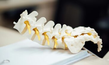
AP thoracic
On AP radiographs, the vertebral bodies, pedicles, and spinous processes in the thoracic and lumbar region form a likeness to a face, sometimes called the “blockhead.” The vertebral bodies form the outline of the head, which should be flat on the top and bottom. The pedicles represent the “eyes” of the face, and both should be visible. Finally, the spinous process represents the nose. By looking for this “face” in each thoracic and lumbar segment, the chiropractor may quickly determine potential abnormalities.
Procedure:
1. Check vertebral end plates for compression fractures, Schmorl’s nodes sclerosis, or other irregularities.
2. Check vertebral bodies for osteopenia and hemangioma.
3. Check pedicles for evidence of osteolytic activity.
4. Check spinous processes.
5. Check ribs.
6. Check clavicles.
Selected Findings:
Osteolytic metastatic carcinoma
Osteopenia
Multiple myeloma
Compression fracture
Hemivertebra
Spina bifida
Schmorl’s node
Scoliosis
Hemangioma
Fractured ribs
Fractured clavicle
Lateral thoracic
Procedure:
1. Check vertebral end plates for compression, Schmorl’s nodes, Scheuermann’s disease, sclerosis, or other irregularities.
2. Check vertebral bodies for osteopenia, hemangioma, or other abnormalities of density.
3. Check pedicles for evidence of osteolytic activity.
4. Draw George’s line (mentally, if not on the film).
5. Draw an anterior body line (mentally, if not on the film).
6. Check the disc spaces.
Selected Findings:
Compression fracture
Schmorl’s node
Scheuermann’s disease
Persistent notochord
Degenerative joint disease
Ankylosing spondylitis
Osteopenia
Infection (discitis, osteomyelitis, Pott’s disease)
AP lumbar and pelvis
Procedure:
1. Check vertebral end plates for compression, Schmorl’s nodes, sclerosis, osteophytes, or other abnormalities.
2. Check vertebral bodies for osteopenia, blastic foci, hemangioma or other irregularities.
3. Check pedicles for osteolytic or osteosclerotic activity.
4. Check facet facings for tropism or degeneration.
5. Check pars for evidence of spondylolysis. Check for “inverted Napoleon hat” sign.
6. Check spinous processes.
7. Check transverse processes.
8. Draw Shenton’s and iliofemoral lines (mentally).
9. Check hip joints and femora.
10. Check pelvic brim for Paget’s disease and Otto’s pelvis.
11. Check soft tissues for stones, calcification or aneurysm of the abdominal aorta.
12. Check psoas shadow.
13. Look for abnormal gas patterns (ileus, etc.).
Selected Findings:
Osteolytic metastatic carcinoma
Osteoblastic metastatic carcinoma
Paget’s disease
Spina bifida
Knife-clasp deformity
Spondylolisthesis (Brailsford’s bowline) Sacralization
Sacroilitis
Ankylosing spondylitis
Reiter’s syndrome
Psoriatic arthritis
Osteitis condensans ilii
Tropism
Laminectomy
Fractures (hip, pelvis, seat belt, compression, transverse process)
Degenerative joint disease
Abdominal aortic aneurysm
Kidney stones
Gallstones
Adynamic ileus
Psoas abscess
In hip of a child:
Legg-Calve-Perthes disease
Fibrous dysplasia
Congenital hip dysplasia
Slipped epiphysis
Lateral lumbar
Procedure:
1. Check vertebral end plates for compression, sclerosis, osteophytosis, Schmorl’s nodes, and other abnormalities.
2. Check vertebral bodies for osteopenia, osteosclerotic foci, hemangioma, or other changes in shape or density.
3. Check pedicles.
4. Check disc spaces.
5. Draw George’s line (mentally).
6. Check disc spaces.
7. Check abdominal aorta for calcification and aneurysm.
Selected Findings:
Spondylolisthesis
Compression fracture
Osteolytic metastatic carcinoma
Osteoblastic metastatic carcinoma
Hemangioma
Limbus
Avulsion fracture
Abdominal aortic aneurysm
Retrolisthesis
Ankylosing spondylitis
Lumbar obliques
Procedure:
1. Check pars interarticularis
2. Check pedicles
3. Check articular surfaces
Selected Findings:
Spondylolysis
Osteolytic metastatic carcinoma
Osteoblastic metastatic carcinoma
Degenerative joint disease
Conclusion
The chiropractor is encouraged to expand and modify these lists to suit the needs of individual patients and specific techniques.
References
- Erhardt R. “Roentgenology of the spine and pelvis notes.” Privately published, Atlanta, GA. No date.
- Kent C: “Protocols for the analysis of plain skeletal radiographs.” Digest of Chiro Economics 31(3): 70, 1988.
- Macrae J: “Roentgenometrics in Chiropractic.” Privately published. 1983.
- Resnick D, Niwayama G: “Diagnosis of Bone and Joint Disorders.” W. B. Saunders, Philadelphia, PA. 1988.
- Yochum T, Rowe L: “Essentials of Skeletal Radiology.” Williams and Wilkins, Baltimore, MD. 1987.