By Dr. Christopher Kent
Accurate spinal analysis is the keystone of successful chiropractic practice. To fulfill the mission of chiropractic in the contemporary health care environment, the chiropractor must be skilled in locating vertebral subluxations. In addition, the chiropractor is responsible for communicating these findings in terms which can be understood by the scientific community and third party payors.
VSC components
Classic chiropractic philosophy defines the vertebral subluxation in terms of four criteria:
- Loss of juxtaposition
- Occlusion of an opening
- Impingement of a nerve
- Interference with mental impulses [1]
- Contemporary chiropractic philosophy acknowledges five components of the vertebral complex:
- Level of the subluxation (such as C-5)
- Connective tissue involvement, including disc, other ligaments, fascia, and muscles
- The neurological component, including nerve roots and spinal cord
- Altered biomechanics
- Advancing complications in the innervated tissues and/or the patient’s symptoms [2]
Both concepts address biomechanical and physiological manifestations of vertebral subluxation. Structural and biomechanical components of the VSC can be objectively demonstrated using radiography and postural analysis. The soft tissue components of the VSC, however, often rely on subjective criteria which may be difficult to duplicate.
For example, most orthopedic tests require the execution of a mechanical maneuver designed to elicit pain or reduplication of symptoms. The examiner must depend upon subjective reports of pain or other symptoms to interpret such tests. This makes traditional orthopedic testing procedures vulnerable to charges of subjectivity.
Similarly, most neurological examination procedures are highly subjective. Reflex findings and manual muscle tests are vulnerable to examiner bias and interpretation. Sensory examinations rely upon patient reports. Such reports may be colored by the patient’s desire to please the examiner, or a desire to produce abnormal findings in substantiation of an injury or illness.
Muscular changes in the VSC
For decades, chiropractors have palpated the soft tissue structures surrounding the spine for evidence of “taut and tender” fibers. Such areas are thought to be due to vertebral subluxation. According to Stephenson, motor activity is one of the nine primary functions of life. [1]
B.J. Palmer stated, “Life is motion; motion is life. The absence of motion is death… In human beings, motion is produced by muscles… That which moves muscles is nerve force.” [3] Abnormal muscle function is still acknowledged as a clinical manifestation of vertebral subluxation.
B.J. Palmer was responsible for overseeing the development of instruments designed to record the activity of nerves, and locate the anatomical sites where nerve interference occurred. The electro-encephaloneuromentimpograph was developed in the B.J. Palmer research clinic in an attempt to evaluate changes in nerve impulse propagation. These changes were thought to be caused by vertebral subluxations. [4]
Electromyography
Electromyography is a technique of recording the electrical activity of muscles. Changes in EMG activity have been used to document the presence of nerve root irritation, muscle, spasm, and the neurological manifestations of disc lesions. [5,6,7,8,9]
Although electromyography has been used as a research tool in chiropractic, its application in clinical practice was limited. [10,11,12]
This was due to the expense and complexity of conventional electromyographic equipment and techniques.
Chiropractic EMG scanning
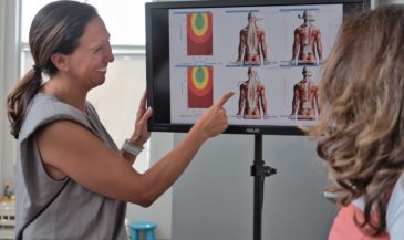
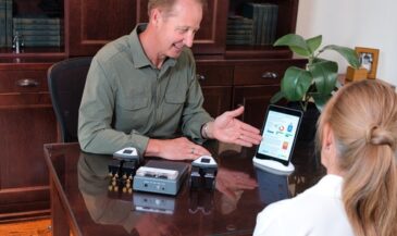
Contemporary computer technology has made objective documentation of the soft tissue component of the VSC a practical and cost-effective reality. Chiropractic EMG scanning involves the use of a small, hand-held pickup which is applied to the skin overlying the paravertebral muscles.
The electrical signal produced by the paravertebral muscles is amplified, filtered, fed to a display, and recorded. A printed report can be generated by computer. The technique is completely non-invasive. There is no exposure to ionizing radiation, and no electricity is introduced into the body. The equipment simply records the electrical activity which is always there. [13,14]
Normative data
The results of the paraspinal EMG scans can be compared to normative data gathered at the University of California-Irvine and the Swedish Hospital Pain Center. [15] In patients with vertebral subluxations, abnormal patterns are often dramatic.
Equally impressive are the EMG changes which follow chiropractic adjustment. [16] Hartfield Instruments has developed software which performs complex statistical calculations, and produces data collection and analysis techniques, a full spine EMG scan can be completed in minutes. The actual scan can be performed by a chiropractic assistant, saving valuable doctor time.
Spinal range of motion studies using EMG scanning
In addition to static EMG readings, computerized EMG scanning can be used to evaluate the dynamics of the spine. By monitoring paravertebral EMG potentials as the spine is taken through ranges of motion, specific areas of abnormality may be detected and evaluated. Dynamic EMG scanning provides the chiropractor with a “motion picture” of spinal kinematics. [17]
VSC documentation in court
Documenting the soft tissue component of the VSC in personal injury cases poses a challenge to chiropractors and attorneys. Many cases are built around subjective findings which may lead to accusations of malingering or exaggeration. At best, serious doubt may be raised in the mind of the judge or jury concerning the reality or severity of the injury.
Electromyography has a distinguished record in courts of law. There are 29 recorded cases where electromyographic evidence was admitted at the appellate or state supreme court level. In one instance, electromyography was accepted as a standard to judge thermography. [18] In other cases, electromyographic findings were used to substantiate injury or reveal malingering. [19]
Third party payors and electromyography
Insurance carriers generally cover chiropractic EMG scans, provided that clinical need is documented. Clinical need can be documented on the basis of subjective findings such as muscle spasm, tenderness, limited range of motion, etc. The objective data produced by EMG scanning can be used to demonstrate the efficacy of chiropractic care, and the need for continued care. If insurance carriers question the need for continued chiropractic services, it is possible to objectively document the existence of a problem, and the fact that improvement is taking place.
Conclusion
The modern chiropractor committed to the location and correction of vertebral subluxations is often called upon to communicate clinical findings to persons with little or no understanding of chiropractic philosophy. Electromyography is a technique widely accepted in the scientific and legal community. Computerized paraspinal EMG scanning provides a rapid, cost-effective method of documenting the soft tissue component of the VSC. EMG scanning benefits the patient by providing improved spinal analysis and third party compliance.
It saves valuable time, freeing the doctor to concentrate on adjusting. Finally, it benefits the profession by demonstrating the reality of the VSC to the scientific and medical communities in terms they can understand.
References
1. Stephenson, R: “Chiropractic Textbook.” Davenport, IA, The Palmer School of Chiropractic, 1927.
2. Herfert, R: “Communicating the Vertebral Subluxation Complex.” Detroit, MI, Herfert Chiropractic Clinics, 1986.
3. Palmer, B: “The Law of Life.” Audiotape of address given at the Palmer School of Chiropractic.
4. Motion picture: “The B.J. Palmer Chiropractic Clinic,” no date.
5. Hoyt, W., Hunt, H., et al: “Electromyographic assessment of chronic low back pain syndrome,” JAOA 80: 728-730, 1981.
6. Jebsen, H., Long, F: “Radiculopathy and the electromyogram in disability applicants,” Arch Phys Med Rehabil 54:471-474, 1973.
7. Johnson, E., Melvin, J: “Value of electromyography in lumbar radiculopathy,” Arch Phys Med Rehabil 52: 239-243, 1971.
8. Knuttson, B: “Comparative value of electromyographic, myelographic, and clinical-neurological examination in diagnosis of lumbar root compression syndrome,” Acta Orthop Scan 49 (Suppl): 1-135, 1961.
9. Grabel, J: “Electromyographic study of low back tensions in subjects with and without low back pain,” Dissertations and Abstracts International 34 (B): 2929-2930, 1973.
10. Triano, J: “Objective electromyographic evidence for use and effects of lift therapy,” JMPT 6, 1: 13-16, Mar 1983.
11. Meeker, J., Gahlinger, P: “Neuromusculoskeletal thermography: a valuable diagnostic tool?,” JMPT 9, 4: 257-266, Dec 1986.
12. Kent, C. Simons, V: “Electromyographic evaluation of vertebral challenge analysis,” Digest of Chiro Econ 21, 4: 30-33, 1979.
13. Kent, C., Hyde, R: “Potential applications for electromyography in chiropractic practice,” Digest of Chiro Econ 30, 2: 20-25, 1987.
14. Brody, S: “Practical chiropractic electromyography,” Digest of Chiro Econ 30, 2: 28-31, 1987.
15. Cram, J: “Clinical EMG: Muscle Scanning for Surface Recordings,” Seattle, WA, Biofeedback Institute of Seattle, 1986.
16. Brody, S: “Electromyographic and x-ray evaluation of the lumbar and thoracic spine for the balancing and leveling of the pelvis and correction of scoliosis,” Digest of Chiro Econ 30, 3: 68-71, 1987.
17. Brody, S: “The orthopedic range of motion exam using EMG scanning,” Digest of Chiro Econ 30, 4: 56-57, 1988.
18. Superior Court of New Jersey, 530 A.2d 56.
19. Kent, C., Fitzsimons, W: “Admissibility of electromyographic evidence in court,” submitted for publication: Digest of Chiro Econ.