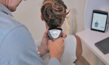
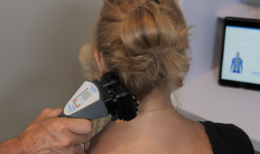
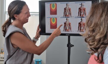
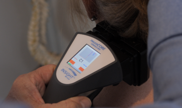
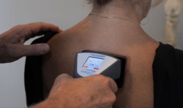
By Drs. Christopher Kent and Patrick Gentempo, Jr. Paraspinal EMG scanning is a popular technique for evaluating and characterizing the muscular dysfunction associated with the vertebral subluxation complex. Some critics of the technology have alleged that it is not a scientifically sound technique. The purpose of this paper is to demonstrate that EMG scanning is a reliable and scientifically sound procedure. Although static and motion palpation are popular techniques, they are only as effective as the examiner is skilled. A review of the literature on the interexaminer reliability of motion palpation of the lumbar spine demonstrated marginaltono reliability. Even in the hands of skilled clinicians, palpatory findings are highly subjective and difficult to quantify. [1-7] Some doctors feel rangeofmotion studies may be used to obtain the same information as that provided by paraspinal EMG studies. Paraspinal muscle dysfunction may result in altered or asymmetrical spinal motion, but is certainly not the only condition which may do so. Arthritic, congenital, metabolic, infectious, and neurological diseases may also present with alterations of spinal motion. These conditions may or may not be accompanied by paraspinal muscle dysfunction. [8,9] These techniques may be useful in clinical practice, but are not substitutes for paraspinal EMG studies. We urge readers to consider the following:
- Paraspinal EMG scanning is a reliable technique. Komi and Buskirk found that the test/retest reliability of surface electrode EMG was superior to that of inserted electrode technique. The average test/retest reliability for surface electrodes was 0.88, compared to 0.62 for inserted electrode EMG. 10 Cram reported a mean reliability coefficient of 0.83 for surface EMG scanning. [11]
Spector conducted a study to determine the reliability of paraspinal surface EMG at New York Chiropractic College. Results of the study yielded correlation coefficients ranging between 0.73 and 0.97. [12] Thompson et al of the Mayo Clinic found EMG scanning technique correlates highly with attached electrode technique. [13] In summary, the reliability of surface EMG has been documented in studies conducted with New York Chiropractic College, the Mayo Clinic and the University of CaliforniaIrvine. A cooperative research program is currently underway at the University of the PacificStockton and Palmer College of ChiropracticWest. Few analytical techniques in chiropractic display such a high level of reliability.
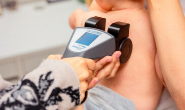
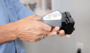
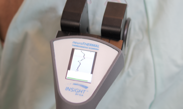
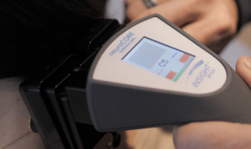
- The technique is completely noninvasive. There is no piercing of the skin. No electrical signal of any kind is introduced into the body. The instrument simply measures the muscular activity that is present in the patient. There are no contraindications to performing the study.
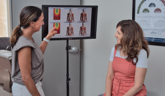
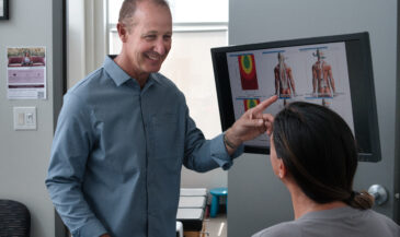
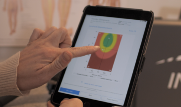
- The technique is directly applicable to the practice of chiropractic. Abnormal paraspinal muscle activity has traditionally been used to detect and characterize vertebral subluxations. Spinal palpation seeks to locate “taut and tender” fibers associated with areas of subluxation. Paraspinal EMG scanning eliminates the subjectivity inherent in palpation. Paraspinal EMG scans, taken in concert with other examination findings assist the chiropractor in determining:
- areas of possible subluxation
- areas of muscle splinting
- severity of the condition
- degree of asymmetry of paraspinal muscle contraction [14-17]
- Chiropractic adjustments alter paraspinal EMG readings. Chiropractors have often observed dramatic palpatory changes in paraspinal muscles pre and postadjustment. Shambaugh [18] conducted a study where surface electrodes were used to measure paraspinal EMG activity before and after chiropractic adjustment. This study was reported in JMPT, a refereed, peerreviewed journal. Shambaugh concluded, “Results of this study show that significant changes in muscle electrical activity occur as a consequence of adjusting. Similar findings were reported in a study conducted by the osteopathic profession. Ellestad et al found that paraspinal EMG activity decreased in patients following osteopathic manipulation. Such changes were not observed in controls. [19]
- The technique has been accepted by courts of law. In their test, “Proving Medical Diagnosis and Prognosis,” Houts and Marmor state: “Properly used, the EMG scanning technique is far more persuasive in the courtroom than is a report of needle EMG. You can present the jury with mathematical, tangible physical evidence which they can see.” [20] A memorandum in support of admission of EMG muscle scans which was filed in superior court, state of Washington, county of Spokane stated, “There is no legal basis for the exclusion of the EMG muscle scan when a proper foundation is laid for the introduction of such scientifically accepted testing. EMGs have been used for many years. Muscle scan testing has been admitted in numerous courts, including this court. [21]
- The technique has been employed under the aegis of accredited chiropractic colleges. Dr. Kent has personally taught the technique in the regular curriculum of Palmer College of ChiropracticWest, and at the postgraduate level for Texas Chiropractic College.
Paraspinal EMG scanning falls within the scope of chiropractic for the following reasons: 1. It has been documented in studies conducted by universities, hospitals, and chiropractic colleges. It is a well established procedure in the healing arts. According to Dr. Jeffrey Cram who developed the technique of muscle scanning, surface EMG scanning is currently utilized in 80% of the pain clinics in the United States. [22] 2. It is completely noninvasive. 3. The technique is useful as an aid in detecting and characterizing vertebral subluxations. 4. EMG scans may be used to determine patient response to chiropractic care. 5. The technique has been taught in accredited chiropractic colleges. 6. EMG data has been accepted in courts of law. 7. The technique is well documented in chiropractic literature, including chiropractic trade publications and refereed journals. 8. The most vocal critics of the technique are those who have no formal training in the procedure, have not utilized it in clinical practice, and have not documented their disparaging remarks with references. Paraspinal EMG scanning has a solid foundation in the medical, chiropractic, and scientific literature. Properly used, it is an invaluable technique in the evaluation of the vertebral subluxation complex. References 1. Keating J: “Interexaminer reliability of motion palpation of the lumbar spine: a review of the quantitative literature.” Proceedings of the Scientific Symposium on Spinal Biomechanics, International Chiropractors Association, May 1921, 1989. 2. Cassidy J, Potter G: “Motion palpation of the lumbar spine.” JMPT 1979; 2(3):151. 3. Gonella C, Paris S, Kutner M: “Reliability in evaluating passive intervertebral motion.” Phys Ther 1982; 62(4):436. 4. Bergstrom E, Courtis G: “An inter and intraexaminer reliability study of motion palpation in lateral flexion in the seated position.” Euro J Chiro 1986;34:121. 5. Love R, Brodeur R: “Inter and intraexaminer reliability of motion palpation for the thoracolumbar spine,” JMPT 1987 10(1):1. 6. Juli G, Bullock M: “A motion profile of the lumbar spine in an aging population assessed by manual examination.” Physiotherapy Practice 1987:3:70. 7. Boline P, Keating J, Brist J, Denver G: “Interexaminer reliability of palpatory examinations of the lumbar spine.” Am J Chiro Med 1988 1(1):5. 8. Calliet R: “Low Back Pain Syndrome.” FA Davis Co., Philadelphia, PA. 1977. 9. Calliet R: “Soft Tissue Pain and Disability.” FA Davis Co., Philadelphia, PA. 1977. 10. Komi P, Buskirk E: “Reproducibility of electromyographic measurements with inserted wire electrodes and surface electrodes,” Electromyography 10:357, 1970. 11. Cram J: Clinical EMG: “Muscle Scanning for Surface Recordings.” Biofeedback Institute of Seattle. Seattle, WA 1986. 12. Spector B: “Surface electromyography as a model for the development of standardized procedures and reliability testing.” JMPT 2(4):214, 1979. 13. Thompson J, Erickson R, Offord K: “EMG muscle scanning: stability of handheld electrodes.” Biofeedback Self Regul 14(1):55, 1989. 14. Kent C: “Documenting the vertebral subluxation complex with electromyography.” The Chiropractic Journal, April 1988. 15. Kent C. Gentempo P: “Computed tomography and electromyography in the evaluation of lumbar subluxation.” Today’s Chiropractic, September/October and November/December 1988. 16. Gentempo P: “Evaluating soft tissue injuries with electromyography: case studies.” Today’s Chiropractic, May/June 1988. 17. Gentempo P: “Characterizing the vertebral subluxation complex with paraspinal electromyography.” Submitted for publication. International Review of Chiropractic. 18. Shambaugh P: “Changes in electrical activity in muscles resulting from chiropractic adjustment: a pilot study.” JMPT 10(6):300, 1987. 19. Ellestad S, Nagle R, Boesler D, Kilmore M: “Electromyographic and skin resistance responses to osteopathic manipulative treatment for low back pain.” JAOA 88(8):991, 1988. 20. Houts M, Marmor L: “Proving Medical Diagnosis and Prognosis.” Matthew Bender, Times Mirror Books, 1989. 82A20. 21. Johnson v Carbon No. 862038064. Memorandum in support of admission of EMG muscle scans. Superior Court, State of Washington, County of Spokane. 22. Cram J: Personal communication.