Suboccipital tension is one of the most common yet misunderstood patterns chiropractors see in practice. A patient comes in talking about a headache that starts at the base of your skull, creeps into the back of your head, and sometimes settles behind your eyes. Another mentions neck pain, neck stiffness, or a feeling of constant tightness at the top of your neck that never quite goes away. Others describe tension headaches, migraine, or even migraine headaches without ever being told why those patterns keep returning. What ties these stories together is not coincidence. It is a predictable response from the nervous system when the upper cervical area is under sustained load.

In today’s world, suboccipital tension rarely develops overnight. It builds slowly through poor posture, forward head posture, long hours of screen time, and daily habits that place constant demand on the back of your neck. Add stress, neck injuries such as whiplash, or prolonged head forward positioning, and the suboccipital region becomes a control point the body relies on to maintain stability. When that control system starts to struggle, the symptoms get louder. Headaches appear. Range of motion shrinks. Muscle spasms show up. And patients begin cycling through different treatment options without lasting clarity.

For chiropractors, this topic matters because suboccipital tension is rarely just about muscle tension. It is about how the head and neck are being organized neurologically. Symptoms fluctuate, but the underlying pattern often remains. When you can identify that pattern and explain it clearly, you change the entire conversation. Instead of chasing headache relief alone, you guide patients toward understanding nervous system performance and why consistent care matters.

Defining Suboccipital Tension Through a Chiropractic Lens

Suboccipital tension refers to increased tone, tightness, or spasm in the small muscles located at the base of the skull, right where the head meets the cervical spine. Patients often point to the base of the skull or the suboccipital area and describe suboccipital pain that radiates upward. Sometimes it feels like pressure. Other times it feels sharp, especially with rotation of the head or extension and rotation. This is where suboccipital tension becomes closely linked to headache patterns that chiropractors recognize every day.

From a chiropractic perspective, this is not simply a sore muscle problem. The suboccipital region plays a key role in head and neck coordination. When neurological distress increases, the body often responds by tightening this area to protect balance and control. That response may temporarily stabilize the system, but over time it contributes to pain and tension, stiffness, and reduced range of motion. Patients notice headaches and neck discomfort, but the driver is deeper than the symptom.

Several types of headaches commonly overlap with suboccipital tension:

  • Suboccipital headache that begins at the base of your skull and travels into the back of your head
  • Tension headaches that feel like pressure or a band around the head
  • Cervicogenic headaches originating from the cervical region
  • Common headaches that patients may label as migraine without understanding the source

The important clinical point is that labels matter less than patterns. When headaches consistently involve the occipital region, the back of your neck, and the top of your neck, chiropractors should be thinking about how the head and neck system is adapting to stress. Suboccipital headache treatment becomes far more effective when it is grounded in neurological understanding rather than symptom suppression alone.

Functional Anatomy and Neurology of the Suboccipital Region

The suboccipital region is small, but it is neurologically dense. This group of muscles sits beneath the occiput and along the occipital bone, forming a control hub for head and neck positioning. The suboccipital muscle group includes rectus capitis posterior major, rectus capitis posterior minor, obliquus capitis superior, and obliquus capitis inferior. Together, this group of muscles manages subtle movements and stabilizes the upper cervical spine during everyday activity.

These small muscles coordinate movement between C1 and C2, the atlas and axis, which connect the head to the spine. Because they influence extension and rotation and fine-tune rotation of the head, even minor dysfunction can have outsized effects. When these muscles become overloaded, trigger points develop, protective tone increases, and patients report stiffness and pain at the base of the skull. Chiropractors often palpate tight suboccipitals and immediately recognize the pattern.

Neurologically, this area is closely associated with the occipital nerve and greater occipital nerve. Irritation in this region can contribute to symptoms that feel disproportionate to the size of the tissue involved. Patients may report sharp pain, discomfort behind your eyes, dizziness, or even visual disturbances. In some cases, the symptom pattern resembles occipital neuralgia, though not every patient with suboccipital tension fits that category. What remains consistent is that the nervous system is on alert.

This region does not work in isolation. Suboccipital tension often exists alongside involvement of the trapezius, deeper neck muscles, and the entire cervical spine. When the head rides forward, the back of your neck becomes a constant stabilizer. Over time, this contributes to muscle spasms, pain and discomfort, and reduced adaptability. Understanding this relationship helps chiropractors move beyond local tissue work and toward evaluating nervous system performance.

Causes, Contributing Factors, and Patterns Chiropractors Commonly See

The causes of suboccipital tension are rarely singular. Most patients present with layered contributors that build over time. Poor posture is at the top of the list. Forward head posture, a habitual slouch, and prolonged screen use place sustained load on the suboccipital region. Poor ergonomics compounds the problem, turning the back of your neck into a structural support rather than a mobile, adaptable system.

Neck injuries also play a significant role. Whiplash and other neck injuries often lead to protective guarding at the top of your neck. The suboccipitals tighten to protect the head and cervical spine, resulting in spasm, muscle spasms, and tenderness at the base of the skull. Patients may feel sharp pain during movement or describe that their neck feels locked. These patterns frequently coexist with headaches and neck complaints that resist simple solutions.

Stress and daily strain contribute to muscle tension as well. Chronic stress shifts the nervous system into a state where tone remains elevated. Jaw clenching, eye strain, and prolonged focus can feed tension into the neck and shoulder region. Over time, this contributes to pain and tension that settles into the suboccipital area. Patients may also report visual disturbances or nausea when symptoms flare.

For chiropractors, recognizing these patterns is essential. Symptoms fluctuate, but the neurological footprint often remains. This is why relying solely on how the patient feels on a given day can be misleading. Identifying the underlying pattern allows chiropractors to address the true drivers of suboccipital tension rather than repeatedly reacting to symptoms.

Conservative Management, Chiropractic Strategies, and Patient Education

Most patients with suboccipital tension have already explored conservative treatments before arriving at a chiropractic office. Massage therapy, physiotherapy, and advice from a physical therapist are common stops along the way. Self-care strategies such as a suboccipital stretch, heat, or self-massage using a peanut tool may offer temporary relief. These approaches can help calm local tissue response and restore some range of motion.

In chiropractic care, the focus expands beyond local symptom management. Adjustments aim to improve function in the cervical spine and restore coordination between the head and neck. This approach supports better movement, reduced guarding, and improved adaptability. Chiropractors often discuss posture, ergonomics, and sleeping positions as part of a broader strategy to prevent suboccipital flare-ups.

Patient education is critical. When patients understand that suboccipital tension reflects nervous system strategy rather than isolated muscle tightness, expectations change. Headache treatment becomes about improving performance rather than chasing symptoms. Suboccipital release techniques may help release tension temporarily, but lasting change comes from addressing the underlying pattern. When patients grasp this concept, they become more engaged in their care plan and less focused on short-term pain relief alone.

Using INSiGHT Scanning Technology to Understand and Track Suboccipital Tension

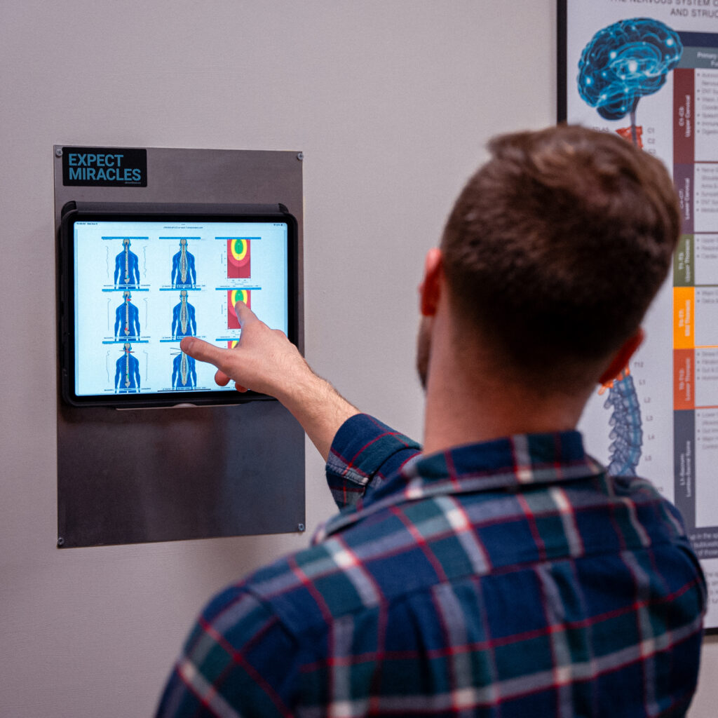
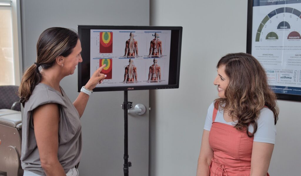
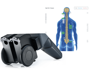
Suboccipital tension is well known for fluctuating. One day the headache is quiet. The next day it returns with intensity. This is where neurological scanning becomes invaluable. INSiGHT scanning technology provides objective exam data that helps chiropractors understand nervous system status beyond symptom reporting.

neuroTHERMAL scanning allows chiropractors to analyze segmental autonomic stress patterns that often correlate with upper cervical involvement. neuroCORE sEMG evaluates postural tension and motor tone reactions, offering insight into how the cervical region and surrounding muscle group are managing load. neuroPULSE HRV assesses adaptability and resilience, especially in patients whose symptoms are influenced by stress.

INSiGHT neuroTECH and Synapse software transform complex neurological data into clear scan views that patients can understand. These scans do not create care plans. Chiropractors interpret the data, combine it with examination findings, and design care plans based on their expertise. Progress scans provide proof your care is making a difference by documenting changes over time, even when symptoms fluctuate.

Clarity, Confidence, and a Better Conversation About Care

Suboccipital tension does not have to remain confusing for patients or chiropractors. When you recognize it as a neurological pattern rather than a simple muscle issue, the entire care conversation shifts. Patients begin to understand why headaches recur, why posture matters, and why consistency in care supports long-term change.

By combining chiropractic principles with INSiGHT scanning technology, chiropractors move from guessing to knowing. Objective analysis supports clearer communication, stronger patient confidence, and better tracking of nervous system performance. In a world where patients are overwhelmed with information, clarity is powerful.

Suboccipital tension is common, but it does not have to be limiting. When chiropractors focus on the nervous system, use objective data, and guide patients with confidence, they help transform recurring headaches and neck patterns into an opportunity for better performance, resilience, and long-term stability.

Dysautonomia is one of the most misunderstood nervous system disorders seen in modern clinical practice. For many people with dysautonomia, the frustration is not only the symptoms themselves but the lack of clarity around why the body seems unable to regulate basic functions. Dysautonomia is closely tied to the autonomic nervous system, the part of neurology responsible for automatic processes like blood pressure, heart rate, digestion, circulation, temperature regulation, and sweat responses. When that system is not regulating efficiently, the body becomes unpredictable.

In chiropractic offices, dysautonomia often appears long before the term is ever used. Dysautonomia patients may describe dizziness, palpitations, orthostatic lightheadedness, fatigue, brain fog, exercise intolerance, or digestive disruption. These dysautonomia symptoms may fluctuate daily, creating an array of symptoms that seem unrelated at first glance. Dysautonomia can affect how patients stand, move, recover, and function, even when they appear outwardly fine.

This matters deeply for chiropractors because dysautonomia is not rare. Estimates suggest that nearly 70 million people worldwide are affected by various forms of dysautonomia. Yet there remains a significant lack of awareness, leaving many people with dysautonomia struggling to find providers who understand autonomic nervous system dysfunction. Chiropractic, when grounded in neurological analysis and objective data, is uniquely positioned to help bring clarity to this complex condition.

Understanding Dysautonomia Through a Neurological Lens

Dysautonomia is an umbrella term used to describe disorders of the autonomic nervous system. In other words, dysautonomia is an umbrella term that encompasses multiple nervous system disorders where involuntary regulation breaks down. Dysautonomia is not a single diagnosis, nor is it a single disorder with one predictable presentation. Instead, dysautonomia can range from mild and intermittent to severe and disabling.

The autonomic nervous system governs processes the body performs without conscious thought. These include heart rate, blood pressure, breathing rhythms, digestion, circulation, pupil response, and sweat production. When autonomic nervous system disorders develop, these functions may fluctuate or fail to adapt appropriately to stress, posture, or activity. Dysautonomia have trouble regulating these processes, which explains why patients often experience different symptoms across multiple systems.

From a chiropractic perspective, it is critical to understand scope. Chiropractors do not diagnose dysautonomia. Diagnosis and treatment decisions, including medical testing such as a tilt table test, fall within medical neurology and cardiology. However, chiropractors can recognize patterns of autonomic dysfunction and autonomic nervous system dysfunction, especially when patients present with orthostatic complaints, blood pressure instability, and nervous system fatigue.

Dysautonomia may also be invisible during short clinical encounters. A patient’s blood pressure and heart rate may appear normal at rest, yet become unstable later in the day. This variability is why dysautonomia depends so heavily on pattern recognition and trend tracking rather than single data points. Understanding dysautonomia through a neurological lens allows chiropractors to focus on nervous system performance rather than chasing isolated symptoms.

Dysautonomia Symptoms and Common Clinical Patterns Chiropractors Observe

One of the defining characteristics of dysautonomia is the sheer breadth of symptoms. Dysautonomia include cardiovascular, neurological, gastrointestinal, thermoregulatory, and energy-related traits. These symptoms of dysautonomia often fluctuate, making diagnosis of dysautonomia challenging for both patients and providers.

Common symptoms include dizziness, lightheadedness, palpitations, fatigue, brain fog, headaches, abnormal sweat responses, temperature intolerance, and difficulty with upright posture. Orthostatic intolerance is a hallmark feature, describing difficulty tolerating standing due to abnormal regulation of blood pressure and heart rate. Orthostatic symptoms may worsen with heat, dehydration, stress, or prolonged standing.

Cardiovascular and Orthostatic Patterns

Cardiovascular dysautonomia symptoms are among the most disruptive. Patients with dysautonomia frequently report unstable blood pressure, rapid or irregular heart rate, and palpitations. Orthostatic hypotension is one presentation, where blood pressure drops when standing. Another common form of dysautonomia is postural orthostatic tachycardia syndrome, sometimes shortened to POTS or pot, where heart rate rises excessively upon standing without a significant drop in blood pressure.

Postural tachycardia syndrome highlights how blood pressure and heart rate regulation is central to autonomic nervous system dysfunction. These orthostatic patterns contribute directly to fatigue, exercise intolerance, and cognitive difficulty. Patients with dysautonomia have trouble standing still, standing in lines, or transitioning from sitting to standing.

Neurological and Systemic Symptoms

Neurological symptoms such as brain fog, light sensitivity, headaches, and difficulty concentrating are frequently associated with dysautonomia. These autonomic symptoms reflect disrupted cerebral blood flow and stress adaptation rather than isolated neurological disease.

Gastrointestinal symptoms are also common. Dysautonomia may include nausea, bloating, constipation, diarrhea, or reflux, reflecting impaired digestion and motility. Thermoregulatory issues such as abnormal sweat patterns, heat intolerance, or frequent chills further point to autonomic nervous system disorders. Many people with dysautonomia experience many symptoms simultaneously, creating debilitating symptoms that impact daily life.

Types and Causes of Dysautonomia Relevant to Chiropractic Care

There are various forms of dysautonomia, and understanding this variability helps chiropractors communicate clearly without oversimplification. Dysautonomia can range from mild to severe depending on the underlying cause and nervous system reserve.

A useful framework distinguishes between primary and secondary dysautonomia. Primary forms occur when dysautonomia itself is the main issue. Secondary dysautonomia develops in association with another condition affecting the nervous system.

Primary Dysautonomia

One form of dysautonomia often referenced is familial dysautonomia, a rare inherited condition affecting autonomic and sensory nerve development. Familial dysautonomia is uncommon but illustrates that some autonomic disorder patterns have genetic origins. Other primary presentations may be idiopathic, meaning no clear cause is identified despite thorough evaluation.

Secondary Dysautonomia and Associated Conditions

Secondary dysautonomia is far more common in chiropractic practice. Dysautonomia may be associated with conditions such as Ehlers-Danlos syndrome, diabetes, autoimmune processes, post-viral syndromes, neuropathy, and autonomic dysfunction or autonomic neuropathy. Neurodegenerative conditions such as multiple system atrophy are also associated with severe autonomic nervous system dysfunction.

Dysautonomia can also be associated with connective tissue instability, chronic stress, and inflammatory processes. Patients often arrive informed by organizations such as Dysautonomia International or resources from the National Institutes of Health, including the National Institute of Neurological Disorders and Stroke information page. This reflects growing public awareness, but also highlights how fragmented care can be.

Diagnosis, Medical Testing, and Where Chiropractic Fits

Diagnosing and treating dysautonomia can be challenging because symptoms may fluctuate and span multiple systems. Many people with dysautonomia experience delays before providers attempt to diagnose dysautonomia in a meaningful way. Diagnosis and treatment often involve collaboration across specialties.

Medical diagnosis may include a tilt table test to assess orthostatic responses, cardiovascular testing to evaluate heart rate and blood pressure, and autonomic testing to assess sweat and reflex responses. These tools help clinicians identify postural orthostatic tachycardia syndrome, orthostatic hypotension, and other autonomic nervous system disorders.

Chiropractors do not diagnose dysautonomia or provide a cure for dysautonomia. However, chiropractors play a valuable role by recognizing autonomic nervous system dysfunction patterns, supporting diagnosis and treatment conversations, and contributing objective neurological analysis. In chiropractic practice, this includes:

  • Recognizing orthostatic patterns and frequent symptoms tied to posture or stress
  • Encouraging appropriate medical referral when symptoms that need urgent care are present
  • Documenting triggers, timelines, and increase in symptoms
  • Using objective analysis to track nervous system trends over time

This approach supports appropriate treatment while staying within scope.

Neurological Scanning and Dysautonomia Conversations in Chiropractic Practice

Dysautonomia patients experience variability. Symptoms may improve one day and worsen the next, even when underlying regulation is slowly changing. This is why neurological scanning is essential in dysautonomia in patients. Objective analysis allows chiropractors to track nervous system performance rather than relying on symptoms alone.

INSiGHT scanning technology provides exam data that supports diagnosing and treating dysautonomia conversations without overstepping scope. neuroPULSE evaluates heart rate variability, offering insight into autonomic nervous system balance, adaptability, and reserve. For patients with dysautonomia who struggle with heart rate and blood pressure regulation, this data helps explain patterns and guide communication.

The neuroTHERMAL analyzes segmental autonomic patterning through skin temperature differentials. This is especially relevant for patients experiencing thermoregulatory issues, abnormal sweat responses, and regional stress patterns associated with dysautonomia.

The neuroCORE sEMG evaluates motor tone reactions and energy expenditure, completing the neurological picture. Dysautonomia can also involve postural fatigue and exercise intolerance, making motor system analysis an important part of understanding the full nervous system story.

Together, these tools help chiropractors track trends, support managing dysautonomia conversations, and explain why symptoms are manageable even when they fluctuate.

Making Sense of Dysautonomia Through Nervous System Performance

Living with dysautonomia is challenging because the condition is unpredictable. People with dysautonomia may feel dismissed when tests appear normal, yet their symptoms persist. Dysautonomia patients experience frustration not because symptoms are imaginary, but because regulation is inconsistent.

Chiropractic contributes clarity by focusing on nervous system performance. Rather than promising a cure or oversimplifying complex nervous system disorders, chiropractors use objective analysis to support appropriate treatment and improve symptoms over time. INSiGHT scanning technology helps make the invisible visible, supporting communication, confidence, and collaboration.

Dysautonomia is complex, but chiropractic does not need to be complicated. When care is grounded in neurological insight, pattern recognition, and objective analysis, chiropractors can lead with certainty while supporting patients through one of the most misunderstood autonomic conditions today.

Frequently Asked Questions

What is the disorder called dysautonomia and how does it relate to the autonomic nervous system?

Dysautonomia is an autonomic disorder in which the autonomic nervous system (ANS) does not function properly, causing disruptions in involuntary processes such as heart rate, blood pressure, digestion, temperature regulation, and sweating. Chiropractors should understand that dysautonomia is a broad category of conditions rather than a single disease, and that these dysfunctions can produce specific symptoms in multiple body systems.

How can chiropractors help diagnose autonomic disorder and what are common diagnostic approaches?

Chiropractors do not typically make definitive medical diagnoses of autonomic disorder, but they can identify red flags and refer patients for formal testing. Common diagnostic approaches include tilt-table testing for orthostatic intolerance, heart rate variability, sudomotor testing, and autonomic reflex screens performed by neurologists or cardiologists. 

What are the common symptoms of dysautonomia that chiropractors should recognize?

Common symptoms include lightheadedness or fainting on standing, rapid or irregular heart rate, blood pressure fluctuations, chronic fatigue, gastrointestinal disturbances, temperature intolerance, and sweating abnormalities. People with dysautonomia have trouble regulating everyday autonomic functions, so chiropractors should note patterns such as orthostatic intolerance, exercise intolerance, and multisystem complaints that suggest referral for further evaluation.

What type of dysautonomia might a chiropractor encounter in practice?

Chiropractors may encounter several types of dysautonomia, including postural orthostatic tachycardia syndrome (POTS), neurogenic orthostatic hypotension, familial dysautonomia (rare), and small-fiber neuropathy with autonomic involvement. Recognizing the type of dysautonomia helps determine when to suggest specialist referral, and what conservative measures or accommodations might be helpful alongside a formal treatment plan.

How is orthostatic intolerance related to dysautonomia and why is it important for patient care?

Orthostatic intolerance—a core feature of many autonomic disorders—manifests as symptoms like dizziness, palpitations, or fainting when standing up. It is important because it increases fall risk and limits daily activities. Chiropractors should screen for orthostatic intolerance during assessment, advise immediate safety measures, and coordinate with medical providers for diagnostic testing and management strategies.

What treatment options and treatment plan components are used for the treatment of dysautonomia?

Treatment options are usually multidisciplinary and individualized, including hydration and salt-loading, compression garments, graded exercise and physical therapy, medication to stabilize heart rate and blood pressure, and interventions targeting specific symptoms (e.g., GI or pain management). A good treatment program or treatment plan combines medical management, lifestyle modifications, and rehabilitative therapies; chiropractors can contribute to conservative care (manual therapy, exercise guidance) while liaising with the treating physician.

How can chiropractors advise patients on living with dysautonomia and daily self-management?

Chiropractors can educate patients on energy conservation, activity pacing, hydration, salt intake (when appropriate), gradual exercise programs, compression stockings, and strategies to avoid triggers of orthostatic intolerance. They can also help create a practical treatment program that complements medical care and provide resources such as a dysautonomia information page or referrals to dysautonomia project groups for peer support.

What causes dysautonomia and how do the causes influence treatment of autonomic dysfunction?

Causes vary and may include autoimmune processes, genetic conditions, nervous system injury, viral triggers, metabolic diseases, or idiopathic origins. Identifying the cause influences the treatment of autonomic dysfunction—autoimmune cases may require immunotherapy, neuropathic causes focus on nerve regeneration and symptom control, and secondary dysautonomia targets the underlying illness. Accurate cause identification guides specific symptoms management and long-term planning.

When should a chiropractor refer a patient to specialists for further evaluation of dysautonomia?

Refer promptly when patients present with recurrent syncope, severe orthostatic intolerance, rapidly progressive autonomic symptoms, signs of cardiac involvement, or when conservative measures fail. Early referral to cardiology, neurology, or autonomic specialists ensures comprehensive testing (tilt-table, autonomic reflex screens) and formulation of an effective treatment plan. Collaboration with the medical team improves outcomes for people with dysautonomia who have trouble managing complex multisystem symptoms.

Few patient conversations derail confidence faster than vertigo and tinnitus. One moment, a patient feels steady. The next, the room spins, their balance disappears, and a high-pitched ringing takes over their awareness. Vertigo and tinnitus are disruptive, unpredictable, and deeply unsettling. For chiropractors, they also demand a higher level of clinical thinking because these symptoms rarely exist in isolation. They sit at the crossroads of balance and hearing, neurological signaling, and how well the body adapts under neurological distress.

What makes vertigo and tinnitus especially challenging is that they sometimes occur together, but not always. Tinnitus and vertigo are both common individually, yet when these two conditions show up at the same time, the clinical implications change. Vertigo isn’t as common as general dizziness, and when patients with dizziness also experience tinnitus, it raises important questions about the inner ear and the brain. The chiropractor’s role is not to guess, label, or promise outcomes, but to bring clarity, structure, and objective insight to a situation that often feels chaotic to the patient.

This is where Neurologically-Focused Chiropractic Care matters. By understanding how hearing and balance are regulated, recognizing red flags, and using objective neurological scanning, chiropractors can guide patients toward proper diagnosis while supporting nervous system performance.

Understanding Vertigo and Tinnitus Through a Neurological Lens

Vertigo is not a vague complaint. It is a specific sensation of spinning, tilting, or motion when the body is not moving. Vertigo symptoms can be brief or prolonged, may start without warning, and vertigo can cause nausea, fear, and a real loss of balance. Dizziness may describe lightheadedness or faintness, but vertigo refers to a disturbance in spatial orientation. That distinction matters, especially for chiropractors evaluating patients with dizziness.

Tinnitus is different, but no less impactful. Tinnitus symptoms include ringing, buzzing, or other perceived sounds that have no external source. Patients may experience tinnitus in one or both ears, and tinnitus may fluctuate with stress, sleep, posture, or neurological distress. Tinnitus can also coexist with hearing problems, and hearing loss may also be present in some cases. While tinnitus may feel like an ear issue, it is ultimately a neurological perception problem involving signals to the brain.

The key clinical takeaway is this: neither vertigo nor tinnitus is a diagnosis. They are expressions of altered signaling. Patients often ask whether vertigo is caused by tinnitus or whether tinnitus is caused by vertigo. In reality, vertigo is not caused by tinnitus, and tinnitus is not caused by vertigo. Instead, these two conditions are related because they often share an underlying cause. The relationship between tinnitus and vertigo reflects how closely linked balance and hearing systems are within the nervous system.

For chiropractors, this means slowing the process down. The focus should be on symptoms and causes, recognizing when tinnitus and vertigo sometimes occur together, and guiding the patient toward proper diagnosis. A doctor may recommend referral to an ENT specialist when the pattern suggests a disease is an inner ear condition or when red flags appear. Chiropractic leadership shows up not in claiming answers, but in helping patients navigate uncertainty with confidence and structure.

  • Vertigo refers to a spinning sensation and vertigo can cause instability and fear.
  • Tinnitus involves perceived sound and tinnitus can also fluctuate over time.
  • Vertigo and tinnitus may occur together, but neither directly causes the other.
  • Clear differentiation supports proper diagnosis and safer care decisions.

The Inner Ear, the Brain, and Why These Two Conditions Are Often Linked

To understand why vertigo and tinnitus are linked, chiropractors must look past the idea of the ear as a simple sensory organ. The inner ear is a neurological hub responsible for balance and hearing. The cochlea processes sound, while the vestibular system manages motion and orientation. Together, they regulate balance and hearing by converting mechanical input into nerve impulses. When this system is disrupted, the brain receives distorted information, and normal balance and hearing signals break down.

The connection deepens at the nerve level. Hearing signals between the inner ear and the brain travel along the vestibulocochlear nerve. This nerve carries both auditory and vestibular information, meaning the same pathway delivers hearing and balance data. When signals to the brain are altered along this route, patients may experience vertigo, tinnitus, or both. This explains why vertigo and tinnitus sometimes occur together and why having both is less common than having either one alone.

Fluid dynamics inside the inner ear also matter. Within the labyrinth is a fluid called endolymph, sometimes referred to as fluid called endolymph or called endolymph in patient education. Changes in pressure or regulation of this fluid can disrupt sensory hair cells and alter nerve signaling. Episodes of vertigo may follow, often accompanied by a feeling of fullness in the ear. These changes are commonly discussed in relation to ménière disease and similar conditions, but the chiropractor’s role is not to diagnose fluid disorders. It is to understand the pattern and recognize when the disease may require medical evaluation.

Balance is not managed by the inner ear alone. Vision and proprioception contribute to stability, and the head and neck play a role in how patients experience movement. Guarding, postural tension, or altered motor tone can amplify dizziness, cause dizziness, or make vertigo attacks feel more intense. This does not mean spinal issues are the cause of vertigo. It means the nervous system functions as an integrated whole, and disruptions anywhere in that loop can influence how patients feel.

  • The inner ear called the labyrinth manages hearing and balance.
  • The vestibulocochlear nerve carries signals to the brain.
  • Endolymph pressure changes can influence vertigo symptoms.
  • Balance integrates input from the inner ear and the brain, vision, and proprioception.

Common Conditions Where Vertigo and Tinnitus Occur Together

When vertigo and tinnitus present together, chiropractors must think broadly while staying grounded. Tinnitus and vertigo are not rare individually, but their combination narrows the field of possible causes. The most commonly discussed condition in this context is ménière disease, also referred to as meniere disease, meniere’s disease, ménière’s disease, or ménière’s disease. These spelling variations reflect how frequently patients research this condition on their own.

People with ménière’s disease often report episodic vertigo attacks, tinnitus, hearing problems, and a feeling of fullness, usually affecting one ear. Ménière’s disease symptoms may appear suddenly, may start without warning, and can worsen over time. Hearing loss may also fluctuate and, in some cases, progress toward permanent hearing loss. Ménière’s disease is an inner ear condition, and meniere’s disease is an inner ear disorder that is disease diagnosed based on a characteristic pattern rather than a single test. Causes of ménière’s disease are still debated, but inner ear fluid imbalance is frequently discussed.

Benign paroxysmal positional vertigo is another common cause of vertigo, though it does not always include tinnitus. Benign paroxysmal positional vertigo typically causes brief spinning sensations triggered by head movement. Exercises that help and repositioning maneuvers are commonly used, and treatment can help restore stability. If tinnitus is strongly associated with these episodes, chiropractors should remain alert to other possible causes.

Other considerations include labyrinthitis and vestibular neuritis, which are often inflammatory in nature, and less common but critical issues such as acoustic neuroma. Acoustic neuroma involves a benign growth affecting the nerve connecting the inner ear to the brain and may present with tinnitus, vertigo, and hearing loss. Middle ear problems, medication effects, prior neck surgery, and systemic factors can also play a role. The goal is not to memorize every condition, but to recognize patterns and guide patients toward diagnosis and treatment appropriately.

  • Ménière disease commonly includes vertigo, tinnitus, and hearing loss.
  • Benign paroxysmal positional vertigo causes brief positional vertigo.
  • Labyrinthitis and vestibular neuritis involve inner ear inflammation.
  • Acoustic neuroma and middle ear issues require medical evaluation.

Diagnosis, Co-Management, and the Chiropractor’s Clinical Role

Patients dealing with vertigo and tinnitus want answers, but what they need first is a clear process. Proper diagnosis protects everyone involved. Chiropractors are often the first clinicians patients consult, which places responsibility on how the case is triaged. A doctor may recommend ENT evaluation when symptoms suggest a disease is an inner ear disorder or when hearing loss may also be present.

Medical providers may use hearing tests, balance tests, and imaging to clarify the underlying cause. Diagnosis and treatment may involve medication, vestibular rehabilitation, or, in rare cases, surgery. Treatment options depend entirely on what is found. Chiropractors should be especially alert to red flags such as sudden hearing loss, severe vertigo attacks, neurological deficits, or drop attacks.

The chiropractor’s role is not diminished by referral. It is strengthened. Chiropractors support patients by documenting neurological patterns, helping them understand what is happening, and maintaining continuity of care. Patients with dizziness often feel dismissed elsewhere. Objective assessment and thoughtful communication can reduce anxiety and improve outcomes through better coordination.

  • Proper diagnosis is essential before care decisions are made.
  • A doctor may recommend ENT evaluation or imaging.
  • Diagnosis and treatment depend on the underlying cause.
  • Chiropractors contribute through observation, communication, and co-management.

Where INSiGHT Neurological Scanning Fits in Vertigo and Tinnitus Cases

Vertigo and tinnitus cases are often inconsistent from visit to visit. That is why objective neurological assessment is so valuable. INSiGHT scanning technology allows chiropractors to analyze nervous system performance without claiming diagnosis of inner ear disease. It creates stability in clinical decision-making when symptoms fluctuate.

The neuroPULSE HRV scan provides insight into adaptability and resilience, particularly in patients experiencing ongoing neurological distress. neuroTHERMAL scan views help identify autonomic patterning that supports conversations about dysregulation. neuroCORE sEMG reveals postural tension and motor tone reactions, especially involving the head and neck. Together, these tools help chiropractors understand how the nervous system is responding over time and may help reduce dizziness and reduce symptoms through smarter co-management.

INSiGHT scanning does not claim an effective treatment for tinnitus or promise to control vertigo directly. It provides objective exam data that strengthens communication, supports referral decisions, and helps patients understand what may be causing your symptoms. That clarity is often the turning point for patients navigating complex vertigo and tinnitus cases.

Clarity Where Patients Need It Most

Vertigo and tinnitus test patience, confidence, and clinical judgment. These symptoms may be frightening, unpredictable, and deeply disruptive. Chiropractors serve these patients best by staying grounded, informed, and collaborative. When tinnitus and vertigo appear together, the priority is not speed, it is clarity.

By understanding the relationship between tinnitus, balance, and neurological signaling, chiropractors can guide patients toward proper diagnosis while supporting nervous system performance. INSiGHT scanning technology strengthens that role by making neurological patterns visible and measurable. In cases where uncertainty dominates, certainty becomes care.

Most people still walk into a chiropractic office believing their problem lives in one spot. A stiff neck. Low back tension. Lingering neck pain that will not fully resolve. They expect a structural explanation and a structural fix. But as chiropractors, we know the deeper truth. The spine is not the story. It is the gateway. What truly determines function, recovery, and long-term wellness is the nervous system, and that is exactly where neurological scans change the conversation.

Neurological scans allow chiropractic to move beyond guesswork and into clarity. They give us a way to evaluate how the nervous system is functioning, how it is adapting to stress, and where patterns of neurological interference may be developing along the spinal regions. Instead of relying solely on how a patient feels, neurological scans provide objective scan results that reflect nervous system function in real time. For the chiropractor, this creates certainty. For the patient, it creates trust.

In today’s chiropractic practice, where families are stressed, children are overstimulated, and adults are constantly adapting to pressure, neurological scans are no longer optional. They are essential. They allow us to practice chiropractic as nervous system based care, grounded in objective analysis and focused on function, adaptability, and long-term performance.

What Neurological Scans Are and Why They Matter in Chiropractic

A neurological scan is a non-invasive assessment that evaluates how well the nervous system is functioning. Unlike X-rays, which focus on structure, neurological scans measure function. They assess how the brain and nervous system are communicating with the body, how stress is being regulated, and how efficiently the system is adapting to internal and external demands.

This distinction matters. The nervous system controls movement, posture, digestion, immune responses, emotional regulation, and recovery. When neurological function is compromised, symptoms may appear anywhere, or sometimes not at all. A patient may say they feel fine, yet their nervous system is stuck in fight or flight. Another may report discomfort, but their scan shows strong adaptability and resilience. Without neurological scans, these differences are easy to miss.

In chiropractic, neurological scans give us a functional lens. They allow us to assess imbalance, nerve interference, and subluxation patterns as expressions of nervous system stress rather than isolated mechanical issues. They also give us a baseline, something we can return to again and again to evaluate whether chiropractic care is truly improving nervous system performance.

From a clinical standpoint, neurological scans help us move from reactive care to proactive care. They help us design a care plan based on how the nervous system is functioning, not just on symptoms. They help us explain why consistency matters. And they help patients understand that chiropractic adjustments influence more than joints. They influence the nervous system itself.

The Three Core Neurological Scan Categories Used in Chiropractic

The nervous system is complex, and no single scan captures the entire picture. That is why modern chiropractic relies on three neurological scan categories that work together. Each evaluates a different part of our nervous system, and together they provide insight into stress, regulation, and adaptability.

These three neurological scanning technologies assess motor output, autonomic regulation, and overall adaptability. When interpreted together, they give us a clearer understanding of how the nervous system is functioning along the spine and throughout the body.

Surface EMG Scans and Neuromuscular Function

An EMG scan, also known as surface electromyography, evaluates neuromuscular system activity along the spinal region. Muscles reflect nervous system output. When the nervous system is under neurological distress, muscles often respond with excessive tension, fatigue, or asymmetry.

An EMG scan helps us identify patterns of postural tension and energy expenditure. It shows where the body is working too hard and where coordination is inefficient. This is not simply a muscle issue. It reflects how the nervous system is organizing motor tone against gravity.

For chiropractic care, EMG scans give us a way to track change. When chiropractic adjustments support better neurological function, we often see improvements in symmetry and efficiency. These scan measures give patients visible proof that their nervous system is responding, even when symptoms fluctuate.

Thermal Scans and Autonomic Stress Patterns

A thermal scan evaluates temperature patterns along the spine, focusing on left and right symmetry. Temperature regulation is controlled within the autonomic nervous system. When asymmetry appears, it suggests imbalance in sympathetic regulation along the spinal nerves.

Thermal scan patterns help us identify areas where neurological interference may be affecting autonomic control. Neurothermal scans are especially useful for assessing stress patterns that do not always produce clear symptoms. They allow us to evaluate nervous system regulation along the spine with speed and consistency.

Heart Rate Variability and Adaptability

Heart rate variability measures how effectively the autonomic nervous system shifts between activation and recovery. An HRV scan provides insight into adaptability and the nervous system’s ability to adapt to stress.

Low heart rate variability often reflects reduced reserve. High variability suggests a more adaptable and resilient system. HRV gives us insight into whether the nervous system is stuck in fight or flight or able to recover efficiently.

In chiropractic, an HRV scan helps us assess adapting to stress over time. It allows us to evaluate whether chiropractic adjustments are supporting better regulation and recovery within the autonomic nervous system.

How Neurological Scans Improve Clinical Certainty and Patient Communication

One of the biggest challenges in patient care is maintaining clarity when symptoms fluctuate. Neurological scans solve this problem by shifting the focus from how a patient feels to how the nervous system is functioning.

When we establish a baseline and repeat scans, scans give us objective feedback. They show whether neurological function is improving, stabilizing, or struggling. This removes guesswork and supports effective care.

  • Scans help us track imbalance and organization over time.
  • Scans give measurable evidence that chiropractic adjustments are influencing nervous system function.
  • Scans allow us to explain subluxation as a functional issue rather than a structural debate.
  • Scans help us communicate progress without relying on symptoms alone.

This is where scans help us build trust. A scan and a quick explanation often changes how a patient views chiropractic care. Instead of counting visits, they start valuing progress. They understand why consistency matters and why a care plan exists.

Neurological scans also support neurologically-focused chiropractic adjustments by showing how tension throughout the neurospinal system responds over time. They give us insight into overall neurological tone and nervous system function, making conversations clearer and more grounded.

Neurological Scans Across the Lifespan: Family and Pediatric Chiropractic

Family chiropractic is uniquely positioned to benefit from neurological scans. Children, adults, and seniors all express stress differently, but the nervous system is the common denominator. Neurological scans allow us to evaluate function consistently across every age group.

Pediatric chiropractic highlights this value clearly. A child’s nervous system is still developing. A child’s scan may reveal patterns long before symptoms appear. This is especially helpful when a child is likely struggling with emotional dysregulation or behavioral changes. Scans allow us to evaluate the child’s nervous system objectively and track progress without relying on verbal feedback.

For parents, this clarity matters. Seeing scan results builds confidence and understanding. It helps families see chiropractic care as support for nervous system performance and wellness, not just symptom relief. This approach supports long-term optimal health and wellness for the entire family.

How INSiGHT Neurological Scanning Technology Supports Modern Chiropractic Care

INSiGHT scanning technology was designed specifically for chiropractic. It integrates the three neurological scan categories into one cohesive system that supports clinical certainty and communication.

The INSiGHT includes EMG scanning, neurothermal scans, and heart rate variability analysis. Together, these neurological scanning technologies provide objective examination data that reflects how the nervous system is functioning along the spine and throughout the body.

It is important to be clear. INSiGHT does not create a care plan. INSiGHT provides scan results and analysis. The chiropractor interprets that data and designs care based on neurological priorities. This distinction preserves clinical integrity while enhancing certainty.

Using INSiGHT scanning allows chiropractors to monitor progress, support retention, and explain chiropractic care with confidence. Insight™ scanning gives us insight into nervous system performance and supports chiropractic practice growth through clarity, not hype.

Where Neurological Scans Are Taking Chiropractic Next

Neurological scans represent a return to chiropractic’s core purpose. They help us focus on nervous system function, adaptability, and performance rather than chasing symptoms alone.

As stress continues to shape modern life, the ability to assess neurological function will only become more important. Neurological scans allow chiropractic to stand confidently as the profession focused on the nervous system truly.

When scans allow us to see how the nervous system is actually functioning, chiropractic care becomes easier to explain, easier to trust, and easier to commit to. That is not the future of chiropractic. It is the standard that neurological scans are helping us reclaim today.

Most patients walk into a chiropractic office believing the story starts and ends with symptoms. If it hurts, they come in. If it stops hurting, they disappear. That mindset has shaped how chiropractic is perceived for decades, even though chiropractors have always known the real story lives in the nervous system, not just how someone feels on a given day.

This is exactly why a thermography scan has become such an important part of modern chiropractic. A thermography scan gives chiropractors a way to see functional stress patterns that patients cannot feel and that hands alone cannot always confirm. It shifts the conversation away from guessing and toward measurable performance. Instead of relying on symptom reports that change from week to week, you can observe how the body is regulating itself in real time.

When patients see their own thermography scan results, something changes. They stop asking whether chiropractic works and start asking how their nervous system is adapting. That shift is powerful, not just clinically, but for long-term care, retention, and trust.

What a Thermography Scan Measures in Chiropractic Practice

A thermography scan is a functional scan that evaluates skin temperature patterns using infrared technology. In chiropractic, this matters because skin temperature is closely tied to autonomic regulation, particularly the sympathetic nervous system and its control over blood flow. When that control is balanced, temperature readings tend to be symmetrical from left to right. When the system is under neurological distress, measurable differences often appear.

This is why thermography is used in chiropractic as part of a neurological conversation rather than a structural one. Paraspinal thermography focuses on temperature readings along the spine, allowing the chiropractor to observe how well the nervous system is regulating vascular tone. A consistent temperature difference between the left and right side of the spine can suggest nerve interference and altered nerve function.

It is important to be clear about what a thermography scan is not. It is not an X-ray. It is not imaging bones or discs. It is not diagnosing disease. Thermographic imaging is functional, not structural. It is pattern analysis based on temperature data collected at the skin surface. That distinction helps patients understand why thermography can be repeated safely and why it complements, rather than replaces, other chiropractic analysis methods.

In the early days of chiropractic, chiropractors talked about tone, adaptability, and the neurological component of the vertebral system long before modern tools existed to visualize those ideas. Today, thermography allows chiropractors to observe the component of the vertebral subluxation that relates to autonomic regulation. By identifying temperature variations and thermal pattern consistency, chiropractors gain insight into physiological regulation along the spine.

When reviewing a thermography scan, chiropractors commonly evaluate:

  • Asymmetry between left and right temperature readings
  • Temperature changes across adjacent spinal segments
  • Differences in skin and skin surface temperature over time
  • Consistent temperature variations that repeat on follow-up scans

This approach keeps thermography grounded in chiropractic analysis rather than isolated data points.

How Chiropractors Use Thermography Scans to Assess Neurological Patterns

The true value of a thermography scan comes from how it is used over time. A single scan can be informative, but a series of scans tells a neurological story. Chiropractors typically integrate thermographic scanning during baseline exams, progress checks, and scheduled re-evaluations. This allows changes in temperature and pattern organization to be tracked in a repeatable way.

During an initial scan, chiropractors look for bilateral symmetry and clear temperature differentials. Healthy regulation tends to show relatively balanced readings, while neurological stress often appears as consistent left-right imbalance. Segmental patterns are especially meaningful. A single irregular reading may be insignificant, but a consistent paraspinal thermal pattern spanning multiple segments often points toward deeper neurological interference.

A proper scanning procedure supports repeatability and accuracy. Thermography can be used efficiently in busy chiropractic offices, but consistency matters. Controlling room conditions, allowing brief acclimation time, and performing the scan the same way each visit all contribute to reliable temperature measurement. When protocols are followed, thermographic scanning can accurately measure physiological regulation patterns and support meaningful comparisons over time.

Many chiropractors use a simple framework when interpreting a thermography scan:

  • Location of the pattern along the spine, such as upper cervical or lumbar regions
  • Laterality, noting whether imbalance is unilateral or bilateral
  • Magnitude of the temperature difference
  • Pattern shape and consistency across scans
  • Change in temperature organization over time

This practical approach keeps thermography focused on function and neurology rather than chasing isolated numbers.

Why Thermography Scans Matter More Than Symptoms

Symptoms are an unreliable scoreboard. They fluctuate with sleep, stress, activity, and countless other factors. A patient can feel fine while their nervous system remains under strain, or feel uncomfortable while meaningful physiological changes are taking place. When chiropractic relies only on symptoms, care decisions become reactive and inconsistent.

A thermography scan provides objective insight into part of your nervous system that does not depend on how a patient feels that day. Thermography is used to assess skin temperature patterns along the spine, revealing differences in temperature that may reflect autonomic imbalance. These findings often appear before symptoms and can remain visible even after symptoms temporarily resolve.

Thermal scanning helps patients understand why chiropractic care continues beyond short-term relief. When patients see temperature changes organizing over time, they stop measuring progress solely by pain. They begin to understand chiropractic as a process of improving nerve function and adaptability. This is where certainty replaces guessing.

Thermography can be used alongside other assessments to reinforce this message. X-rays show structure. Thermographic imaging shows function. Infrared thermography is non-invasive, repeatable, and safe and effective for ongoing progress monitoring. This clarity builds trust and encourages long-term engagement.

The Role of Thermography Scans Within Neurological Scanning Systems

Thermography scanning becomes even more powerful when it is integrated into a comprehensive neurological scanning approach. Rather than viewing thermography in isolation, chiropractors increasingly use it alongside other neurological assessments to build a complete picture of nervous system performance.

INSiGHT scanning technology brings this integration together. The INSiGHT neuroTHERMAL uses digital infrared imaging to capture paraspinal thermographic data with high precision. This paraspinal thermographic approach highlights asymmetry, thermal patterns, and temperature differentials along the spine in a way that is easy to repeat and easy to explain.

Within the INSiGHT neuroTECH and Synapse software ecosystem, thermography works alongside neuroCORE sEMG and neuroPULSE HRV. Together, these tools evaluate autonomic regulation, motor tone, adaptability, and physiological changes. Synapse organizes the scan data into visual reports that help patients understand what is happening without overwhelming them.

It is important to clarify that INSiGHT provides objective examination data. It does not generate care plans. Chiropractors interpret the findings and design care plans based on their clinical judgment. What INSiGHT scanning offers is clarity. It supports chiropractic theories with measurable data and helps chiropractors communicate why care continues even when symptoms fluctuate.

Safety, Reliability, and Practical Integration of Thermography Scans

One of the major benefits of thermography is safety. Thermography is used in chiropractic because it is non-invasive, involves no radiation, and can be repeated as often as needed. Infrared thermal technology simply observes skin surface temperature. Nothing is introduced into the body, making it appropriate for patients of all ages, including pediatric and family care settings.

The reliability of paraspinal thermography has been examined in available research and peer-reviewed journals. Studies commonly focus on test-retest reliability and reliability analyses, often reporting high reliability when protocols are followed. Measures such as the intraclass correlation coefficient, sometimes abbreviated as ICC, are used to quantify repeatability. This research supports thermography as an adjunctive assessment tool when used consistently.

Practical integration is straightforward. Many chiropractors incorporate thermographic scanning at baseline, during scheduled re-evaluations, and periodically throughout care. When patients understand that scans are monitoring physiological regulation rather than chasing symptoms, they become more engaged. The ability to accurately measure temperature variations and observe change over time strengthens both clinical decision-making and patient confidence.

Seeing the Nervous System Clearly Changes Everything

Chiropractic has always focused on the nervous system. The challenge has been helping patients see what chiropractors have understood all along. A thermography scan bridges that gap. It provides a functional, repeatable way to observe temperature data and understand neurological regulation along the spine.

When thermography scanning is combined with INSiGHT neurological scanning systems, chiropractic moves beyond explanation into demonstration. Patients see their own thermogram results, understand the significance of temperature differences, and recognize why ongoing chiropractic care supports long-term performance. That clarity builds trust, retention, and referrals naturally.

In a profession rooted in adaptability and function, the ability to visualize physiological patterns may be one of the most meaningful advancements chiropractic has embraced. A thermography scan does not replace hands-on care. It strengthens it by making the invisible visible.

In practice, patients do not walk in saying, “My nervous system is dysregulated.” They walk in saying they feel anxious, they are overwhelmed, they cannot sleep, their digestion is unpredictable, and their body feels like it is always locked up and fatigued.

Those everyday complaints are often the signs of dysregulated nervous system function spread across multiple systems at once. And that is the key point for chiropractors: nervous systems do not break in one place. Nervous systems adapt everywhere, all at once. When the adaptive load is high and recovery is low, dysregulation can manifest as physical symptoms, emotional symptoms, and cognitive symptoms that seem unrelated until you focus on nervous system performance.

If you can move the conversation from “Where does it bother you?” to “How is your nervous system adapting today?” you shift the conversation. And when you add objective scanning using INSiGHT technology, you do not have to rely on opinions to prove change. You can measure what is happening and re-check it as nervous systems improve their ability to regulate.

Why “Dysregulated” Matters in Chiropractic Practice

In chiropractic, recognizing the signs is not a trendy label. It is pattern recognition. A dysregulated nervous system is often a nervous system that has trouble shifting gears. It either stays revved up in fight-or-flight, or it drops into a shutdown pattern. Many nervous systems swing between those two states, which is why patients can feel wired at night and drained in the morning.

This is what nervous system dysregulation looks like in real life: the nervous system’s ability to switch from demand to recovery gets inconsistent. The patient’s ability to regulate becomes unreliable. Their sleep fluctuates. Their emotional regulation feels thin. Their physical well-being and emotional health feel fragile. And because nervous systems are adaptive, the patient may still look “fine” while they are actually running in sympathetic overdrive or drifting into shutdown.

Here is the chiropractic advantage. Nervous systems are measurable. Nervous systems leave clues in posture, tone, breathing, and adaptability. Nervous systems often reveal patterns long before a patient has the words to describe them. When you understand the common signs of a dysregulated pattern, you can build a care plan conversation that is calm, specific, and grounded in data, not fear.

  • Hyperarousal pattern: the system is stuck “up.” Patients may feel anxious, vigilant, reactive, and tense. Their stress response does not downshift well.
  • Shutdown pattern: the system is stuck “down.” Patients may feel flat, foggy, disconnected, low energy, or “stuck.”

In both patterns, you will hear the same underlying theme: nervous systems are trying to protect. Your job is not to label the person. Your job is to identify the signs of dysregulated nervous system function, explain the pattern in simple language, and help regulate nervous systems with measurable re-checks.

Physical Signs Chiropractors Commonly Associate with Nervous System Dysregulation

Most patients describe the signs of dysregulated nervous system function through their bodies first. They tell you symptoms like “I’m always tight,” “I’m exhausted,” “My stomach is off,” or “My heart rate spikes over small things.” These are common indicators of a dysregulated nervous system, especially when they occur together and repeat during periods of chronic stress.

Postural tension is one of the most consistent physical signs. The patient who lives with their jaw clenched, shoulders elevated, and neck and back braced is often showing you nervous systems that are recruiting tone for protection. That can show up as persistent tightness, guarding, headaches, trembling, and the feeling that the body cannot “let go.” The point is not to blame muscles. The point is to recognize that an imbalanced nervous system can drive patterns that keep returning.

Sleep is another big category of physical symptoms. Some patients struggle to fall asleep. Others wake repeatedly. Others wake unrefreshed. A chronically dysregulated nervous system can produce “tired but wired” nights and sluggish mornings. When nervous systems are imbalanced, recovery becomes inconsistent, and that affects energy, resilience, and physical health over time. This is often where a patient’s overall well-being starts to feel unstable, even if their day-to-day responsibilities have not changed.

And then there is the gut. Patients may describe digestive issues, digestive problems, or changes in digestion that flare with stress. Bloating, constipation, diarrhea tendencies, and stomach discomfort often travel with stress and nervous system dysregulation. Add in sensory sensitivity (light, sound, touch) and fatigue that does not match the day, and you have a familiar clinical picture. Here are physical signs chiropractors commonly hear when nervous systems become dysregulated:

  • Persistent postural tension in neck, shoulders, and back
  • Sleep disruption (falling asleep, staying asleep, waking unrefreshed)
  • Fatigue, low stamina, and slower recovery
  • Gut reactivity and unpredictable digestion
  • Sensory overload and heightened sensitivity
  • Headaches, bracing, and tension patterns that return quickly

These physical symptoms matter because they often show up together as the signs of dysregulated nervous system function. When you focus on nervous system performance, you stop treating each complaint as a separate fire. You start seeing one pattern in nervous systems, and that changes your clinical certainty.

Emotional, Behavioral, and Cognitive Signs

Once you start focusing on nervous system patterns, emotional signs stop feeling “mysterious.” Patients may report they feel anxious, overwhelmed, irritable, or constantly on edge. They may describe persistent worry, mood swings, or being easily triggered. These emotional symptoms are often part of nervous system dysregulation, not a character flaw. Many patients also describe that they do not feel safe in their body, even when their life is stable. That is one of the clearest signs of dysregulated nervous system function in a hyperarousal pattern.

Behaviorally, some patients push harder when nervous systems are stressed. They become driven, restless, and reactive. Others withdraw. They go quiet, numb, or disconnected. They may appear “fine” while describing low motivation and a sense of being stuck. Without stepping outside your scope, you can acknowledge that physical and mental health are connected through nervous systems, and that a patient may have a history of mental health conditions or mental health issues that complicate how they experience stress. The important part is staying grounded: you are observing patterns in nervous systems and helping nervous systems improve their ability to regulate.

Cognitively, patients often describe brain fog, scattered focus, or memory lapses. These cognitive symptoms are commonly listed as symptoms of nervous system dysregulation because attention and memory can suffer when the system stays in neurological distress. Some patients feel sped up and scattered. Others feel slow and spaced out. That “either too much or too little” pattern is a practical clue that nervous systems are not switching gears cleanly.

  • Hyperarousal signs: worry, restlessness, irritability, reactivity, sensory overload
  • Shutdown signs: low energy, numbness, disconnection, withdrawal, feeling stuck

This is why objective data matters. Emotional signs and cognitive signs can be minimized or normalized by patients. If you can show the pattern in scan reports, you help patients understand that their experience is real, common, and measurable. That supports nervous system regulation and helps patients stay consistent long enough for nervous systems to move toward a regulated nervous system over time.

Root Causes and Common Contributors Chiropractors Should Consider

When you see the signs of dysregulated nervous system function, the next question is “why.” The cleanest framework is load versus adaptability. When the demands of life repeatedly exceed recovery, nervous systems can become dysregulated. This is rarely one dramatic event. It is often stacked inputs over time, especially during chronic stress.

From a systems perspective, the autonomic nervous system is a useful lens. The sympathetic nervous system is the accelerator. The parasympathetic nervous system supports recovery. In resilient people, the system flexes between the two. In dysregulation, that flexibility fades, and the patient starts living in an imbalance. In other words, the sympathetic and parasympathetic relationship becomes less adaptive. You may see this as a stuck stress response, or as shutdown. Many nervous systems oscillate, which is why long-term dysregulation can feel unpredictable for the patient.

So what are the common causes and primary causes chiropractors should listen for? Not to diagnose a life story, but to understand what may contribute to nervous system dysregulation. Sleep debt. Overstimulation. Recovery gaps. Routine instability. Relentless urgency. High sensory environments. Overtraining. Under-recovering. These are real-world inputs that stack up and affect mental and physical well-being. When the load is high, nervous systems can lose adaptability, and dysregulation can manifest across sleep, digestion, mood, and posture. That is why the phrase stress and nervous system dysregulation belongs in your case history vocabulary.

  • Sleep debt and inconsistent sleep rhythm
  • Overstimulation and constant screen exposure
  • Recovery gaps (work, training, parenting load)
  • Routine instability (meals, hydration, schedule)
  • Ongoing worry, urgency, and emotional load

Here is the hopeful part: nervous systems can rebuild adaptability. A well-regulated nervous system is not “perfect.” It is flexible. It can ramp up when needed and recover when the demand passes. Your role is to help regulate patterns, measure the trend, and keep your nervous system conversation anchored in observable change.

From Recognizing the Signs to Proving Change with INSiGHT Scanning Technology

Hey Doc, you can educate patients all day, but what changes behavior is clarity. When patients are living with the signs of dysregulated nervous system function, they often feel confused because their symptoms fluctuate. That is exactly why scanning belongs in this conversation. INSiGHT scanning technology does not build care plans. It provides objective exam data and scan reports that help you interpret what nervous systems are doing, then you use your clinical judgment to shape a care plan and adjustments that match the findings.

This is where the INSiGHT neuroTECH and Synapse software becomes a clinical advantage. It turns “I think your nervous system is dysregulated” into “Here is what we can measure today, and here is what we will re-check.” That is how you keep your nervous system conversations grounded in data and how you create proof your care is making a difference without hype. Here is how each technology supports the interpretation:

  • neuroPULSE (HRV): supports interpretation of adaptability and resilience using heart rate variability. It helps you discuss nervous system imbalance and how the system responds under stress. It also gives you a way to talk about the sympathetic and parasympathetic systems in a simple, patient-friendly way.
  • neuroTHERMAL: supports interpretation of segmental stress patterns along the spine by analyzing thermal patterning. It gives scan views that help patients see where stress patterns concentrate and how those patterns fluctuate under care.
  • neuroCORE (sEMG): supports interpretation of postural tension and motor tone reactions by evaluating paraspinal muscle activity patterns. For the patient who is always braced, this can connect the story of tension to measurable findings.

This is how you move from recognizing the signs to changing the trajectory. You identify the signs of dysregulated nervous system function, you measure baseline patterns, you deliver a care plan and adjustments, and you re-check. That is how you help regulate nervous systems over time. That is how you restore balance. That is how you support a regulated nervous system instead of chasing the loudest symptom of the week. And when patients see their nervous systems in living color, it clicks. They stop counting visits and start valuing results.

Focus on nervous system performance and re-check it. INSiGHT scanning technology gives you a repeatable, objective way to show whether nervous systems are less imbalanced, whether the pattern is stabilizing, and whether your care plan is helping a patient move toward a more healthy nervous and a more healthy nervous system state over time. Learn more about our technology here.

From a chiropractic perspective, learning how to “decompress the lower back” means understanding that compression is rarely just a structural issue. It is often a neurological pattern that shows up in the low back when the body is under sustained stress. When pressure on your spine increases, the nervous system responds by bracing, tightening, and limiting motion. That is why people can stretch, traction, or rest and still feel compressed again the next day.Beforte any lasting changes can occur the nervous system must regain control. 

Understanding Spinal Compression in the Lower Back

Before discussing how to decompress the lower back, it is essential to understand what compression actually means. Spinal compression refers to increased load and force placed on the spine, particularly through the lumbar spine where gravity, posture, and daily movement accumulate. Over time, compression can irritate spinal joints, stress spinal discs and spinal disks, and involve sensitive nerve roots within the spinal canal.

Compression is not only mechanical. It is also neurological. When the nervous system perceives instability or threat, it increases tone in the back muscles to protect the area. This protective strategy can create uneven force through the vertebra, contributing to spinal compression that does not resolve on its own. Poor posture, repetitive bending, prolonged sitting, and unresolved stress patterns all increase pressure on your spine.

Common clinical presentations linked to compression include herniated disc, spinal stenosis, and sciatica. These conditions are often associated with symptoms that travel into the hips or legs and can be leading to pain when nerve roots are irritated. Chiropractors recognize that two people with the same imaging findings can present very differently depending on nervous system adaptability.

  • Load: how much force the spine is managing daily
  • Control: how the nervous system stabilizes the low back
  • Capacity: how well the body adapts and recovers

Understanding spinal compression through this lens helps chiropractors identify the true cause of your pain instead of chasing symptoms alone.

How Chiropractors Think About Decompression of the Lower Back

In chiropractic, decompression is not viewed as a single technique. It is a strategy. When patients ask how to decompress their lower back, chiropractors evaluate whether the nervous system can tolerate change without escalating symptoms. Decompression is used to reduce pressure, restore motion, and calm protective guarding, but only when the body is ready.

There are many terms used in the industry, including spinal decompression, spinal decompression therapy, lumbar decompression, and spine decompression. While these phrases are often used interchangeably, chiropractors differentiate between passive approaches like traction and active approaches that help stabilize your spine. Passive decompression may create short-term relief, but without control and posture support, compression often returns.

Decompression must be paired with stabilization. Helping decompress the lower back today without addressing posture and movement tomorrow creates a cycle of recurring symptoms. Chiropractors emphasize maintaining change so the spine does not repeatedly collapse into compression.

Clinical decision-making also requires safety. In cases of acute pain, conservative strategies are prioritized. In more complex cases, referral to a healthcare provider, clearance from your doctor, or consultation with a healthcare professional may be necessary. Decompression surgery and spinal decompression surgeries are considered only after conservative care fails, especially in cases involving spinal fusion or advanced spinal condition patterns.

Decompression Techniques Chiropractors Commonly Recommend or Modify

When teaching how to decompress the lower back, chiropractors often begin with low-force strategies that reduce pressure without overwhelming the nervous system. Decompression at home can be effective when done correctly and consistently.

One common recommendation is to lie on your back with a pillow under your knees. This position reduces lumbar arching and allows the back muscles to relax. Patients are encouraged to breathe slowly and allow the spine to settle. This is a simple form of at-home spinal decompression.

Movement-based spinal decompression exercises such as Cat-Cow allow patients to gently stretch and improve motion. These movements help decompress the spine while maintaining control. In some cases, bar hangs or supported hangs are used because gravity elongates your spine, but only when appropriate. These decompression methods must be carefully selected to avoid flare-ups.

In-office care may include traction, standard decompression, or use of a spinal decompression table. Non-surgical spinal decompression is often used alongside chiropractic adjustments to help decompress your back and relieve pressure on irritated tissues. These approaches can provide back pain relief when matched to the patient’s tolerance and neurological pattern.

In severe cases involving herniated disc, spinal stenosis, or pain and injury that do not respond to conservative care, spinal surgery may be discussed. Chiropractors play a critical role in guiding patients through spine injuries and post-surgical stabilization.

Why Decompression Without Neurological Assessment Falls Short

Many people learn how to decompress their lower back but still suffer from back pain because decompression alone does not address nervous system control. Compression patterns often return when adaptability is low and posture remains unchanged.

Without neurological assessment, clinicians miss whether compression is driven by motor tension, autonomic stress, or reduced reserve. This is why decompression techniques can temporarily reduce pain but fail to stabilize the spine long term. Pain levels fluctuate, and symptoms may return even after short-term relief.

Sleep habits matter. Spine while sleeping influences compression. If a sleeping position isn’t supportive, decompression efforts during the day are undone at night. Back sleeping with a pillow under your neck or back with a pillow under the legs can help decompress your spine at night. Small changes help muscles relax and reduce spinal compression.

Understanding spinal compression from a neurological perspective allows chiropractors to identify the true cause of pain patterns, manage pain effectively, and intervene before pain persists or becomes chronic back dysfunction.

Using INSiGHT Scanning Technology to Guide Lower Back Decompression Decisions

This is where INSiGHT scanning technology elevates the conversation about how to decompress the lower back. INSiGHT provides objective data that helps chiropractors determine whether decompression will help decompress or worsen guarding. neuroCORE sEMG reveals how the back muscles manage gravity and whether motor tone patterns are contributing to compression.

neuroTHERMAL scanning identifies segmental stress patterns along the spine that may drive protective responses. neuroPULSE HRV measures adaptability, showing whether the nervous system can tolerate decompression techniques. Together, these tools explain why some patients find relief while others flare.

INSiGHT does not create care plans. It provides the data chiropractors interpret to design precise strategies via spinal decompression. Follow-up scans allow clinicians to verify the benefits of spinal decompression, show whether pressure patterns are changing, and ensure care is making a difference.

From Temporary Relief to Long-Term Stability

Knowing how to decompress the lower back is valuable, but knowing when and how to apply decompression makes the difference. Decompression is not a shortcut. It is a clinical skill that works best when guided by neurological assessment and reinforced with stabilization.

When chiropractors combine decompression methods with posture correction, movement education, and objective scanning, patients find relief and build resilience. This approach helps decompress the lower, stabilize your spine, and move beyond repeated flare-ups.

By making the invisible visible and focusing on nervous system performance, chiropractors lead patients out of cycles of compression and into sustainable improvement.

Most patients come in with a simple goal: relief of back pain. Chiropractors know it is rarely that simple. The paraspinal system is where stability, protection, and movement options meet. When those options shrink, people brace. When people brace, they fatigue. When they fatigue, the same flare-ups keep returning, even when imaging looks “not that bad.”

Paraspinal muscles matter because they are often the first place you can see how the nervous system is adapting. If you can make that adaptation measurable and visible, you lead with certainty instead of guesswork.

Why Paraspinal Matters in Chiropractic Practice

In chiropractic today, the paraspinal muscles are often treated like a side note: “tight on the left,” “spasm in the low back,” “guarding around L4.” Useful observations, sure. But if we stop there, we miss what those observations are trying to tell us. Paraspinal tone is frequently a protective output, and protective output is a nervous system decision.

The paraspinal muscles in the control of posture and segmental stability do more than hold you upright. They help the body decide how to distribute load, how to brace, and how to move without collapse. That is why patients with the same back pain condition can look completely different in movement. One hinges cleanly. One braces like a plank. One can twist. One cannot. Their spine-related pain might be similar, but the strategy is not.

This is also why you will see paraspinal patterns show up across the whole spine. A patient may complain about low back symptoms, but you observe altered alignment and movement from the pelvis into the thoracic spine and even the cervical spine. The body does not compartmentalize the way we label regions. It adapts globally. When you treat paraspinal as a system, your evaluation gets sharper and your communication gets simpler.

If you want a practical way to frame it for a patient or team member, use this line: the paraspinal muscles allow the spine to be stable and still move. That is the target. Not just less guarding today, but more options tomorrow.

Anatomy and Function Chiropractors Need to Know

Paraspinal is not one muscle. It is a group of muscles that run vertically along the spinal column on both sides, nested between the vertebrae and ribs, spanning from upper regions down toward the pelvis. That placement is strategic. It lets the body support the spine, fine-tune movement, and protect segments under load.

From a clinical lens, two layers matter most. First, the erector spinae muscles, including iliocostalis, longissimus, and spinalis. These are the workhorses for extension and general trunk support, and they function like a long extensor muscle system that helps maintain posture over time. Second, the deep segmental stabilizers, including multifidus and other transversospinalis fibers, provide fine control and stabilize the motion segment when the patient bends, twists, or transitions from load to movement. This is the layer that quietly decides whether a patient moves smoothly or braces through every task.

And none of this happens in isolation. Paraspinal function depends on coordination with abdominal muscles and other core muscles, the pelvis, and hip stabilizers. That is why relationships like paraspinal and psoas muscle and paraspinal and quadratus lumborum muscles show up constantly in clinical discussion. When deep stabilizers underperform, other muscles try to “save the day.” It can look like strength, but it is often compensation.

This is where the phrase hold up the spine becomes more than a metaphor. The paraspinal system is managing load, timing, and coordination while the body is moving. When you ask a patient to arch your back, rotate, or hinge, you are watching the nervous system choose a strategy. If the strategy is rigid, we do not just blame flexibility. We ask why stability is being bought with stiffness.

Common Paraspinal Problems Chiropractors See Every Day

Most paraspinal complaints in practice follow a familiar pattern. A patient has a back injury, a sudden flare-up, or a slow-building fatigue problem, and the next thing you see is protective guarding and reduced tolerance. Over time, repeated episodes create predictable movement habits that can turn acute events into chronic back pain. You will also see unilateral back pain patterns where one side of the paraspinal system becomes the primary stabilizer, and the patient loses symmetry and endurance.

In acute cases, you often see spasm, strain, and a clear “don’t go there” response when they try to move. In chronic cases, the more interesting story is paraspinal muscle quality. This is where research discussions around fatty infiltration of muscles become relevant. When tissue composition shifts, performance shifts. Authors will describe fat content and weakness, and they will debate how fatty infiltration of muscles affects stability, endurance, and motor control in long-standing cases. That does not mean every chronic patient has the same tissue changes, but it does mean the paraspinal story is not always solved by rest, stretching, or “just getting adjusted.”

Clinically, you will see this show up as changes in the paraspinal that alter movement and load tolerance. In the literature, you might see terms like paraspinal muscle changes, structural changes of lumbar muscles, and chronic paraspinal muscle patterns tied to low back disability and pain and disability discussions. You will also see broader concepts like damage and chronic low back and endplate damage and chronic low show up as part of chronic low back conversations. Some papers describe how changes contributed to the back and contributed to the back pain, but in daily practice, the question remains the same: can the patient stabilize, adapt, and move without bracing?

One of the most common combinations chiropractors observe is a strong-looking back with poor control. The quadratus lumborum muscles were still overactive, the lumborum muscles were still weak in endurance and coordination, and the patient is stuck in a protection loop. That is why finding the right exercise matters so much. You are not just building strength. You are restoring strategy.

And if you want a fascinating reminder of how quickly the system can change, look at back pain astronauts and what back pain astronauts experience after unloading in microgravity. It is a vivid example of how spine musculature can change when normal loading is removed. You do not need space travel to see the concept. You see it anytime a patient avoids movement long enough that their stabilizers lose endurance and their body shifts into compensation.

Assessment and Clinical Interpretation Without Guesswork

The best paraspinal assessment is layered. Start with what you can observe: posture, hinge mechanics, rotation tolerance, and how the patient transitions under load. This is the beginning of an honest assessment of back function, and it tells you whether the patient is moving with options or moving with protection. Stabilizing the spine is not just a strength question. It is a coordination question.

Then add your hands-on findings in a way that supports the story instead of replacing it. Palpation, tissue tone, and segmental motion are part of the chiropractic treatment, but they are not the whole picture. Tie them to what the patient can do and what you can document. If the patient braces through rotation, note that. If they cannot tolerate extension, note that. If their ability to move your spine is limited by guarding, note that. You are building a functional narrative, not a list of tender spots.

When imaging is available, it can add context, especially in chronic cases. MRI and CT are commonly used in research to look at the cross-sectional area of these muscles and the presence of atrophy or fatty infiltration. You will see authors discuss using the cross-sectional area as a way to quantify change, and you may see a longitudinal magnetic resonance imaging study design used to track changes over time. You may also see discussions that include lumbar intervertebral disc degeneration, degenerative lumbar findings, atrophy after posterior procedures, and anatomy of the lumbar spine as part of the broader picture. Keep the messaging steady: imaging can describe structure, but it does not directly measure symptoms.

This is where objective muscle activity data can be a practical bridge. Surface EMG helps you measure motor output patterns that can otherwise be hard to communicate. It is not a standalone diagnostic tool, and it is not a symptom meter. But it can support your assessment of back when interpreted responsibly and compared against appropriate norms. In the end, muscles are the muscles, but the nervous system decides how they fire. That is what you are tracking.

How Neurological Scanning Guides a Paraspinal-Centered Strategy

If you want paraspinal care to feel modern, precise, and easy to communicate, you need objective data. This is where neurological scanning changes the tone of the conversation. Instead of explaining everything as “tight” or “weak,” you can show patterns that explain why the patient is stuck, and you can document how those patterns change over time. That is the difference between a vague plan and a chiropractic treatment plan to strengthen function with confidence.

In the INSiGHT neuroTECH and Synapse software, each technology supports a different layer of the paraspinal story. neuroCORE sEMG supports the motor tone conversation. It helps you document patterns that relate to muscle weakness, asymmetry, and guarding, and it supports tracking your efforts to strengthen the paraspinal system over time. neuroTHERMAL supports a segmental stress pattern conversation, giving you a visual map that can match what you are seeing in motion. neuroPULSE HRV gives you a read on adaptability and resilience, which matters when a patient is stuck in sympathetic overdrive and cannot downshift. None of these instruments create the care plan. They provide objective exam data and reports that you interpret and use to guide decisions.

On the soft tissue side, you will also see approaches that include treating paraspinal tissue directly. That may include treatment of paraspinal muscles via manual work, and in some practices, paraspinal muscles via fascia manipulation is used to support improved movement options. You will even see phrases like muscles via fascia manipulation goes and via fascia manipulation goes along in summaries because clinicians are trying to describe sequencing. In real-world terms, the goal is coordination: fascia work supports motion, and the adjustment supports function. You may also see language like along with spinal manipulation treatment when the plan includes both components. Use what is accurate for your office and your documentation standards.

When you build the plan around capacity and measurement, you can give patients a simple path. Start with movement confidence, add control, then add endurance. Use exercises as part of the plan, not as an afterthought. This is where chiropractic recommends exercises as part of restoring stability and tolerance. You can include exercises to strengthen, a specific form of exercise early, then progress to a higher-demand type of exercise, and eventually high intensity or other type loading only when control is stable. That progression matters because strength or lack thereof changes how the body protects. And when the patient asks whether paraspinal muscles can be beneficial to focus on, you can answer clearly: yes, because critical paraspinal muscles help stabilize and adapt. The goal is the strength of these critical paraspinal stabilizers, not just soreness from a workout.

Make Paraspinal Measurable, Not Mysterious

Paraspinal care works best when it is simple, honest, and measurable. You are not trying to “fix a tight spot.” You are guiding the nervous system and the motor system back toward options. That means you respect stability, you respect endurance, and you build a plan the patient can follow. That is how you help relieve pain and also relieve back pain in a way that holds up over time.

Use practical language and stay grounded. Explain that the spine and allow movement depends on coordinated stabilizers, not just flexible tissue. Remind them that the spine as you must manage it is a system, and the whole spine adapts together. If they ask why one side keeps taking over, explain the lack thereof of your paraspinal control and the thereof of your paraspinal muscles endurance, then show them what you mean with objective data.

  • Use a consistent assessment each visit so progress is visible, not assumed. If you have INSiGHT scanning tech in your practice, we use a progress report called the CORESCORE.
  • Build movement confidence first, then layer in muscle exercises and progressive loading.
  • Choose the lowest dose that restores control, then progress. Do not skip the basics.
  • Remember the broader context: thoracic spine and cervical spine mechanics influence lumbar strategy, and neck and back patterns often travel together across the global spine. You will see authors debate an association with low outcomes, but your job is to evaluate function and capacity today.

The neuroCORE sEMG helps you document motor tone patterns and track change. neuroTHERMAL helps you visualize segmental stress patterns. neuroPULSE HRV helps you explain adaptability and resilience when neurological distress is driving protection. When patients can see that story, they follow it. When your team can see it, they communicate it. That is how you make paraspinal care feel clear, modern, and worth sticking with.

Hey Doc, if there is one area of the spine that quietly tells the truth about how the nervous system is coping, it is the paraspinal muscles in chiropractic. These spine muscles sit alongside the vertebra from the cervical spine through the thoracic spine and into the lumbar spine, and they rarely lie. When they are overworking, underperforming, or asymmetrical, they are signaling how the body is stabilizing, adapting, and protecting itself under neurological distress.

Most people walk into a chiropractic appointment thinking their lower back or neck and back issue is “just a joint problem.” They feel symptoms and assume something is out of place. What they do not realize is that the back muscle system along the spine is often the clearest indicator of how the nervous system is managing stress, load, and movement. This is why paraspinal muscles in chiropractic are never just about tightness. They are about control, coordination, and the ability to move your spine as you must in real life.

When chiropractors shift the conversation away from isolated pain and toward nervous system performance, everything changes. Neurological scanning helps make that shift possible. It moves the focus from what a patient feels to what their nervous system is doing, and that is where long-term clarity, confidence, and retention are built.

What Are the Paraspinal Muscles? A Chiropractic Perspective

The paraspinal muscles in chiropractic refer to a group of muscles that run parallel to the spine, supporting posture, movement, and stability. This muscle group is responsible for helping hold up the spine, guiding movement, and providing constant feedback to the nervous system. Chiropractors pay close attention to these muscles because they reflect how well the spine is being controlled, not just how far it can move.

Anatomically, the paraspinal muscle system includes layers of skeletal muscle designed for endurance and coordination. Some fibers create larger movements like extension and rotation, while others fine-tune segmental motion. This is why the paraspinal musculature functions differently from many other muscles in the body. Its job is not just power. Its job is precision.

Key paraspinal muscle groups often discussed in chiropractic include the erector spinae, which act as a primary extensor muscle system, and the deeper stabilizers such as the multifidus muscle. The lumbar multifidus and lumbar multifidus muscle are especially important in the lumbar spine because they contribute to segmental control and stabilization. Together with other paravertebral muscles, these tissues form a coordinated system that supports alignment and movement throughout the whole spine.

Structure and Function of the Paraspinal Musculature

The structure and function of the paraspinal muscle system explain why chiropractors care so deeply about how these muscles behave. These spine muscles are built to work all day, every day. They provide postural support, guide movement, and help stabilize the spine under constant load. When the system is efficient, movement feels smooth and controlled. When it is not, the nervous system often substitutes tension for stability.

The erector spinae muscles act as the larger movers, assisting with extension and upright posture, while deeper stabilizers like the multifidus muscle provide fine-tuned control. If that balance is lost, the lumbar paraspinal muscle system often compensates by recruiting larger back muscle fibers, leading to fatigue and stiffness. Over time, this can affect muscle function, muscle strength, and physical function.

Paraspinal muscles do not work in isolation. They coordinate with abdominal muscles and other core muscles to manage load and motion. When one part of the system underperforms, another part overworks. This imbalance can affect alignment and movement across the global spine, from the upper back to the lower back.

Paraspinal Muscles as the “Eyes of the Spine”

The phrase “eyes of the spine” resonates because it captures the role of proprioception. The paraspinal muscles in chiropractic are rich in sensory receptors that constantly inform the brain about spinal position and movement. These signals help the nervous system decide how much muscle activity is needed to support and reposition your spine.

When proprioceptive input is clear, the spine moves efficiently. When it is distorted, the nervous system responds with postural tension and protective strategies.

Neurological distress can alter how paraspinal muscles in the control of spinal movement behave. Instead of fluid coordination, the system braces. This affects the ability to move your spine smoothly and can contribute to unilateral back pain, reduced flexibility, and fatigue.

What Happens When Paraspinal Muscles Are Not Functioning Well

When paraspinal muscles in chiropractic are not functioning efficiently, the body adapts. Patients may experience paraspinal muscle pain, muscle weakness, or a constant sense of tightness. Over time, chronic protective strategies can lead to changes in paraspinal muscle quality.

These changes are often associated with low back pain and low back disability, but they are better understood as part of a broader neurological strategy rather than isolated tissue failure.

Chiropractic Assessment of Paraspinal Muscles

An effective assessment of paraspinal muscle function blends observation, palpation, movement analysis, and objective neurological data. Traditional assessment of back function remains valuable, but it can miss compensation patterns that develop over time. Soft tissue approaches, including paraspinal muscles via fascia manipulation, may be part of the chiropractic treatment. Exercise also plays a role. Chiropractic recommends exercises as part of care plans to build endurance, coordination, and control, with the understanding that finding the right exercise matters.

Objective neurological scanning supports a more complete assessment of back and spine musculature, especially in patients with acute and chronic presentations where symptoms alone do not tell the full story.

Why Neurological Scanning Changes the Paraspinal Muscle Conversation

This is where the INSiGHT becomes central. INSiGHT scanning technology provides objective exam data that helps chiropractors interpret how the paraspinal muscle system is functioning. neuroCORE sEMG evaluates muscle activity patterns, neuroTHERMAL identifies segmental autonomic stress, and neuroPULSE HRV measures adaptability and resilience.

INSiGHT does not generate care plans. It provides data. Chiropractors interpret that data to design care plans and adjustments. When patients see scan views, the conversation shifts from chasing symptoms to understanding nervous system performance.

Tracking Changes in Paraspinal Muscle Function Over Time

Paraspinal muscle changes occur gradually. Baseline scans establish a starting point. Follow-up scans demonstrate changes in the paraspinal system over time, offering proof your care is making a difference.

This objective tracking helps patients understand why consistency matters, even when symptoms fluctuate.

Seeing the Spine Differently

Paraspinal muscles matter because they reflect how the nervous system is managing the spine. When chiropractors understand and track these patterns, chiropractic becomes clearer and more valuable.

This is chiropractic today. Objective data. Clear communication. Better outcomes. And it all starts with understanding the paraspinal muscles in chiropractic.

If you have been practicing for any length of time, you already know this truth: patients do not walk in asking for a new spine. They walk in because something feels off. It might be neck pain, back pain, poor sleep, fatigue, or a general sense that their body is not keeping up with life. The challenge for the chiropractor is translating those concerns into something objective, understandable, and actionable. That is exactly where thermography in chiropractic continues to earn its place.

Thermography in chiropractic is not new, but it is often misunderstood. When used properly, it is one of the most practical ways to observe physiological changes related to spinal nervous system regulation. Rather than guessing or waiting for symptoms to escalate, thermography in chiropractic allows doctors of chiropractic to visualize how the body is adapting through measurable temperature variations. That shift, from feeling towards function, changes the entire clinical conversation.

In today’s chiropractic, patients expect clarity. They want to see what you see. Thermography in chiropractic care provides a visual bridge between the chiropractor’s clinical reasoning and the patient’s lived experience. It helps move care away from short-term symptom chasing and toward long-term wellness and nervous system performance.

Why Thermography Still Matters in Today’s Chiropractic

Thermography in chiropractic remains relevant because it focuses on function rather than structure alone. While traditional methods like x-ray imaging have their place, thermography measures how the body is regulating in real time. A patient may show stable findings on structural imaging, yet still display ongoing physiological stress.

Thermography is used in chiropractic as a non-invasive way to observe skin surface temperature and temperature patterns. These patterns reflect how the spinal sympathetic nervous system is regulating blood flow. When regulation is balanced, side-to-side temperature symmetry is typically present. When regulation is strained, a measurable temperature difference may appear.

Used appropriately, thermography is an adjunctive assessment rather than a stand-alone diagnostic tool. It strengthens chiropractic analysis by adding functional context to structural and postural findings.

What Thermography Is (and What It Is Not)

Thermography, also referred to as digital infrared imaging or infrared thermography, uses infrared technology to detect radiant heat emitted from the skin. This information is converted into a thermogram, a visual display of skin temperature measurement across the body.

Thermography measures function, not anatomy. Structural tools such as x-rays, ultrasound, or magnetic resonance imaging show anatomy. Thermography measures temperature variations and temperature changes that reflect physiological output. That distinction is critical in chiropractic.

According to the FDA, thermography is considered investigational and complementary. In chiropractic, thermography can be used responsibly as a functional screening method that supports, but does not replace, other evaluations.

The Neurological Basis of Thermography

Thermography in chiropractic is grounded in neurology. Skin temperature is regulated primarily by the sympathetic nervous system through vascular control. When nerve function is balanced, blood flow produces symmetrical temperature patterns along the skin.

When neurological distress is present, regulation can falter. This may appear as asymmetry, increased temperature, or cooling along the spine. These findings represent a measurable change in temperature, reflecting altered nerve function rather than structural damage.

This neurological foundation explains why thermography allows chiropractors to detect nerve-related physiological changes before symptoms become severe.

What Is Paraspinal Thermography?

Paraspinal thermography focuses on temperature measurement along the spine. By comparing left and right temperature output, chiropractors can identify patterns that may indicate nerve interference or altered autonomic regulation.

In chiropractic, paraspinal thermography is used to identify areas of interest that may correlate with vertebral involvement, vertebral subluxation, or subluxations. The scan itself does not diagnose a condition. It supports interpretation and chiropractic analysis.

When chiropractors use paraspinal thermography consistently, they can track trends over time and visually demonstrate changes in regulation.

How Thermography Scans Are Performed in a Chiropractic Setting

Proper thermography scanning begins with environmental control. Skin temperature is influenced by recent activity, ambient temperature, and airflow. Allowing acclimation ensures accurate temperature measurement.

Thermography scans are quick and non-contact. Modern systems use infrared technology rather than older thermocouple or liquid crystal devices. This improves accuracy and reproducibility in clinical practice.

Interpretation focuses on consistent thermal abnormalities rather than single data points. Over time, trends matter more than isolated findings.

Common Clinical Applications of Thermography in Chiropractic

Thermography in chiropractic is commonly applied to musculoskeletal and spinal assessments. Areas of inflammation may present as increased temperature, while chronic dysregulation may appear as cooling.

Clinically, thermography is often used when evaluating cervical, upper cervical, thoracic, and lumbar regions. It can support assessment of postural strain, nerve interference, and adaptation patterns.

Thermography can also be discussed as an adjunctive screening approach in contexts such as breast health, always alongside traditional methods and appropriate referrals.

Thermography, Chiropractic Analysis, and Clinical Research

The medical literature includes decades of discussion on thermography as a functional assessment. Clinical research and peer-reviewed journals have demonstrated that thermography can produce reliable data when protocols are followed.

This evidence supporting thermography reinforces its role as a complementary diagnostic tool rather than a replacement for other evaluations.

Why Thermography Fits the Chiropractic Philosophy

From the early days of chiropractic, the profession has emphasized nerve function and adaptation. Thermography fits this model by focusing on regulation rather than structure alone.

It aligns with chiropractic theories that prioritize function, adaptability, and overall health.

Integrating Thermography with INSiGHT Neurological Scanning Technology

Thermography reaches its highest clinical value when integrated with comprehensive neurological scanning. INSiGHT scanning technology was designed with this exact purpose in mind.

The INSiGHT neuroTHERMAL performs precise paraspinal thermography using advanced infrared technology. Through INSiGHT neuroTECH and Synapse software, thermography measures are combined with other objective scans to support a complete view of nervous system function.

Importantly, INSiGHT does not create care plans. It provides objective data that chiropractors interpret to design care plans grounded in clarity and confidence.

Seeing the Nervous System Story More Clearly

Thermography in chiropractic helps chiropractors detect nerve-related physiological changes, identify areas needing attention, and communicate findings clearly. When combined with neurological scanning, thermography supports better decisions and stronger patient engagement.

When patients can see how their nervous system is responding, chiropractic stops being a mystery and becomes a measurable, trusted path toward better overall health.

+