A patient can sit across from you, look composed, and still tell you that something feels off. They are tired but cannot settle. Their sleep is lighter, their focus is poorer, and ordinary demands take more out of them than they once did. Their physiology may be telling a bigger story about chronic stress and the nervous system.

Stress is not only an emotion. It is a coordinated neurological response involving the brain, spinal cord, endocrine signaling, cardiovascular activity, breathing, digestion, posture, and recovery. Acute stress can prepare the body to meet a short-term challenge. Chronic stress puts that same protective response on repeat, often after the immediate demand has passed.

For chiropractors, the better question is not simply, “How much stress do you have?” It is, “How well is this nervous system responding, and how efficiently does it return toward baseline?” That question changes the conversation around chronic stress and the nervous system. Symptoms and history matter, but objective neurological scanning can help show how the patient is organizing under demand.

How the Stress Response Changes the Nervous System

The stress response is one of the body’s most intelligent protective systems. When the brain perceives a threat, challenge, or urgent demand, the body’s stress response activates the sympathetic nervous system. The release of stress hormones like adrenaline, norepinephrine, and cortisol helps prepare the body for action. Heart rate rises, blood pressure increases, airways open, energy becomes more available, and attention narrows.

This physical response to stress is often called the fight-or-flight response. It developed to help people respond quickly to danger, but the same system can activate during traffic, work pressure, financial worry, poor sleep, family conflict, illness, or a difficult conversation. The nervous system responds to stress according to the meaning the brain assigns to the situation.

Acute Stress Is Not the Enemy

Acute stress is usually brief and useful. A short stress reaction can sharpen focus, increase energy, and improve reaction time. Once the challenge passes, the parasympathetic nervous system should help the body return toward digestion, restoration, and recovery.

The concern begins when repeated acute stress becomes normal. High levels of stress, poor sleep, physical strain, and emotional demand can keep the stress response system active for too long. A resilient system can rise when action is required and settle when the demand has passed.

The Nervous System Must Be Able to Shift Gears

The central nervous system includes the brain and spinal cord. The peripheral nervous system carries information between the central nervous system and the rest of the body. Within that network, the autonomic nervous system regulates heart pacing, blood vessel tone, digestion, temperature, and recovery.

  • Sympathetic activation: Mobilizes the body and increases alertness.
  • Parasympathetic recovery: Supports digestion, restoration, and repair.
  • Neurological flexibility: Allows movement between activation and recovery without becoming trapped in either pattern.

When chronic stress and the nervous system remain locked into a protective rhythm, the patient may move toward sympathetic overdrive. The problem is not activation itself. The problem is losing the ability to return.

How Chronic Stress Affects the Brain and Body

Stress affects the nervous system from the top down and the bottom up. The brain interprets demand, organizes the response, and receives continuous feedback from the body. When this loop activates repeatedly, stress can affect brain function, autonomic regulation, immune signaling, sleep, digestion, cardiovascular activity, and energy use.

The impact of stress is not identical in every patient. Early life stress, current stress levels, sleep, movement, relationships, illness, and prior exposure to stress all influence the response. Still, chronic stress leads to more protective physiology and less recovery.

Stress Effects on Brain Function

The amygdala helps detect threats and organize emotional responses. Prolonged demand may increase its activity and sensitivity, making ordinary challenges feel larger. The hippocampus supports learning, memory, and context. Elevated levels of stress hormones have been associated with structural and functional changes in this region, which may affect memory and learning.

The prefrontal cortex supports focus, planning, judgment, emotional regulation, and self-control. Chronic stress can make these tasks more difficult, leaving a patient overwhelmed by basic decisions. The brain may become more practiced at protection while flexible recovery becomes harder.

Stress Affects the Body Through Multiple Systems

The physical response to stress changes heart rate, vascular tone, breathing, glucose availability, and postural control. Those effects on the body make sense during a genuine threat. When activation persists, chronic stress can lead to sustained cardiovascular demand, disrupted sleep, altered digestion, reduced recovery, and a growing sense that the body and mind cannot switch off.

People often hear that stress causes muscles to tighten. In chiropractic language, we can be more precise. Neurological distress may maintain postural tension and protective motor patterns because the environment is still being interpreted as demanding. The body may spend more energy stabilizing, guarding, and preparing than it should need for ordinary movement.

  • Cardiovascular activity: Stress may affect heart rate, blood pressure, and blood vessel tone.
  • Digestion: Stress can disrupt motility, appetite, and digestive regulation.
  • Immune regulation: The suppressive effects of stress depend on timing and intensity, but prolonged strain can interfere with coordinated immune responses.
  • Sleep and recovery: Stress can result in lighter sleep, fatigue, and brain fog.
  • Reproductive system: Prolonged demand may affect function when immediate protection is repeatedly prioritized.

The statement “stress weakens the immune system” is too broad by itself, but chronic stress may also contribute to altered immune regulation. Stress can exacerbate symptoms or complicate management for some people with asthma and chronic obstructive pulmonary disease, but it is not the sole cause. The same caution applies to anxiety, depression, a stress disorder, and posttraumatic stress disorder. These situations deserve appropriate diagnosis and support.

Neuroplasticity and the Effects of Chronic Stress

Neural pathways change according to what they repeatedly practice. Chronic stress causes no single inevitable outcome, but repeated vigilance may strengthen patterns associated with threat detection. Sleep, movement, supportive relationships, breathing practices, mental health care, and appropriate chiropractic care may help the system practice a different response.

Chronic stress and the nervous system should never be reduced to one symptom or one scan. The chiropractor needs context, objective analysis, and trend. The deeper question is whether the system can meet demand without staying stuck there.

Why Chronic Stress and the Nervous System Belong in Chiropractic

Patients usually arrive with language for what they feel. They say they are tense, exhausted, restless, foggy, or unable to sleep. Those signs matter, but they do not tell the whole story. A patient may feel better before regaining enough reserve to handle the same demands that challenged them in the first place.

Another patient may feel fine while using considerable energy to maintain posture and internal stability. Chronic stress can also lead to a widening gap between how someone feels and how efficiently the brain and the body are regulating. Neurologically-Focused Chiropractic Care brings the conversation back to adaptability.

The Better Question Is Adaptability

The goal is not to diagnose every effect of stress or promise that an adjustment removes every source of neurological distress. The chiropractor is asking how sensory input, spinal function, motor output, and autonomic regulation are working together. That is a responsible way to look at chronic stress and the nervous system.

A healthy nervous system is better described as a resilient nervous system. It should have enough flexibility and reserve to respond, recover, and reorganize. In practice, that means asking:

  • Autonomic balance: Is the patient showing signs of sympathetic overdrive or reduced recovery?
  • Postural energy: How much energy is being used for stabilization and compensation?
  • Adaptive reserve: Is there enough capacity for the next demand?
  • Trajectory: Are objective patterns becoming more organized over time?

These are not disease diagnoses. The chiropractor interprets the history, examination, and neurological scan findings, then builds the care plan from the complete clinical picture.

Manage Stress Without Losing Sight of Performance

Sleep, exercise, breathing practices, nutrition, social support, and mental health care may help manage stress. They may reduce stress and support recovery, but general stress management advice does not show how the nervous system is responding.

Stress may affect one patient through sleep and digestion, while another carries the effects through postural tension and reduced focus. The amount of stress matters, but so do timing, recovery, and reserve. Rather than saying, “You should feel calmer by now,” the chiropractor can establish a baseline and evaluate adaptability.

Scope, Referral, and Responsible Care

A neurological scan does not diagnose depression, anxiety, posttraumatic stress disorder, endocrine disease, cardiovascular disease, or autonomic conditions. Significant, progressive, or concerning signs require appropriate referral and collaboration. That responsibility allows chiropractors to speak confidently about performance without claiming that every concern is caused by stress or vertebral subluxation.

Making Chronic Stress and the Nervous System Visible With INSiGHT Scanning

Chronic stress and the nervous system can be difficult to explain because so much of the process is invisible. Patients cannot see autonomic balance, adaptive reserve, postural energy, or temperature regulation along the spinal region. They can only describe what they notice.

INSiGHT scanning technology provides objective exam data and patient-friendly scan views. It does not create the care plan, diagnose a stress disorder, or replace clinical judgment. The chiropractor interprets the history, examination, and scans in context.

INSiGHT neuroTECH and Synapse software bring together three views of neurological performance: neuroPULSE for autonomic reserve, neuroCORE for spinal motor energy, and neuroTHERMAL for segmental autonomic patterns. Together, they make chronic stress and the nervous system easier to assess and explain.

neuroPULSE: Adaptability and Reserve

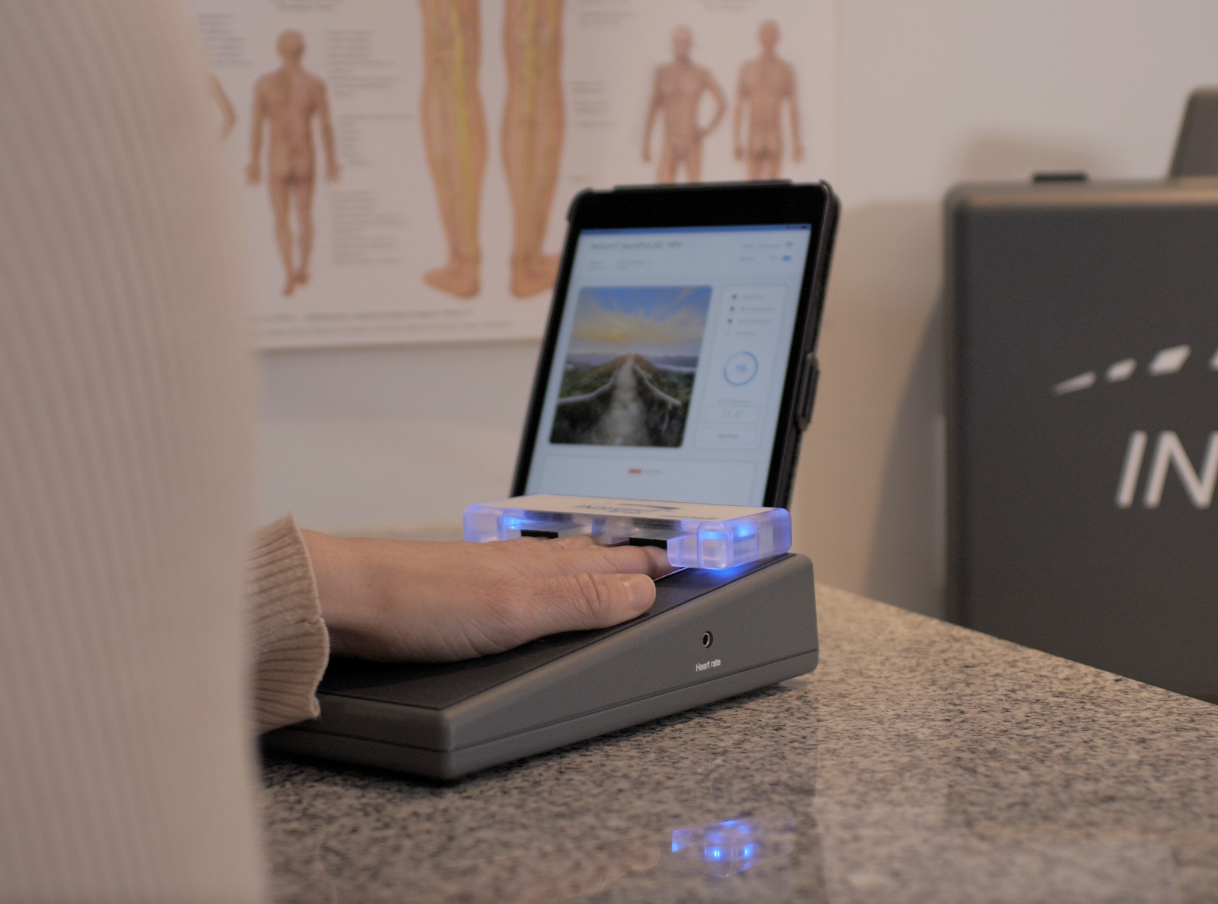
The neuroPULSE analyzes Heart Rate Variability, the beat-to-beat variation in heart pacing. HRV offers insight into autonomic balance, recovery, and adaptive reserve, helping the chiropractor assess whether the system may be operating with reduced reserve.

One result is not a verdict. Baseline, response, and trajectory are more useful than chasing a universal score. A simple patient explanation might be: “We are looking at how well your system can shift gears. Can it respond when demand rises and recover when the demand passes?”

neuroCORE: The Energy Cost of Compensation

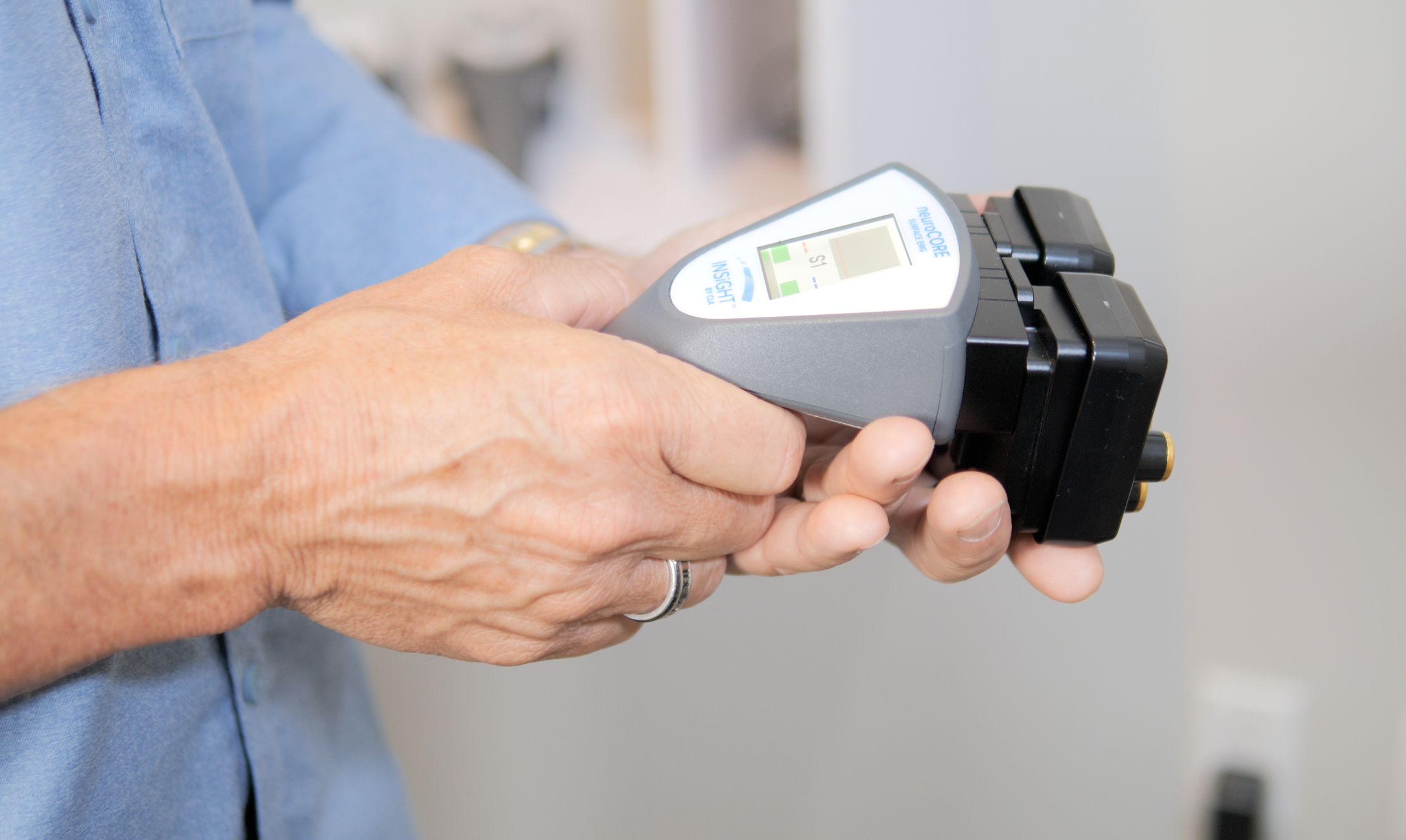
The neuroCORE uses surface electromyography to analyze paraspinal electrical activity. It can reveal postural tension, symmetry, motor tone reactions, and inefficient energy use. Stress impacts the nervous system through more than hormones. It can influence posture, movement, breathing, and stabilization.

neuroTHERMAL: Autonomic Patterns Along the Spine

The neuroTHERMAL performs a full spine nerve system scan in under 30 seconds and analyzes paraspinal temperature patterns connected to autonomic regulation. Bilateral differences provide another window into how chronic stress and the nervous system may be expressed along spinal regions.

Rolling and segmental scan modes help analyze patterns during an exam or before and after an adjustment. Thermal analysis does not diagnose the cause. It identifies functional regions needing attention.

Synapse Software, CORESCORE, and the Scan Rhythm

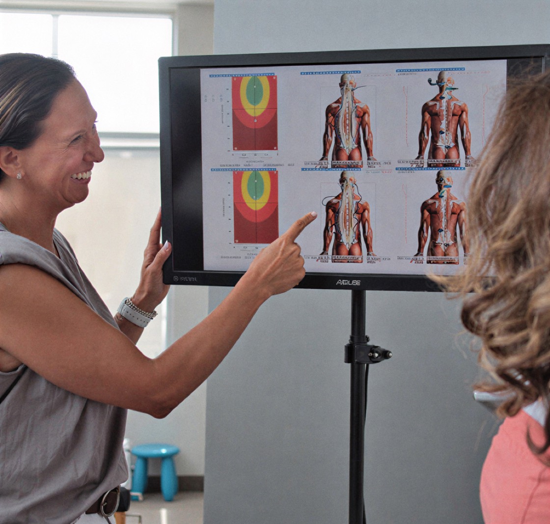
Synapse software organizes neuroPULSE, neuroCORE, and neuroTHERMAL findings into reports patients can understand. CORESCORE brings Reserve, Energy, and Depth together into a patient-friendly neurological efficiency score.

  1. Baseline: Establish the patient’s starting nervous system status.
  2. Response: Re-scan to assess how patterns respond under the care plan.
  3. Trajectory: Compare findings over time to see whether regulation, reserve, and efficiency are moving in a better direction.

When patients see their nervous system in living color, where neurological distress is building, how well they are adapting, and how their care is making a difference, it clicks. The subject stops being abstract and becomes a visible story the doctor and patient can follow together.

Building a Nervous System That Can Meet Life

A life without stress is not the goal. Acute and chronic stress differ because acute stress is usually brief and recoverable, while chronic strain repeatedly asks the body to remain ready. The concern is not that the system responds. It is that it may lose the ability to return.

Chronic stress and the nervous system are connected through brain function, autonomic regulation, postural control, sleep, digestion, cardiovascular activity, and recovery. Yet these patterns can fluctuate. Reserve can improve. Patients can learn the difference between feeling calm and functioning with resilience.

For chiropractors, this is a meaningful place to lead. We can listen carefully, collaborate when needed, and use objective neurological scanning to replace vague assumptions with clearer clinical conversations. We are not promising that chiropractic removes every stressor. We are helping patients understand whether their system is becoming more capable of recovery.

Once patients understand the why behind their care, they stop counting visits and start valuing results. Chronic stress and the nervous system then become more than a wellness topic. They become a measurable story of adaptation, recovery, and performance, and that is a story the chiropractic profession is uniquely prepared to tell.

A patient tells you the room is spinning, yet they are sitting perfectly still. Another says the floor feels unsteady in a grocery store, especially under bright lights or around moving people. They may feel rocked, pulled, or off balance, but there may be no obvious injury and no headache at all.

Most patients think migraine always means severe head pain. Many chiropractors were taught to expect the same familiar pattern: a throbbing headache, nausea, light sensitivity, and perhaps a visual aura. This presentation asks us to widen that picture. The dominant complaint may be vertigo, dizziness, motion sensitivity, or balance problems rather than a migraine headache.

That is what makes this presentation important for the chiropractic profession. The goal is not to label every dizzy patient with vestibular migraine. It is to recognize the pattern, refer appropriately, and examine how the nervous system organizes balance, posture, sensory input, and recovery. Objective neurological scanning can help reveal part of that functional story.

What Is Vestibular Migraine and Why Can It Make Patients Dizzy?

Vestibular migraine is a neurological condition that causes vertigo, dizziness, motion sensitivity, and balance problems. It is generally regarded as a type of migraine presentation, although researchers continue to study the mechanisms of migraine that connect head symptoms with vestibular function. Either way, there is a clear association of migraine disorders with altered balance and motion processing.

The word vestibular refers to the systems that help a person understand where they are in space. The inner ear supplies information about head movement and position, but the vestibular system depends on much more than the ear alone. The brain must combine signals from the inner ear, vision, proprioception, and other sensory pathways. When those signals are not being organized smoothly, a person may feel dizzy even while standing still.

The condition has also been described as migrainous vertigo, migraine-associated vertigo, migraine-related vestibulopathy, or migraine-associated vestibular symptoms. These names reflect a common clinical observation: migraine can affect the parts of the brain involved in balance, movement, and spatial orientation.

  • Vertigo: The patient may feel as though the room is spinning or the body is moving.
  • Imbalance: Walking in a straight line or turning the head may become difficult.
  • Motion sensitivity: Cars, escalators, screens, crowds, or patterned floors may provoke symptoms.
  • Migraine features: Light sensitivity, sound sensitivity, nausea, aura, or headache may occur.

One of the most useful facts for chiropractors to understand is that headache is not required. Some patients with migraine experience head pain during only part of their vestibular attacks. Others have a history of frequent migraine or migraine with aura but experience episodes of vertigo without a regular migraine headache.

The condition may tend to run in families, especially when there is a family history of migraine.

That is where the condition can be missed. A patient may assume the problem is coming entirely from the inner ear, neck, blood pressure, or vision. A careful history can reveal whether the dizziness is positional, visually triggered, spontaneous, or linked with other manifestations of migraine.

Neurological scanning does not diagnose vestibular migraine. Its value is in adding objective information about autonomic regulation, adaptive reserve, postural tension, and compensation. That helps the chiropractor move beyond a symptom-only conversation while keeping the clinical boundaries clear.

Vestibular Migraine Symptoms, Migraine Triggers, and Attack Patterns

The symptoms vary considerably. Even within the same patient, one episode may feel very different from the next. One migraine attack may involve obvious vertigo symptoms and nausea. Another may feel more like visual disorientation, imbalance, or a vague sense that the body is moving when it is not.

Vestibular migraine may occur before, during, or after other migraine symptoms. The relationship between the vestibular symptoms and the migraine features is often more useful than whether they happen at exactly the same moment.

Common Vestibular Symptoms

Patients may use several words for the same experience. They may say they feel dizzy, light-headed, off balance, unstable, or unable to trust their footing. Some episodes of vertigo interfere with driving, working, exercising, shopping, or travelling.

  • Spinning or swaying: The patient may feel as if they or the environment are moving.
  • Dizziness or balance problems: Standing and walking may feel uncertain.
  • Motion sickness: A history of motion sensitivity may begin in childhood.
  • Visual stimulation: Bright lights, flashing lights, traffic, screens, and busy patterns may aggravate the presentation.
  • Associated symptoms: Nausea, vomiting, fatigue, brain fog, visual blurring, or difficulty finding words may occur.
  • Ear complaints: Ear pressure or ringing in the ears may be reported, although meaningful hearing loss deserves closer evaluation.

The vertigo attacks may last from approximately five minutes to 72 hours under commonly used diagnostic criteria. In practical language, the attacks may last for minutes or days. Some patients experience repeated short bursts, while others have prolonged imbalance that continues after the most intense spinning settles.

Persistent postural perceptual dizziness is a separate condition involving ongoing unsteadiness, often worsened by movement or visually complex environments. It may coexist with migraine, but it is not the same situation.

Common Migraine Triggers

Migraine triggers are highly personal. One person may react strongly to sleep disruption, while another notices a relationship with skipped meals, hormones, or weather changes. Some attacks appear to be triggered by stress, while others follow the letdown after a demanding period.

  • Sleep: Too little sleep, too much sleep, or an irregular schedule may become a trigger.
  • Food and hydration: Skipping meals, dehydration, caffeine, chocolate, alcohol, or aged cheeses may contribute.
  • Hormones: Menstrual cycles and other hormonal fluctuations may affect attack patterns.
  • Environment: Bright light, flashing light, weather changes, and barometric pressure may play a role.
  • Neurological distress: Fatigue, emotional demand, and poor recovery may lower tolerance.

Not every attack has an identifiable trigger. A headache journal can record what happened before the episode, which symptoms of vestibular disturbance appeared, how long the attack lasted, and whether aura, light sensitivity, nausea, or migraine pain occurred. Neurological scans add another layer by helping the chiropractor assess postural tension, autonomic balance, and adaptive reserve beyond one particular day.

Diagnosis and Treatment for Vestibular Migraine: Where Chiropractic Fits

Vestibular migraine is a clinical diagnosis. There is no single blood test, imaging study, or neurological scan that confirms it in isolation. Diagnosis and treatment depend on the history, attack pattern, migraine features, examination findings, and exclusion of other reasonable causes.

That distinction matters in chiropractic. A patient may arrive with neck tension, dizziness, and imbalance, but those signs do not automatically establish the diagnosis. They also do not prove that the presentation is being caused by neurological interference in one spinal region. The responsible chiropractor gathers the history, completes an appropriate examination, recognizes referral needs, and stays within the clinical lane.

Diagnostic Criteria and Differential Considerations

The International Classification of Headache Disorders and related diagnostic criteria generally describe at least five vestibular episodes, attacks lasting five minutes to 72 hours, a current or previous history of migraine, migraine features during at least half of the attacks, and no better explanation from another condition.

Several other conditions can cause vertigo. BPPV, or benign paroxysmal positional vertigo, commonly produces brief episodes linked to head position. Disorders such as Meniere’s disease may involve vertigo, ear pressure, ringing in the ears, and hearing loss. Other considerations include peripheral vestibular conditions, cardiovascular causes, medication effects, and other balance disorders.

Depending on the presentation, evaluation may include a neurological examination, hearing assessment, vestibular tests, videonystagmography, electronystagmography, CT, or MRI. Referral to a neurologist, headache specialist, otolaryngologist, or vestibular provider may be appropriate.

New, severe, progressive, or unfamiliar neurological signs deserve timely medical evaluation. The same is true for substantial hearing changes, repeated vomiting, severe loss of coordination, or symptoms that differ sharply from the patient’s usual pattern.

Options for Vestibular Migraine

The care approach is individualized because attack frequency, triggers, migraine features, and associated symptoms vary.

  • Lifestyle strategies: Regular meals, hydration, consistent sleep, and gradual exercise may reduce avoidable triggers.
  • Migraine prophylaxis: A medical provider may recommend a migraine preventive medication when attacks are frequent or disruptive.
  • Acute care: Prescription options may be used during a migraine attack, depending on the patient’s medical history.
  • Vestibular rehabilitation: Specialized exercises may support balance, gaze stability, motion tolerance, and sensory integration.
  • Nutritional support: Some providers discuss magnesium, vitamin B2, or Coenzyme Q10 as part of a broader care plan.

Vestibular migraine treatment should not be reduced to chasing symptoms. The chiropractor’s role is not to replace the physician or promise that adjustments cure the condition. A Neurologically-Focused Chiropractor can still evaluate posture, cervical and spinal motion, proprioceptive input, postural tension, neurological interference, autonomic regulation, and adaptive reserve.

Medical diagnosis asks, “Which condition best explains these attacks?” Neurological scanning asks a different question: “What objective patterns can we observe in nervous system performance, compensation, and adaptability?” Both questions can be useful when they remain in the proper lane.

How INSiGHT Scanning Supports Vestibular Migraine Patients

This condition can be frustrating because the presentation fluctuates. A patient may feel intensely dizzy during one episode and steady at the next appointment. Some vestibular migraine patients begin to question their own experience because the attack has settled by the time they reach the office.

Objective neurological analysis can strengthen the chiropractic conversation. INSiGHT scanning technology does not diagnose vestibular migraine, BPPV, Meniere’s disease, or another vestibular condition. It provides objective exam data that helps the chiropractor assess and communicate nervous system performance.

neuroPULSE and Adaptive Reserve

The neuroPULSE analyzes Heart Rate Variability, or HRV. Heart rate variability reflects the small beat-to-beat differences in heart pacing and provides information about autonomic activity, balance, and adaptability.

A resilient nervous system should increase output when demand rises and return toward recovery when it passes. The neuroPULSE helps the chiropractor analyze sympathetic and parasympathetic activity, adaptive reserve, and recovery capacity.

An HRV finding does not confirm the diagnosis. It gives the doctor another view of how the autonomic nervous system may be responding to life, recovery demands, and the circumstances surrounding the attacks.

neuroCORE and Postural Compensation

The neuroCORE analyzes surface electromyography in the paraspinal muscles. It helps the chiropractor examine postural tension, symmetry, motor tone reactions, and energy expenditure.

A dizzy patient may stiffen the neck, widen their stance, guard against head movement, or use excess muscular effort to feel stable. That does not mean postural tension causes vertigo. It means the body may be spending considerable energy to maintain orientation and balance.

The scan views may reveal postural tension, asymmetrical muscle activity, motor compensation, inefficient energy use, or spinal regions needing attention. Patients understand this when it is explained simply: “Your body may be using more energy than it should just to stay steady.”

neuroTHERMAL, Synapse Software, and CORESCORE

The neuroTHERMAL performs a full spine nerve system scan that analyzes paraspinal temperature patterns associated with autonomic regulation. Since autonomic activity influences blood vessel tone and temperature regulation, thermal findings offer another functional perspective.

INSiGHT neuroTECH and Synapse software bring the neuroPULSE, neuroCORE, and neuroTHERMAL perspectives together. Synapse organizes scan views, comparisons, and patient-friendly reports so the chiropractor can explain complex neurological findings without turning the report of findings into a lecture.

CORESCORE combines the three technologies into a single neurological efficiency score. It is not a migraine severity score. It is a communication metric that helps patients see how different parts of their nervous system profile fit together.

  • Baseline: Establish the patient’s starting nervous system status.
  • Response: Analyze how scan findings fluctuate under care.
  • Trajectory: Compare progress over time rather than relying on one good or difficult day.

The technology does not create the care plan. The chiropractor does. INSiGHT scanning technology provides objective analysis and reporting that support the chiropractor’s interpretation, clinical judgment, and recommendations.

Helping the Dizzy Patient See the Bigger Neurological Story

The condition is more than a headache, and it is more than an inner ear complaint. It involves the relationship between migraine features, motion processing, visual input, proprioception, balance, autonomic regulation, and the parts of the brain responsible for organizing those signals.

That complexity is why the condition deserves thoughtful examination. A patient may have severe vertigo without headache. Another may experience light sensitivity, motion sickness, visual stimulation intolerance, and fatigue before recognizing the pattern as migraine-associated.

The chiropractor’s responsibility is not to force every presentation into a chiropractic explanation. It is to listen carefully, recognize when medical evaluation is needed, complete a strong neurological and chiropractic examination, and contribute useful functional information within scope.

The question is not only, “Are you dizzy today?” A better question is, “How well is your nervous system organizing sensory input, adapting to demand, and recovering between episodes?”

Neurologically-Focused Chiropractic Care has a meaningful place in that conversation because it looks beyond symptoms alone. The chiropractor can assess posture, proprioceptive input, neurological interference, autonomic regulation, and adaptive reserve while collaborating with the providers responsible for diagnosis and treatment.

INSiGHT scanning technology strengthens that process by giving the doctor objective exam data patients can see and understand. It does not label the condition. It does not replace vestibular tests or the judgment of a neurologist. It helps make nervous system performance visible.

When chiropractors combine responsible referral, thoughtful examination, and objective neurological scanning, patients gain a clearer view of what their nervous system may be expressing and why their care plan involves more than chasing the next dizzy spell.

A patient says, “Doc, the room started spinning when I rolled over in bed.” Another says they feel dizzy every time they stand. Both may use the word dizziness, but they are describing very different experiences. That distinction is where a careful chiropractic examination begins.

So, what causes vertigo? Vertigo is the feeling that a person or the environment is moving when no movement is actually occurring. It is a specific type of dizziness and a symptom rather than a diagnosis by itself. The underlying cause is often found in the inner ear, although headache syndromes, medication, injury, circulation, and central neurological conditions can also cause vertigo.

For chiropractors, the better question is not simply, “Are you dizzy?” It is, “What kind of sensation are you having, what triggers it, and which part of the balance system may be sending conflicting information?” Understanding vertigo begins with a disciplined history, a neurological examination, and sound judgment about when referral comes first.

Understanding Vertigo Symptoms and the Vestibular System

Patients often use dizziness as an umbrella term. They may mean light-headedness, faintness, visual instability, imbalance, or a spinning sensation. Before deciding what causes vertigo, the chiropractor needs to clarify what the patient is actually experiencing. That description may shift the case toward an inner ear problem, a cardiovascular issue, a medication effect, or a central neurological concern.

Vertigo is a symptom with a particular quality: a sensation of movement without corresponding physical motion. The patient may feel as though the room is rotating, the floor is tilting, or the body is being pulled to one side. Some people experience vertigo for seconds, while others report vertigo lasting much longer, depending on the cause.

The vestibular system detects head movement, acceleration, and position. The semicircular canals respond to rotation, while other structures in the inner ear detect gravity and linear motion. That information is compared with vision and proprioceptive input from the joints, muscles, and spinal regions. When those signals conflict, vertigo occurs, and eye movement, posture, or balance may become less coordinated.

Common vertigo symptoms may include:

  • Nausea: The spinning may trigger nausea or vomiting.
  • Unsteadiness: Patients may have trouble standing or walking safely.
  • Nystagmus: Rapid, involuntary eye movement may accompany an episode.
  • Hearing changes: Changes in hearing, tinnitus, or ear fullness may point toward certain inner ear issues.
  • Balance difficulty: Dizziness and balance issues may increase fall risk.

These associated signs help narrow the possibilities, but symptoms of vertigo do not provide the full diagnosis. Neurological scanning may add objective analysis of autonomic activity, postural tension, and nervous system status. It belongs beside the history and examination, not in place of vestibular testing, imaging, or referral.

What Causes Vertigo Most Often?

Most cases begin with peripheral causes involving the inner ear or vestibular nerve. These are more common than central causes arising from the brain. Timing, triggers, hearing changes, recent illness, injury, and neurological signs help separate the different types of vertigo.

BPPV and Positional Vertigo

Benign paroxysmal positional vertigo is a common cause of vertigo triggered by a change in head position. BPPV occurs when tiny calcium carbonate particles, sometimes described as a loose crystal, move from the utricle into a semicircular canal. When you move your head, look up, bend down, or roll in bed, the displaced material can send a false motion signal.

The spinning is usually brief but can produce severe vertigo. BPPV is considered benign, yet the phrase vertigo is benign should never dismiss the patient’s experience. Canalith repositioning uses a series of head movements to reposition the particles toward the utricle. The Epley maneuver is the best-known example. These repositioning maneuvers are not interchangeable with a chiropractic adjustment, and a neurological scan does not confirm BPPV.

Inner Ear Fluid Pressure, Vestibular Neuritis, and Labyrinthitis

This fluid-pressure condition is associated with abnormal pressure in the inner ear. People with Meniere’s disease may have a vertigo attack with fluctuating hearing loss, tinnitus, and fullness in the affected ear. Vestibular neuritis involves inflammation of the vestibular nerve and may produce sudden onset, prolonged vertigo, nausea, and instability. Labyrinthitis affects the labyrinth and may add hearing changes or tinnitus.

Migraine, Central Causes, and Other Possibilities

A migraine may cause episodes of vertigo with or without a severe headache. Central vertigo is less common, but stroke, transient ischemic attack, traumatic brain injury, multiple sclerosis, infection, or reduced blood flow to the back of the brain may cause episodes. Other health conditions and circumstances include ear surgery, perilymphatic fistula, acoustic neuroma, medication effects, alcohol interactions, prolonged bed rest, low blood pressure, diabetes, and abnormal heart rhythm.

Balance disorders can create several signs, including vertigo, nausea, and unstable vision. Medications may cause vertigo, and vertigo can sometimes recur even after a quiet period. Vertigo causes dizziness, but dizziness does not always mean spinning. Those differences matter in all cases of vertigo.

Some situations cause light-headedness rather than true spinning. That is why patients need a complete history and appropriate testing to get the correct diagnosis. A scan can add functional context, but it cannot determine what causes vertigo or identify a specific peripheral or central diagnosis.

How Chiropractors Evaluate the Cause of Vertigo Without Guessing

Patients may arrive in a chiropractic office because the spinning began around the same time as neck tension, headache, injury, altered posture, or restricted movement. Those relationships deserve attention, but they do not establish that the cervical spine is the underlying cause of vertigo.

A responsible Neurologically-Focused Chiropractor listens for the pattern, screens for risk, examines the nervous system, and decides whether care, referral, or collaboration is appropriate. When evaluating the presentation, the goal is not to force every case into one explanation. It is to understand the vertigo episode and identify what needs attention first.

A provider should ask about your symptoms in enough detail to separate spinning from faintness. Useful questions include:

  • Duration: Does the episode last seconds, minutes, hours, or most of the day?
  • Trigger: Do rolling in bed, looking up, bending down, or other head movements trigger vertigo symptoms?
  • Recent events: Did it begin after illness, injury, surgery, or prolonged inactivity?
  • Hearing: Is there tinnitus, fullness, or a change in hearing?
  • Posture: Does standing create spinning, or does the patient feel faint?
  • Neurology: Are there new weakness, speech, visual, or walking changes?

New weakness, facial asymmetry, difficulty speaking, double vision, loss of consciousness, severe difficulty walking, sudden hearing loss, chest symptoms, persistent vomiting, or a new severe headache deserve urgent medical attention. This is calm clinical leadership. Vertigo can be caused by many situations, and some require prompt diagnosis and treatment.

The cervical spine provides important proprioceptive information about head placement and movement. That input is integrated with vision and vestibular signals to organize gaze, posture, and orientation. Cervical findings may be relevant without being the sole explanation causing your vertigo. The chiropractor can examine motion, posture, balance, and neurological interference while respecting peripheral, vascular, medication-related, and central causes.

Treatment depends on the underlying cause. Treatment for vertigo may include canalith repositioning for BPPV, vestibular rehabilitation, short-term medication to manage your symptoms, headache care, or medical management for another condition. A patient may receive vertigo treatment from a medical or rehabilitation provider while also receiving chiropractic care for appropriate findings.

How Is Vertigo Treated?

Vertigo treatment is never one-size-fits-all. Certain vertigo presentations respond to a maneuver, while others require rehabilitation, medication, hearing care, or neurological evaluation. Because more than one condition can cause a similar report, patients need to get treatment matched to the diagnosis. When vertigo is treated according to the underlying cause, the care strategy is clearer and safer.

How INSiGHT Scanning Technology Adds a Neurological View

What causes vertigo cannot be answered by one scan. Two patients can describe the same vertigo attack and have entirely different causes. One may have a positional condition. Another may have a central or headache-related presentation. A third may be light-headed rather than experiencing vertigo at all.

INSiGHT scanning technology adds objective exam data to that process. It does not diagnose the cause. It analyzes patterns related to autonomic regulation, postural tension, motor output, and adaptive reserve. The chiropractor interprets those findings alongside the history, examination, vestibular findings, imaging, and referral decisions. The technology supports the care plan; it does not create it.

The three INSiGHT technologies add complementary information:

  • neuroPULSE: Analyzes Heart Rate Variability to provide insight into autonomic balance, recovery, and adaptive reserve. It does not determine whether the patient has a peripheral vestibular condition, but it can support broader conversations about recovery habits intended to reduce stress.
  • neuroCORE: Analyzes surface EMG activity in the paraspinal muscles, including postural tension, asymmetry, guarding, and inefficient energy expenditure. It is not a diagnostic test for balance disorders.
  • neuroTHERMAL: Performs a fast full spine nerve system scan and analyzes bilateral temperature regulation patterns associated with autonomic function. It does not identify a specific ear or central condition.

INSiGHT neuroTECH and Synapse software bring those findings into one reporting process. Synapse organizes scan views, comparisons, and progress reports. CORESCORE combines the three technologies into a patient-friendly neurological efficiency score. This can shift the conversation from “Am I still dizzy today?” to “How is my nervous system performing over time?”

A responsible scan-led workflow clarifies the patient’s language, screens for referral needs, completes the chiropractic examination, establishes objective baselines when appropriate, and interprets the data within the complete picture. Re-scanning then helps follow baseline, response, and trajectory without promising that every presentation will respond the same way.

Neurological scanning shifts the patient’s focus from spinal regions and complaints to nerves and performance. When patients with vertigo can see nervous system status in living color, complex neurology becomes easier to understand. The scan cannot name what causes vertigo, but it can help the chiropractor communicate the broader functional story.

A Clearer Path Through Vertigo and Dizziness

What causes vertigo depends on which part of the balance system is sending inaccurate or conflicting information. Vertigo is often peripheral, but central and systemic causes must remain part of the clinical picture. A positional inner ear condition may create brief spinning after movement. The fluid-pressure condition may combine a vertigo episode with tinnitus and ear fullness. Inflammatory vestibular conditions may produce sudden or prolonged symptoms. Headache syndromes, injury, medication, circulation, and central neurological conditions may also cause episodes.

The chiropractor’s first responsibility is not to force the presentation into a chiropractic explanation. It is to listen carefully, distinguish spinning from faintness, identify timing and triggers, recognize referral signs, and examine how the nervous system is organizing posture, movement, and balance.

The question of what causes vertigo still requires appropriate clinical judgment, testing, and referral when indicated. When neurological scanning is appropriate, neuroPULSE, neuroCORE, neuroTHERMAL, Synapse software, and CORESCORE add objective information without replacing those steps. They help the chiropractor analyze nervous system performance and give the patient a visual reference point for the broader neurological story.

That is where responsible Neurologically-Focused Chiropractic Care can make a meaningful contribution. The goal is not to treat vertigo as though every case has the same origin. The goal is to recognize what is known, identify what still needs evaluation, and guide the next step without overstatement.

When people with vertigo understand what causes vertigo and the reason behind a careful neurological process, they stop hearing disconnected complaints and begin seeing one coordinated system. When chiropractors can show nervous system performance clearly while staying firmly within scope, patients receive something far better than a guess.

One of the most common questions people ask after purchasing a new Heart Rate Variability (HRV) device is why its readings do not match those from another device.

An Oura Ring might report an HRV of 62, while an Apple Watch shows 51, an Elite HRV app displays 74, and a Garmin provides a different result altogether. It is natural to wonder which number is correct, but the answer is that each device may be accurately reflecting the data it collected under its own measurement conditions.

Heart Rate Variability is not a fixed value like the length of a table. It is a dynamic physiological measurement that changes throughout the day in response to factors such as posture, breathing, activity, stress, recovery, and timing. Different devices may also collect data at different moments, use different sensors, or process the information through different algorithms.

Understanding these differences helps reduce confusion and makes it easier to interpret HRV data within the context of the device being used.

HRV Isn’t One Fixed Number

Many people assume HRV works like body weight, where stepping on a scale should produce essentially the same result each time. Heart Rate Variability behaves differently because it reflects an autonomic nervous system that is continuously adapting to changes in stress, activity, recovery, breathing, posture, and the surrounding environment.

Your HRV changes from minute to minute in response to:

  • Breathing
  • Stress
  • Activity
  • Body position
  • Recovery
  • Sleep
  • Temperature
  • Hydration
  • Emotions

Because HRV is always changing, two devices collecting data at different times may naturally produce different values—even if both measurements are accurate.

Different Devices Measure HRV Differently

HRV devices do not all collect data in the same way. Some measure HRV during sleep, while others take a reading shortly after you wake up. Certain devices require a brief one-minute spot measurement, whereas others collect information periodically or continuously throughout the day. They may also differ in how they summarize the data, with some averaging several readings and others reporting a single recording.

Each approach has its own advantages, but these differences in timing, duration, and data processing can lead to noticeably different HRV values.

Different Devices Use Different Sensors

Another reason HRV values differ is the sensor technology.

Many consumer wearables use photoplethysmography (PPG).

Some chest straps use electrocardiography (ECG).

Although both technologies can accurately measure HRV under appropriate conditions, they collect heartbeat information differently.

PPG measures blood volume changes.

ECG measures the heart’s electrical activity directly.

Both approaches have been validated for HRV assessment, but slight differences in signal collection may contribute to variation between devices.

Different Algorithms Produce Different Results

Even when two devices collect exactly the same heartbeat data, they may still calculate HRV differently.

Manufacturers often use proprietary software algorithms that differ in how they:

  • Remove movement artifacts
  • Filter irregular beats
  • Process noisy signals
  • Average recordings
  • Calculate HRV metrics
  • Display results

These algorithms are rarely identical.

As a result, two devices may process the same physiological information differently while still producing clinically reasonable values.

Different HRV Metrics

Sometimes people compare numbers that aren’t even measuring the same thing.

One device may display:

  • RMSSD

Another may display:

  • SDNN

Another may report:

  • A proprietary “Recovery Score”

Or:

These measurements are related, but they are not interchangeable.

Before comparing numbers, it’s important to know exactly which HRV metric your device is reporting.

Timing Makes a Huge Difference

Consider these two measurements.

Measurement One:

Collected while you’re asleep at 3:00 AM.

Measurement Two:

Collected after your morning coffee while answering emails.

Should they match?

Probably not.

Your nervous system is functioning under completely different conditions.

Even if the same device collected both measurements, you would expect different HRV values.

This is why timing plays such an important role in HRV interpretation.

Body Position Changes HRV

Posture also influences HRV.

Lying down.

Sitting.

Standing.

Each position changes cardiovascular demands and autonomic nervous system activity.

If one device measures HRV while you’re asleep and another measures while you’re sitting upright, differences are completely expected.

Neither device is necessarily wrong.

They’re simply measuring different physiological states.

Wearables Are Designed for Trends

One of the biggest misconceptions about HRV is believing that every measurement should match perfectly.

That’s not actually the goal of wearable devices.

Consumer wearables are designed primarily to help you observe trends over time.

For example:

  • Is your recovery improving?
  • Is your HRV consistently declining?
  • How does travel affect you?
  • Does alcohol reduce your overnight recovery?
  • How does exercise influence your nervous system?

These long-term trends provide much more useful information than comparing one isolated HRV value between devices.

Should You Switch Between Devices?

Many people upgrade from one wearable to another.

Or they begin using a smartphone HRV app alongside a smartwatch.

When this happens, they often become frustrated because the numbers don’t match.

Rather than trying to compare them directly, most experts recommend establishing a new baseline with the new device.

Each platform should be viewed as its own measurement system.

The most valuable comparisons occur within the same device over time—not between different devices.

Why Clinical Assessments Focus on Standardization

Consumer wearables prioritize convenience.

Clinical HRV assessments prioritize reproducibility.

Those goals require different approaches.

Rather than measuring HRV during everyday activities, clinical systems standardize factors such as:

  • Body position
  • Arm position
  • Movement
  • Recording conditions
  • Collection procedures

This helps reduce unnecessary sources of variability so clinicians can compare one examination with the next more confidently.

How neuroPULSE Standardizes HRV Collection

The neuroPULSE technology within the INSiGHT scanning technology also uses photoplethysmography (PPG), but it is designed specifically for standardized clinical assessment.

During a neuroPULSE examination:

  • The patient remains seated.
  • The hand is supported throughout the recording.
  • The hand is stabilized to reduce movement.
  • The arm is maintained near heart level.
  • Standardized procedures are followed for every assessment.

By controlling these variables, neuroPULSE helps produce consistent HRV measurements that can be reproduced more reliably over time as part of an objective neurological assessment.

What further distinguishes the neuroPULSE is that it evaluates three autonomic biomarkers before the HRV assessment begins. Alongside the PPG pulse signal, Galvanic Skin Response sensors measure electrodermal activity to confirm that the patient has reached an appropriate resting state, while a temperature sensor monitors the hand and fingertip to verify that blood perfusion at the sensing site is sufficient for clear, valid pulse collection.

The software reviews pulse quality, electrodermal activity, and fingertip temperature together before authorizing the assessment. When these measurements fall outside reasonable standardized ranges, data collection does not begin. A built-in latency period of at least 10 seconds and up to one minute also allows temporary arousal associated with entering a clinical setting, sometimes described as a “white coat” response, to settle before recording starts.

This combination of standardized positioning, simultaneous biomarker monitoring, pre-test validation, and a prescribed settling period creates a level of quality control that consumer wearables are not designed to provide. It supports more reproducible clinical trend tracking by helping ensure that each assessment begins under consistent physiological and sensing conditions.

Which Device Should You Trust?

The better question isn’t:

“Which device is most accurate?”

Instead ask:

“Which device is most appropriate for my purpose?”

If you’re interested in:

  • Daily recovery
  • Sleep quality
  • Fitness
  • Wellness habits
  • Lifestyle trends

A quality wearable can provide tremendous value.

If you’re undergoing a standardized neurological assessment in a clinical setting, reproducibility and controlled testing conditions become much more important.

The two approaches serve different purposes.

Don’t Chase Matching Numbers

Trying to force different devices to agree is usually an exercise in frustration.

Instead:

Use the same device consistently.

Collect measurements under similar conditions whenever possible.

Focus on long-term trends.

Pay attention to meaningful changes over weeks and months rather than individual daily fluctuations.

That’s where HRV becomes most valuable.

Heart Rate Variability (HRV) has become one of the most talked-about health metrics in recent years. Once reserved primarily for research laboratories and clinical settings, HRV is now available to millions of people through smartwatches, fitness trackers, rings, chest straps, and smartphone apps.

Devices such as Oura, WHOOP, Apple Watch, Garmin, Polar, and apps like Elite HRV have introduced consumers to the idea that the time between heartbeats can provide valuable insights into stress, recovery, sleep, and overall nervous system function.

This growing awareness is a positive development. More people than ever are paying attention to how their bodies respond to stress and how lifestyle choices influence their health.

However, as HRV has become more accessible, an important misconception has also emerged: that all HRV modes of measurements and their data presentation are equivalent.

While consumer wearables and clinical HRV assessments may report similar metrics, they are designed for very different purposes. Understanding those differences helps set appropriate expectations for both patients and healthcare professionals.

What Is Heart Rate Variability?

Heart Rate Variability refers to the natural variation in time between consecutive heartbeats. Contrary to what many people assume, a healthy heart does not beat like a perfectly timed metronome.

Instead, the intervals between beats are constantly changing as the autonomic nervous system adapts to internal and external demands.

HRV provides valuable information about how effectively the sympathetic (“fight or flight”) and parasympathetic (“rest and digest”) branches of the autonomic nervous system work together. Because of this, HRV has become a widely studied marker of adaptability, resilience, recovery, and autonomic regulation.

Numerous research studies have demonstrated relationships between HRV and factors such as stress, sleep quality, exercise recovery, cardiovascular health, and overall nervous system function.

The Rise of Consumer HRV Wearables

One of the greatest strengths of today’s wearable technology is accessibility.

Instead of visiting a clinic or using specialized equipment, individuals can monitor HRV every day with devices they already wear while sleeping, exercising, or going about normal daily activities.

For many people, this creates greater awareness of how daily habits influence their nervous system.

For example, users may notice that:

  • Poor sleep lowers HRV.
  • Regular exercise improves recovery over time.
  • Alcohol consumption decreases HRV.
  • Chronic stress reduces resilience.
  • Better nutrition and hydration positively influence recovery.

This type of ongoing feedback can encourage healthier lifestyle choices and greater personal engagement with wellness.

For these reasons, consumer HRV devices have played an important role in bringing nervous system awareness into mainstream healthcare conversations.

Trends can be tracked over time  

HRV devices are most valuable when mapping the data trends over time. When HRV is collected consistently over weeks or months, users can begin identifying patterns that correspond with changes in lifestyle, recovery, training load, sleep habits, travel, illness, or emotional stress.

Looking at these long-term trends often provides more useful information than focusing on any single day’s reading.

For personal wellness tracking, this continuous monitoring offers substantial value.

Why Individual HRV Readings Can Vary

Although HRV is a valuable physiological measurement, it is also highly sensitive.

Many variables influence HRV from one recording to the next, including:

  • Body position
  • Movement
  • Breathing patterns
  • Sleep stage
  • Emotional stress
  • Hydration
  • Recent exercise
  • Illness
  • Ambient temperature
  • Time of day

Even small differences in these factors can produce noticeable changes in HRV measurements.

This does not mean the device is inaccurate. It relates mostly to the stability of the collection as HRV must be calculated to the millisecond of heart beat timing. 

For clinicians making objective comparisons between visits, however, reducing unnecessary sources of variability becomes much more important.

Why Standardization Matters

Imagine stepping onto a bathroom scale every morning.

One day you’re wearing heavy winter boots.

The next day you’re barefoot.

The following day you’re carrying a backpack full of groceries.

The scale itself may be perfectly accurate, but because the conditions keep changing, meaningful comparisons become much more difficult.

HRV works in a similar way.

If posture changes…

If arm position changes…

If breathing differs, like after exercise or meditation…

If movement creates signal interference…

Or if the quality of the pulse signal changes…

Some of the differences seen between recordings may reflect changing measurement conditions rather than meaningful physiological change.

This is why clinical HRV assessments place such a strong emphasis on standardized testing conditions.

Clinical Assessments Have a Different Goal

Consumer wearables are designed to collect frequent measurements throughout everyday life.

Clinical HRV assessments have a different objective.

Their purpose is to produce measurements that can be reproduced consistently from one examination to the next, allowing healthcare providers to compare changes over time with greater confidence.

Rather than maximizing convenience, clinical systems aim to minimize variables that could influence the recording.

That distinction is fundamental.

The goal is not simply collecting HRV.

The goal is collecting HRV under controlled conditions.

Understanding neuroPULSE HRV Technology

The neuroPULSE technology used within the INSiGHT scanning technology is designed specifically for standardized clinical HRV assessment.

Like many modern consumer wearables, neuroPULSE uses photoplethysmography (PPG), an established technology that measures blood volume changes with each heartbeat.

PPG itself is not unique to clinical equipment.

What distinguishes a clinical assessment is how the measurement is collected.

During a neuroPULSE assessment:

  • The patient’s hand is supported and stabilized.
  • The hand remains fixed throughout the recording.
  • The arm is maintained at approximately heart level.
  • The patient remains seated during collection.
  • The testing environment is standardized to reduce unnecessary variability.

These controlled conditions help produce a stable pulse wave while minimizing motion artifact and reducing factors that can influence the accuracy of HRV calculations.

Because the recording conditions remain consistent from one examination to the next, clinicians can compare changes with greater confidence.

The Importance of Signal Quality

Photoplethysmography has become a well-established method for measuring cardiovascular signals.

However, the quality of any PPG measurement depends heavily on the stability of the signal being collected.

Movement, changing body position, poor contact with the sensor, or irregular pulse recordings can all introduce artifacts that algorithms must attempt to correct.

Many consumer devices use sophisticated software to compensate for these challenges, and they continue to improve each year.

Even so, wearable devices are intentionally designed for convenience and everyday use, which means they cannot fully control the environment in which data is collected.

Clinical assessment systems address this challenge by reducing many of these variables before the measurement is even taken.

Rather than relying primarily on software correction after the fact, standardized collection methods help improve signal quality from the beginning.

Clinical Data Supports Clinical Decisions

One of the greatest strengths of standardized HRV assessment is reproducibility.

When patients return for follow-up examinations, clinicians want confidence that observed changes reflect meaningful physiological adaptation rather than differences in how the measurement was collected.

This consistency becomes especially valuable when HRV is one component of a broader neurological assessment.

Within the INSiGHT scanning technology, neuroPULSE contributes objective autonomic nervous system information alongside neuroCORE surface electromyography (sEMG) and neuroTHERMAL scanning to help create a more comprehensive picture of nervous system function. Together, these technologies support objective assessment, patient education, and progress tracking within chiropractic practice.

Consumer Wearables and Clinical Assessments Serve Different Purposes

It is tempting to compare wearable HRV devices directly with clinical assessment systems.

In reality, they are designed to answer different questions.

Consumer HRV Wearables Clinical neuroPULSE Assessment
Designed for daily wellness tracking Designed for standardized clinical assessment
Convenient continuous monitoring Controlled examination environment
Measurements collected during everyday life Measurements collected under standardized conditions
Excellent for observing long-term trends Appropriate for objective comparison between clinical visits
Helps individuals understand lifestyle patterns Helps clinicians evaluate nervous system function consistently

Neither approach replaces the other.

Instead, they complement one another.

Someone may use a wearable device every day to better understand recovery, sleep, and lifestyle habits while also receiving periodic standardized HRV assessments during clinical evaluations.

Each provides valuable information within its intended role.

Using Both Clinical HRV Assessments And Consumer HRV Wearables Together

Consumer HRV wearables have transformed public awareness of nervous system health. They provide convenient access to HRV data and encourage millions of people to pay closer attention to sleep, stress, recovery, and overall wellness. For monitoring long-term trends and building healthier habits, they are valuable tools.

Clinical HRV assessment serves a different purpose.

When healthcare providers need objective measurements that can be compared confidently over time, standardization becomes essential. Controlling patient positioning, minimizing movement, stabilizing the measurement site, and creating consistent testing conditions all help improve the reproducibility of the data.

The neuroPULSE technology was developed with these principles in mind. By combining established photoplethysmography technology with standardized clinical collection methods, it provides chiropractors with consistent HRV information that supports objective neurological assessment, patient communication, and progress tracking as part of the broader INSiGHT scanning technology.

Wearables and clinical HRV assessments are not competitors.

They simply answer different questions.

One helps individuals monitor their nervous system throughout everyday life.

The other provides clinicians with standardized physiological information that supports confident clinical evaluation.

Most patients assume that if the body completes a task, the nervous system must be doing its job well. Chiropractors know there is another question worth asking: how much effort did the system need to produce that result? A person can stand, move, concentrate, and get through the day while using far more neurological energy than the task should require. Function is present, but the cost of that function may be high.

That is the practical promise of neural efficiency. In neuroscience, the term describes the relationship between performance and the neural resources used to achieve it. A more efficient brain may solve the same cognitive task with lower brain activation, more focused recruitment, or less metabolic demand. For a Neurologically-Focused Chiropractor, the concept opens a broader conversation about reserve, energy expenditure, regulation, and the nervous system’s ability to meet demand without spending every resource it has.

The point is not that less activity is always better. Activity should match the job. Neural efficiency asks whether the nervous system is organizing the right response, at the right intensity, with enough reserve left for what comes next.

What Neural Efficiency Means in Neuroscience and Chiropractic

At its simplest, neural efficiency is the ability to produce an appropriate result with an economical use of neural resources. Researchers study this by comparing task performance with electrical activity, blood flow, or metabolism. When two people solve the same problem equally well but one requires lower or more focused activation, neural efficiency may be higher. The concept links a behavioral result to the neural cost of producing it.

The word “may” matters. Lower brain activity by itself does not prove efficient brain functioning. A person who shows lower activation because they are disengaged, confused, or performing poorly is not necessarily efficient. The pattern must be interpreted alongside task difficulty, accuracy, strategy, and the brain regions involved. The better question is not simply, “How active was the brain?” It is, “What did that activity accomplish?”

The Difference Between Activity and Efficiency

Think of two people carrying the same box across a room. One moves smoothly and finishes with energy to spare. The other braces every muscle and uses far more effort to reach the same destination. The outcome looks similar, but the efficiency is not. Cognitive neuroscience applies that same logic to brain function.

This distinction belongs in chiropractic too. A patient may maintain posture while the spinal motor system shows postural tension or inefficient energy distribution. Another may appear calm while autonomic analysis suggests low adaptive reserve. The body is functioning, but it may be paying too much for that function.

  • Performance: What result did the nervous system produce?
  • Effort: How much neural effort was required?
  • Demand: How difficult was the task?
  • Reserve: How much capacity remained afterward?

Chiropractic neurological scanning does not measure human intelligence, cortical activation, or the neural correlates of cognitive performance. EEG, PET, fMRI, and fNIRS answer different questions. Chiropractic scanning looks at autonomic adaptability, paraspinal motor activity, energy expenditure, and temperature regulation patterns along the spinal region.

For chiropractors, the concept becomes useful when it sharpens the exam. Is the response coordinated or compensatory? Is the system adaptable or stuck in sympathetic overdrive? Is ordinary function requiring excessive effort? Neurological scanning gives the doctor objective analysis to explore those questions rather than relying on symptoms or impressions alone.

The Neural Efficiency Hypothesis as a Function of Task Demands

The neural efficiency hypothesis grew from research on intelligence and brain activation. Broadly stated, it proposes that individuals with higher intelligence may require fewer neural resources than individuals with lower intelligence to complete the same cognitive task successfully. Early PET research associated with Haier examined brain glucose metabolism levels, while later functional brain imaging studies explored electrical activity, cortical activation, and functional connectivity.

Haier et al helped draw attention to the relationship between intelligence and brain activity. Later studies by Neubauer, Fink, Grabner, Benedek, and others showed that the neural efficiency phenomenon is more complicated than “smarter brains always work less.” A systematic review by Neubauer and Fink considered the influence of task content, task content and sex, brain region, and research method.

Why Task Difficulty Changes the Pattern

The neural efficiency hypothesis is best understood as a function of task demands. When a task is extremely easy, people at different levels of intelligence may all complete it with little cognitive effort. There may be no meaningful differences in activation because neither group needs to recruit much power.

Moderately difficult tasks are often where intelligence related differences become easiest to see. The task requires real neural processing, but it is not so difficult that everyone reaches their limit. Under those conditions, intelligent individuals may achieve comparable or better performance with lower levels of activation or more focused activity in relevant regions.

A very difficult task may require strong neural activation from everyone, including individuals with higher intelligence. The difference between lower and higher intelligence can narrow when task complexity reaches the limits of current ability. This is why whether neural efficiency appears in a study often depends on how well the task was matched to the participants.

Elsbeth Stern used a helpful car analogy. At low speed, both an efficient and less efficient car use little fuel. At maximum speed, both burn a great deal. At moderate speed, the difference becomes obvious. The analogy suggests that neural efficiency is easiest to see when a cognitive task is challenging but manageable.

Why Performance Must Be Considered With Activation

Research using functional near-infrared spectroscopy, or fNIRS, makes this point well. During laboratory executive-function tasks and flight simulation, oxygenated hemoglobin in the prefrontal cortex increased as tasks became more complex. Yet activation intensity did not clearly predict who performed best. It offered more insight into how hard the participant was working.

That distinction matters in chiropractic. A high or low activity level should not be labeled efficient without context. The doctor must consider baseline, workload, sleep, recovery, neurological distress, stage of care, and trajectory across repeated exams. One scan identifies a pattern. A sequence helps show whether that pattern is becoming more adaptable and organized.

Working Memory, Expertise, and a Systematic Review of Neural Processing

Working memory is one of the clearest settings in which researchers have examined neural efficiency. It allows a person to hold information temporarily, connect it with new information, adjust to changing goals, and filter out what is no longer relevant. These higher cognitive abilities depend on coordinated activity in the frontal cortex and related brain networks.

In research described by Elsbeth Stern and Daniela Nussbaumer, university students completed letter and facial-memory tasks while EEG recorded their brain activity. Participants also completed a conventional intelligence test and were grouped by intelligence scores. The clearest differences in brain activity appeared during moderately difficult working memory tasks, where both groups could succeed but individuals with higher intelligence required fewer resources.

Formal Testing and Research Tools Ask Different Questions

This finding does not turn EEG into an intelligence test. Conventional testing remains the more reliable way to assess individual differences in cognitive abilities. EEG can reveal event-related electrical patterns and timing, but those neural markers are not precise enough to label one person as having high intelligence, superior intelligence, or lower intelligence.

The same caution applies across imaging methods:

  • EEG: Records electrical brain activity and event-related responses during a cognitive task.
  • PET: Analyzes regional energy use, which was important in early Haier research.
  • Functional magnetic resonance imaging: Examines blood-oxygen-related activity and functional connectivity.
  • fNIRS: Analyzes oxygenated and deoxygenated hemoglobin in the cortex, offering insight into mental effort.

Haier, Nuechterlein, and colleagues contributed to early work on intelligence and brain activation. Later work by Neubauer, Fink, Grabner, and Benedek explored differences in intelligence, the influence of task content, creativity, sex on the brain–IQ relationship, and connections between brain regions. These studies of neural performance show why brain activation and intelligence cannot be reduced to one rule.

Expertise Can Make Neural Processing More Focused

Training can influence how the functional brain recruits resources. As a person practises a mental skill or gains experience performing a sport-specific task, the brain may rely more heavily on task-related pathways while reducing activity in areas that are not needed.

The result can be more focused neural processing, better coordination within a brain network, and less conscious effort. That does not mean expertise shuts the brain down. It means experience can refine which connections between brain regions are recruited and when.

Practice effects may remain specific to the trained activity. Someone can improve at one working memory exercise without showing the same advantage on a new, similar task. Higher neural efficiency in one setting does not prove universal efficiency across every cognitive challenge.

Applying Neural Efficiency Through INSiGHT Neurological Scanning

The neuroscience definition of neural efficiency focuses largely on cognitive performance relative to cortical effort. Chiropractic uses the phrase in a different but related functional context. The chiropractic question is not, “How intelligent is this brain?” It is, “How efficiently is this nervous system using energy, maintaining posture, regulating autonomic activity, and preserving enough reserve to adapt?”

INSiGHT scanning technology does not analyze IQ, the sensory and motor cortex, or cerebral metabolism. INSiGHT neuroTECH and Synapse software provide objective exam data about Reserve, Energy, and Depth, then bring those findings together in a patient-friendly neural efficiency index.

neuroPULSE Shows Reserve and Adaptability

The neuroPULSE analyzes Heart Rate Variability, giving the chiropractor a practical view of autonomic balance, activity, adaptability, and reserve. A resilient nervous system should be able to increase output when demand rises and recover when the demand passes. In the RED framework, neuroPULSE represents Reserve.

neuroCORE Reveals the Energy Cost of Compensation

The neuroCORE uses surface electromyography to analyze paraspinal muscle activity, symmetry, pattern efficiency, and energy expenditure. The motor system may use excessive energy simply to maintain posture against gravity, or it may show low activity where better organization is needed. In the RED framework, neuroCORE represents Energy.

Its scan views help the doctor see whether the spinal motor system appears organized, exhausted, asymmetrical, or compensatory. Higher activity is not automatically worse, just as lower activity is not automatically better. The chiropractor interprets the pattern in context.

neuroTHERMAL Adds Depth to the Autonomic Picture

The neuroTHERMAL performs a full spine nerve system scan in under 30 seconds and analyzes bilateral temperature patterns connected to autonomic regulation. In the RED framework, neuroTHERMAL represents Depth. It does not diagnose disease from a thermal pattern. It gives the chiropractor a reproducible view of regional autonomic patterns needing attention.

CORESCORE Turns Complex Neurology Into a Clear Report

Synapse software combines the three analyses into CORESCORE, a 0–100 neurological efficiency score patients can understand. CORESCORE is not an intelligence score, cortical analysis, or diagnosis. It is a report-card-style summary that supports a clearer discussion of overall neural efficiency from a chiropractic perspective.

  • Initial: Establishes the patient’s baseline nervous system status.
  • Progress: Analyzes whether early patterns are changing under the sequence of adjustments.
  • Comparative: Examines the developing trend across multiple exams.
  • Continuation: Tracks ongoing stability and adaptation using recent scans.

The technology does not create the care plan. The chiropractor does. INSiGHT scanning technology provides objective analysis and scan views that the doctor combines with history, examination, goals, and clinical judgment.

A More Precise Way to Think About Nervous System Performance

Efficient nervous system performance is not the absence of activity. It is the appropriate use of neural resources for the demand at hand. A resilient nervous system should be able to increase output for a difficult task, reduce unnecessary effort when the task is easier, and recover with enough reserve for the next challenge.

The tools and conclusions differ between cognitive neuroscience and chiropractic analysis, but the discipline is similar: never interpret activity without context. A single scan, one score, or one day cannot describe the whole nervous system.

From Symptoms to Performance

For the chiropractor, the better questions are practical. Is this nervous system becoming more organized? Is it using energy more economically? Can it respond when demand rises and recover when the demand passes? Is there enough reserve to meet life without remaining in sympathetic overdrive?

Neurological scanning gives chiropractors an objective way to explore those questions. With INSiGHT scanning technology, patients can see Reserve through neuroPULSE, Energy through neuroCORE, Depth through neuroTHERMAL, and the combined pattern through CORESCORE.

When patients see their nervous system status in living color, they stop thinking only about symptoms and begin seeing the bigger story of regulation, adaptability, and performance. The nervous system is always working. Neural efficiency asks whether it is working harder than necessary or organizing its resources well enough to meet life with greater adaptability and reserve.

Most patients walk into a chiropractic office with one question: “What is causing the way I feel?” They may talk about poor sleep, postural tension, low energy, digestion, or a body that never seems to settle. Those signs matter, but they do not always reveal how the nervous system is functioning beneath the surface.

That is why more doctors are asking what is a nervous system scan and what it adds to a modern chiropractic assessment. A nervous system scan is a non-invasive nervous system analysis that helps a chiropractor evaluate functional patterns related to nerve activity, autonomic regulation, muscular output, and adaptive reserve. It does not take a structural picture like an X-ray. It helps show how the system is working today.

When patients understand what is a nervous system scan, chiropractic care becomes more than a response to symptoms. Their attention moves from spinal structure alone toward nerves, regulation, adaptability, and long-term health.

What Is a Nervous System Scan and What Does It Assess?

In chiropractic, what is a nervous system scan usually refers to a group of complementary neurological scans rather than one isolated test. These scans assess electrical activity in the paraspinal muscles, skin temperature patterns along the spine, and Heart Rate Variability. Together, they provide objective scan data that helps the doctor understand how the nervous system is organizing movement, regulating automatic functions, and responding to demand.

Nervous system scans aren’t pictures of the brain, spinal cord, or individual nerves. Surface sensors gather physiological information that the chiropractor interprets alongside the patient’s history, examination, and goals.

A Functional Assessment Rather Than a Structural Image

An X-ray, MRI, or other imaging study answers a structural question. It may show anatomy, degeneration, fracture, alignment, or another finding that requires visual confirmation. Neurological scanning answers a different question: how is the nervous system working right now? Imaging shows structure. A nervous system scan helps assess function.

Structure can remain relatively stable while function fluctuates with sleep, physical load, illness, emotional demand, and recovery. Nervous system scans compare those patterns over time, giving the chiropractor more than a static image or one day of symptom reporting.

The Central Nervous System and the Body’s Communication Network

The central nervous system includes the brain and spinal cord. Peripheral nerves carry information to and from the body. Sensory pathways report internal and external conditions, while motor pathways help coordinate movement, posture, and muscular activity.

The autonomic nervous system controls heart pacing, digestion, blood flow, and temperature regulation. Its sympathetic branch supports action, while the parasympathetic nervous system supports recovery. A resilient system should move between them as demand fluctuates.

What the Scan Does Not Do

A nerve scan does not replace a complete history, physical examination, neurological testing, or imaging when imaging is indicated. It does not diagnose diseases, prove a medical root cause, or create the care plan.

The scans provide objective analysis. The chiropractor combines the scan views with the full chiropractic assessment and decides what the findings mean. The technology supports clinical judgment and helps personalize recommendations in a way that is easy to understand.

The Three Core Technologies Behind a Chiropractic Nervous System Scan

To fully answer what is a nervous system scan, we have to look at the three scans that make up the most useful neurological profile: surface EMG, thermal analysis, and HRV. Together, these scanning technologies help the chiropractor see motor activity, regional autonomic patterns, and global adaptability.

The body does not express neurological distress in only one way. The strongest insights from scans come when the findings are interpreted together.

Surface EMG and the Neuromuscular System

A surface EMG scan analyzes electrical activity in the paraspinal muscles. Skin sensors detect signals associated with muscular output. Unlike needle electromyography, chiropractic neurospinal EMG scans are surface-based and examine postural activity, symmetry, and energy use.

The nervous system controlling muscles must constantly make small adjustments to keep the body upright. When compensation or neurological interference is present, an EMG scan may show elevated output, left-to-right imbalance, low activity, fatigue patterns, or inefficient energy expenditure.

An EMG scan detects patterns patients may have normalized. Someone may feel relaxed while the scan shows postural tension, or appear tight while the scan suggests low activity. The scan helps the chiropractor understand how the spinal motor system is organizing effort.

  • Postural activity: Shows how much electrical output the body is using to maintain posture.
  • Symmetry: Compares activity on the right and left sides of the spine.
  • Energy use: Helps reveal overactivity, low activity, or inefficient compensation.

Thermal Scans and Autonomic Regulation Along the Spine

Thermal scans use infrared sensors to analyze skin temperature patterns along the spine. Skin temperature is influenced by autonomic control of blood vessels and regional blood flow. Side-to-side temperature differences may provide information about regional autonomic nervous activity.

A neurothermal scan does not diagnose inflammation, organ dysfunction, or a medical condition. It analyzes patterns that may be associated with autonomic imbalance, nerve tension, or neurological interference, including how those patterns fluctuate across exams.

The neurothermal scan can be completed quickly as a full spine nerve system scan. It is gentle, radiation-free, and practical for family and pediatric settings. Neurothermal scans help chiropractors explain the chiropractic connection between spinal regions and autonomic regulation without turning the visit into a neurology lecture.

HRV and Heart Rate Variability

Heart Rate Variability, or HRV, analyzes the variation in time between consecutive heartbeats. It is not simply a reading of whether the heart rate is high or low. An HRV scan provides insight into autonomic activity, adaptability, recovery, and reserve.

The sympathetic nervous system prepares the body for action, while the parasympathetic nervous system supports recovery. Neither is the enemy. The goal is flexibility: responding when demand rises and recovering when it passes.

An HRV scan helps assess whether that flexibility is present. Lower variability may suggest reduced adaptive reserve, poor recovery, or sympathetic overdrive. Scans like HRV give the doctor a practical way to discuss how well your nervous system can shift gears.

  • EMG scan: Analyzes motor activity, symmetry, and energy use.
  • Thermal scan: Analyzes regional autonomic patterns and skin temperature.
  • HRV scan: Analyzes global adaptability, recovery, and reserve.

How Chiropractors Use Nervous System Scans in Clinical Practice

In a real chiropractic office, the answer to what is a nervous system scan becomes practical when the scan is built into the exam, report of findings, re-exam, and care plan conversation. The goal is not to collect data for its own sake. The goal is to help the chiropractor make clearer decisions.

A nervous system scan today is most useful within a consistent rhythm. The first scan establishes a baseline, follow-up scans assess response, and comparative reports show trajectory. That gives the doctor and patient something more reliable than memory.

Establishing an Objective Baseline

The baseline scan records nervous system status at the beginning of care. It may show postural tension, asymmetry, thermal imbalance, reduced HRV, sympathetic overdrive, or low adaptive reserve. These findings do not tell the whole story, but they give the chiropractor a starting point.

Scans provide the greatest value in context. The history and examination explain what the scan cannot, including whether referral is appropriate. The scan adds objective neurological information.

Supporting the Chiropractic Assessment of Subluxation

From a chiropractic perspective, subluxation is not simply a spinal region that appears out of position. The concern includes altered motion, neurological interference, nerve tension, and changes in how the body processes sensory input and organizes motor or autonomic output.

A nervous system scan helps analyze patterns associated with subluxation. Surface EMG may show altered motor activity, thermal analysis may reveal autonomic asymmetry, and HRV may suggest reduced adaptability. None diagnoses subluxation alone, but together they strengthen interpretation.

Using Re-Scans to Assess Response and Trajectory

One scan is a snapshot. A series of scans tells a story. Nervous system scans compare current findings with the baseline so the chiropractor can see whether patterns are improving, fluctuating, plateauing, or still needing attention.

  • Baseline: Shows where the patient is starting.
  • Response: Helps assess how the system is responding under chiropractic care.
  • Trajectory: Shows whether patterns are moving toward greater stability and adaptability.

Repeated scans give the chiropractor proof your care is making a difference when the data supports it. They can also show when a strategy needs reconsideration.

Strengthening the Report of Findings

Patients have language for symptoms. They rarely have language for adaptive reserve, autonomic balance, or postural energy expenditure. Scan views close that gap. Instead of delivering a long explanation, the chiropractor can show what the scans reveal and connect it to the patient’s goals.

The scan helps us explain why feeling better does not always tell the whole story. When patients can see how the nervous system is functioning, the care plan becomes a clinical conversation rather than a sales conversation.

How INSiGHT Scanning Technology Brings Neurological Assessment Together

Once neurological scanning becomes part of the examination, consistency matters. The scans should be accurate, reproducible, efficient, and easy to explain.

This is where INSiGHT scanning technology fits. INSiGHT neuroTECH and Synapse software bring EMG, thermal analysis, HRV, comparative reporting, and patient education into one workflow. The chiropractor still provides the examination, interpretation, adjustment, and care plan. The technology supplies objective analysis and scan views that support those decisions.

neuroCORE, neuroTHERMAL, and neuroPULSE

The neuroCORE analyzes surface EMG to assess paraspinal activity, symmetry, postural tension, and energy expenditure. The neuroTHERMAL performs a fast full spine nerve system scan using infrared technology to analyze bilateral temperature patterns associated with regional autonomic regulation. The neuroPULSE analyzes HRV to assess autonomic activity, adaptability, recovery, and reserve.

INSiGHT scans look at three connected dimensions of nervous system performance:

  • neuroCORE: Motor activity and energy expenditure.
  • neuroTHERMAL: Regional autonomic regulation along the spine.
  • neuroPULSE: Global autonomic balance, adaptability, and reserve.

INSiGHT scans allow us to look beyond one isolated finding. They help the chiropractor see how the nervous system is working across multiple functional layers without pretending that the scan alone creates the clinical answer.

Synapse Software and CORESCORE

With INSiGHT neuroTECH and Synapse software, complex neurology becomes visual and easier to understand. Synapse organizes scan views, comparisons, reports, and progress data across the patient journey.

The CORESCORE combines information from neuroPULSE, neuroCORE, and neuroTHERMAL into one patient-friendly neurological efficiency score. It does not replace detailed findings or define overall health, but it gives patients an accessible starting point.

Pediatric Nervous System Scans and Family Chiropractic

The exciting thing about pediatric chiropractic is the opportunity to assess function during an important developmental period. A child’s nervous system is still learning, organizing, and adapting. That makes objective neurological scanning useful when it is applied carefully and interpreted within the proper scope.

A child’s EMG scan may help the chiropractor study activity patterns in the child’s neuromotor system. An HRV scan may offer information about adaptability and reserve. A neurothermal scan of a child may show regional temperature patterns connected to autonomic regulation.

A scan of a child struggling with sleep, digestion, regulation, or coordination does not diagnose the cause. It does not measure overall brain function and coordination, Autism Spectrum Disorder, Sensory Processing Disorder, immune dysfunction, or developmental delay. It provides objective exam data that supports the chiropractor’s assessment and helps parents understand what is being monitored.

The scans are gentle, radiation-free, and practical for repeated analysis. For families interested in health and wellness, this creates a clear conversation without fear or exaggerated claims.

Bringing Nervous System Performance Into the Center of Chiropractic Care

So, what is a nervous system scan? It is a functional neurological assessment that helps a chiropractor analyze motor activity, autonomic regulation, and adaptive reserve. It provides information about how the nervous system is functioning rather than relying only on symptoms, palpation, or structural imaging.

An EMG scan shows how the spinal motor system is using energy. A thermal scan shows regional autonomic patterns. An HRV scan helps assess how well the system can respond, recover, and move between sympathetic and parasympathetic activity.

When patients ask what is a nervous system scan, the most useful answer is that it makes function visible. The scan does not replace the chiropractor’s hands, examination, or care plan. It gives the doctor objective information that can be interpreted, compared, and explained clearly.

That is why what is a nervous system scan belongs at the center of the modern chiropractic conversation. It moves the focus away from symptoms and structure alone and brings it back to nerve function, regulation, adaptability, and performance.

When patients see their nervous system status in living color, where neurological distress is building, how well they are adapting, and how their care plan is making a difference, it clicks. That is the real promise behind what is a nervous system scan and the reason INSiGHT scanning continues to have such an important role in chiropractic.

A patient rolls over in bed and the room seems to spin. Another looks up toward a shelf and suddenly reaches for the wall. Someone else feels unsteady after a neck injury but cannot tell whether the problem is coming from the eyes, the inner ear, or the neck. These patients often arrive with the same question: can chiropractic help vertigo?

The honest answer is yes, chiropractic care may help with certain forms of vertigo, but the cause determines the care. Benign paroxysmal positional vertigo requires a different approach than cervicogenic dizziness. Vestibular migraine does not behave like loose crystals inside an ear canal, and a central neurological or cardiovascular concern should never be reduced to a neck adjustment.

That distinction is where good clinical judgment begins. When a patient asks, “Can chiropractic help vertigo?” the better question is what is creating the dizziness, which balance systems are involved, and what form of care fits the findings. A thoughtful chiropractor brings those pieces together before deciding whether chiropractic care can help.

What Causes Vertigo and Dizziness?

Vertigo is a common reason people seek help, but the word is often used too broadly. Some patients use it to describe a spinning sensation. Others mean they feel faint, foggy, off-balance, or as if they are walking on a moving dock. Before deciding whether chiropractic care may help, the doctor must first understand the type of dizziness the patient is describing.

True vertigo is the false sense that the person or environment is moving. Disequilibrium feels more like unsteadiness. Lightheadedness may point toward cardiovascular, medication-related, metabolic, or autonomic factors. These differences matter because dizziness and vertigo are not interchangeable.

Balance Depends on the Eyes, Inner Ear, and Cervical Spine

Balance is a neurological team effort. The visual system tells the brain what the eyes see. The vestibular system in the inner ear reports head movement, acceleration, and position. Proprioceptive input from the cervical spine, muscles, joints, and the rest of the body tells the brain where the head and body are in space.

When those messages agree, the brain organizes movement with confidence. When they do not, a sensory mismatch may leave the patient feeling dizzy, unsteady, or disoriented. This is why the neck can be relevant in some cases, but should never be blamed automatically.

Common Causes of Vertigo

  • Benign paroxysmal positional vertigo: BPPV occurs when a small crystal or group of crystals moves into a semicircular canal of the inner ear and causes brief positional episodes.
  • Cervicogenic dizziness: Altered sensory input from the neck may contribute to unsteadiness, especially when vertigo symptoms occur with neck pain, restricted motion, or a previous neck injury.
  • Vestibular neuritis or labyrinthitis: Inflammation involving the balance nerve or nearby inner-ear structures can produce significant dizziness and imbalance.
  • Vestibular migraine: A migraine can cause vertigo, motion sensitivity, or imbalance with or without a typical headache.
  • Meniere’s disease: This condition can involve hearing changes, tinnitus, ear pressure, and episodes of peripheral vertigo.
  • Concussion or central neurological causes: These presentations require careful assessment and, when indicated, medical collaboration or urgent referral.

No single symptom, palpatory finding, or scan can identify all causes of vertigo. Neurological scanning can add objective information about nervous system performance, postural tension, autonomic patterns, and adaptive reserve, but it does not show displaced inner-ear crystals or diagnose the source of vertigo.

Can Chiropractic Help Vertigo? It Depends on the Cause

Can chiropractic help vertigo? In the right case, yes. Chiropractic may help when altered cervical function contributes to the presentation, when a trained doctor identifies BPPV, or when vestibular rehabilitation falls within the doctor’s training. But no responsible chiropractor should promise to treat vertigo before understanding why the patient is dizzy.

People searching for “chiropractor help with vertigo” deserve a cause-specific answer. One patient may benefit from an Epley maneuver, another may need gentle chiropractic care directed at cervical function, and a third may need medical evaluation or coordinated vestibular care.

Chiropractic Care and Cervicogenic Dizziness

Cervicogenic dizziness is generally considered when unsteadiness occurs alongside altered neck function. The patient may report restricted cervical movement, headache, postural tension, or dizziness after whiplash or another neck injury. Some notice that turning or holding the head in certain positions makes them feel less steady.

The neurological explanation centers on proprioception. If cervical input about head position does not match information from the eyes and vestibular system, the conflict may contribute to feelings of dizziness. This does not mean every patient with neck pain and dizziness has cervical vertigo. The full pattern still requires examination.

When cervical findings appear relevant, chiropractic care may include gentle chiropractic adjustments, mobilization, exercises for cervical control, and soft tissue approaches for postural tension. The goal is not simply neck alignment. The goal is to improve movement quality and sensory input while observing whether the patient’s balance and vertigo symptoms respond.

Other Forms of Vertigo Need Different Care

Vestibular neuritis, labyrinthitis, migraine-related vertigo, Meniere’s disease, and concussion-related imbalance do not share one simple course of treatment. Some patients need vestibular exercises to help the brain recalibrate. Others need medical care, hearing assessment, medication review, or coordinated rehabilitation.

That is the responsible answer to “Can chiropractic help vertigo?” Chiropractic care for vertigo may help in selected cases, but not every patient should receive the same approach. Sometimes the best route is collaborative care.

Chiropractic Techniques and Examination for Vertigo Relief

Patients with dizziness or vertigo often arrive with several labels but no clear explanation. A thoughtful chiropractor brings order to that story by determining whether the presentation appears positional, cervical, inner-ear, migraine-related, autonomic, or potentially central. The value is not in how quickly an adjustment is delivered. It is in the quality of the clinical decision.

When a patient asks, “Can chiropractic help vertigo?” a good history often provides the first useful clues. The duration, trigger, associated signs, and onset pattern usually tell the doctor more than the word dizziness alone.

Begin With the Patient’s Description

  • Clarify the sensation: Is it a spinning sensation, rocking, imbalance, or lightheadedness?
  • Identify the trigger: Does rolling over, looking up, bending, standing, or turning the neck bring it on?
  • Ask about associated signs: Are there headaches, nausea, tinnitus, ear pressure, hearing loss, weakness, or vision changes?
  • Review the history: Did the patient experience vertigo after illness, concussion, whiplash, or a medication change?
  • Define the timeline: Does each episode last seconds, minutes, hours, or remain constant?

Short positional bursts point in a different direction than continuous dizziness. Hearing changes, neck-related triggers, headache patterns, or neurological signs also reshape the examination and referral decision.

Examine the Systems That Maintain Balance

Depending on training and scope, the doctor may assess cervical range of motion, joint function, posture, neurological findings, eye movements, gaze stability, balance, coordination, and tolerance to head movement. Orthopedic examination may be relevant after a neck injury. Neurological scanning becomes an essential part of the exam. 

Each procedure should answer a clinical question. Is the complaint reproduced by head position or cervical movement? Can the eyes remain stable during head motion? Do the findings fit a peripheral pattern? One restricted joint, balance test, or scan cannot establish the cause alone.

Match the Chiropractic Techniques to the Findings

  • Gentle chiropractic care: Adjustments or mobilization may be used when altered cervical function and sensory input appear relevant.
  • Cervical exercises: These may improve movement quality, head-position awareness, and confidence with motion.
  • Vestibular rehabilitation: A chiropractor may use gaze-stability, habituation, balance, and head-movement exercises when appropriate.
  • Canalith repositioning: This may be used for an appropriate BPPV presentation rather than as a general vertigo treatment.
  • Collaborative referral: Hearing, neurological, cardiovascular, or complex migraine findings may require another provider.

Patients who see a chiropractor deserve an approach that matches the problem. Some may experience vertigo relief quickly after repositioning, while cervical, concussion, or vestibular cases may progress more gradually. Long-term relief cannot be promised, but balance, movement tolerance, and objective findings can be followed.

Know When Not to Adjust

A patient with sudden severe vertigo and new neurological signs needs urgent medical attention. Warning signs include:

  • Speech or facial changes: New difficulty speaking or facial weakness
  • Limb findings: Weakness or numbness in an arm or leg
  • Vision changes: Double vision or sudden vision loss
  • Loss of coordination: An inability to stand, walk, or control movement
  • Major neurological signs: Fainting, confusion, collapse, or a severe unfamiliar headache
  • Hearing or trauma concerns: Sudden hearing loss or significant recent head or neck trauma

Responsible chiropractic care includes knowing when to adjust, when to rehabilitate, and when to refer.

How INSiGHT Scanning Supports Chiropractic Care for Patients With Dizziness

Vertigo is invisible. The patient can describe the room spinning, the uneasy feeling after turning the head, or the difficulty walking through a busy store, but those words do not show the broader nervous system status. This is where INSiGHT scanning technology supports a Neurologically-Focused Chiropractor without pretending to diagnose the cause of vertigo.

The purpose is not to label BPPV, Meniere’s disease, vestibular neuritis, or cervicogenic dizziness. INSiGHT neuroTECH and Synapse software provide objective exam data about nervous system performance that the doctor considers alongside history, cervical function, balance testing, and patient goals.

neurological scanning tech

neuroCORE and Cervical-Postural Patterns

The neuroCORE analyzes surface electromyography in the paraspinal muscles. It helps the chiropractor see patterns of postural tension, asymmetry, motor output, and energy expenditure along the cervical and thoracic regions.

This can be useful when the examination also points toward altered cervical function or compensation after a neck injury. neuroCORE does not prove that postural tension causes vertigo. It provides objective analysis that supports the doctor’s interpretation of how the postural motor system is functioning.

neuroPULSE and Adaptive Reserve

The neuroPULSE analyzes Heart Rate Variability, giving insight into autonomic balance, adaptability, recovery, and reserve. This can be relevant when patients with dizziness also report fatigue, poor recovery, or lightheadedness that does not behave like classic positional vertigo.

HRV helps the doctor ask whether the nervous system can respond to demand and return toward baseline, or whether it appears to be operating in sympathetic overdrive with limited reserve. It does not diagnose a cause of vertigo.

neuroTHERMAL and Autonomic Patterns

The neuroTHERMAL completes a full spine nerve system scan in under 30 seconds and analyzes paraspinal temperature patterns associated with autonomic regulation. Rolling and segmental modes help the chiropractor view patterns along specific spinal regions or across the full spine.

The neuroTHERMAL cannot see inner-ear debris, confirm cervical vertigo, or replace positional, neurological, or medical testing. Its value lies in showing another dimension of nervous system performance.

Synapse Software and Progress Reporting

Synapse software brings neuroCORE, neuroPULSE, and neuroTHERMAL findings together through scan views, comparisons, and patient-friendly reports. CORESCORE can simplify the broader neurological picture into an understandable efficiency score, but it should never be described as a vertigo score.

  • Establish a baseline: Complete appropriate screening and examination before interpreting the neurological scans.
  • Explain the findings: Show patients what the scans suggest about postural, autonomic, and adaptive patterns.
  • Build the care plan: Combine the objective data with the history, examination, and patient goals.
  • Re-scan over time: Follow response, trajectory, and areas needing attention.
  • Refer when appropriate: Reconsider the working explanation when the patient is not progressing as expected.

The technology does not create the care plan. The chiropractor does. INSiGHT scanning technology provides objective analysis and reporting that make the neurological conversation clearer.

Can Chiropractic Help Vertigo? Start With the Right Question

Can chiropractic help vertigo? Yes, chiropractic care can help selected patients when the care matches the cause. A patient with BPPV may respond to an appropriate repositioning procedure. Someone with cervicogenic dizziness may benefit from gentle chiropractic care, cervical rehabilitation, and exercises designed to improve balance. Another person may need specialized vestibular care, migraine management, or medical evaluation.

Patients may want to learn how chiropractic care can help reduce dizziness and improve balance, but the doctor’s first responsibility is not to promise relief from vertigo. It is to understand the presentation. Is the patient describing true vertigo symptoms or another type of dizziness? Do the findings point toward the inner ear, cervical spine, visual system, migraine, autonomic regulation, or a central neurological concern?

That is the best answer when someone asks, “Can chiropractic help vertigo?” Chiropractic care may help vertigo when the chiropractor identifies a factor within scope and selects an appropriate care plan. Neurological scanning strengthens that process by clarifying nervous system performance, not by replacing the examination or diagnosing the condition.

The goal is not simply to treat vertigo or stop one spinning episode. The goal is to understand why the patient feels unsteady, address the factors that can be helped through chiropractic, improve balance where possible, and follow nervous system performance with greater clarity. That is how a chiropractor can help with vertigo without overpromising, oversimplifying, or losing sight of the bigger neurological story.

Heart rate variability has moved rapidly into the consumer mainstream. Smartwatches, rings, phone cameras, and mobile apps now provide daily HRV readings alongside polished labels such as “readiness,” “recovery,” “stress,” and “body battery.”

These scores can make complex physiology feel simple. They can also create a level of certainty that the underlying data may not support.

A new study published in JMIR Cardio on July 17, 2026, offers one of the most comprehensive examinations of this issue to date. Titled “Mobile Apps for Heart Rate Variability: App Store Search and Content Analysis”, the study reviewed how consumer apps collect, analyze, interpret, and communicate HRV.

Its findings reinforce an important principle for chiropractors using HRV clinically: the value of an HRV measurement depends heavily on how consistently it is collected and how carefully it is interpreted.

That principle is central to the highly controlled approach used by the neuroPULSE.

A Polished Score Does Not Guarantee A Transparent Process

The researchers initially identified 746 apps and ultimately found 206 that met the eligibility criteria for recording, analyzing, or providing feedback on HRV.

However, sufficiently complete information about HRV measurement and analysis could be obtained for only 93 apps. That represents just 45.1% of the eligible market.

In other words, the researchers could not adequately determine how more than half of the apps measured HRV, processed the data, or produced the feedback shown to users.

The contrast became even more significant when the researchers looked at the guidance these apps provided. Among the 93 apps with enough information for content analysis:

  • 86% offered contextual guidance such as readiness, recovery, or stress scores.
  • Many of those scores were produced using proprietary algorithms.
  • The derivation and validation of those algorithms were frequently undisclosed.
  • Only 31.2% described standardized measurement conditions.

The result is a considerable transparency gap. A user may see a highly polished score presented with impressive charts and confident language while knowing very little about the measurement protocol, metric selection, signal processing, or evidence supporting the final interpretation.

Raw HRV Metrics And App-Generated Labels Are Different Things

HRV refers to the variation in time between consecutive heartbeats. Raw HRV analysis may include established measurements such as:

  • RMSSD, or the root mean square of successive differences
  • SDNN, or the standard deviation of normal-to-normal intervals
  • Frequency-domain measurements such as low-frequency and high-frequency power

The study found that RMSSD and SDNN were the most frequently reported metrics among the apps reviewed.

These established metrics should be distinguished from branded labels such as:

  • Readiness
  • Recovery
  • Stress
  • Body battery
  • Coherence
  • Resilience

Those labels may combine HRV with sleep, activity, heart rate, respiratory data, temperature, and other information. The app then applies its own weighting and algorithm to produce a simplified score.

That score may be useful for general self-monitoring, but its meaning depends on what was measured, when it was measured, how the information was processed, and whether the algorithm has been independently validated.

A “readiness” score is therefore an interpretation layered on top of the underlying data. It is not the same thing as the raw HRV measurement itself.

PPG Can Be Useful When The Conditions Are Controlled

Electrocardiography remains the gold standard for HRV measurement. Consumer devices commonly use photoplethysmography, or PPG, to estimate the timing between pulse waves by detecting changes in blood volume through the skin.

The study offers a balanced view of PPG. It does not dismiss the technology. In fact, previous research cited in the paper suggests that PPG-derived measurements can demonstrate reasonable agreement with ECG under resting or sleeping conditions.

The challenge emerges when PPG is used during active, uncontrolled, real-world conditions.

Movement, posture, sensor contact, environmental conditions, breathing, and the timing of the recording can all influence the signal. A measurement collected while a person is seated quietly may not be directly comparable with one collected while they are moving, working, exercising, talking, or recovering from physical activity.

The paper also notes that automatically collected measurements during random daily activities may not carry the same validation as measurements deliberately initiated under defined conditions.

This leads to a critical distinction:

PPG is not inherently the problem. Measurement variability becomes the problem when the collection process is inconsistent.

Why Standardization Changes The Value Of An HRV Measurement

HRV is sensitive to context. Two readings from the same person may differ because of changes in:

  • Body position
  • Breathing pattern
  • Recent exercise
  • Sleep quality
  • Illness
  • Hydration
  • Caffeine or alcohol intake
  • Medication use
  • Emotional stress
  • Recording duration
  • Movement during measurement
  • Sensor type and placement

When these variables change from one reading to the next, it becomes harder to know whether a difference reflects a meaningful physiological trend or simply a different collection environment.

This is why repeatability matters.

A reliable HRV assessment process attempts to reduce avoidable variability by collecting measurements under similar conditions each time. The patient’s posture, level of movement, sensor placement, recording procedure, and surrounding environment should remain as consistent as practical.

Without this consistency, a score may still be interesting, but its value for comparison becomes limited.

How The neuroPULSE Creates A More Controlled HRV Assessment

The neuroPULSE also uses PPG technology, although its purpose and collection process differ from passive consumer monitoring.

During a neuroPULSE examination:

  • The patient remains seated.
  • The hand is supported throughout the recording.
  • The hand is stabilized to reduce movement.
  • The arm is maintained near heart level.
  • ElectroDermal Activity and temperature are recorded to assure arousal limits are controlled 
  • A standardized procedure is followed for each assessment.
  • The recording is collected intentionally rather than incidentally during unrelated daily activity.

These controls help reduce some of the variables that can affect PPG-derived HRV.

The goal is to create a repeatable clinical assessment that can be compared more meaningfully with previous scans. When the same patient is measured under similar conditions over time, changes in the data can be discussed within a clearer context.

This does not turn HRV into a standalone diagnosis. It creates a more disciplined framework for using HRV as one part of an objective neurological assessment.

Personal Trends Are More Meaningful Than Isolated Scores

One of the strongest findings from the study was that 81.7% of the transparent apps presented HRV as a personal trend or individualized range.

This reflects an important feature of HRV: there is substantial variation between individuals.

A number that is typical for one patient may be unusual for another. Age, fitness, physiology, health status, sleep, medications, and many other factors can influence an individual’s HRV range.

For that reason, an isolated reading should be interpreted carefully. The stronger clinical conversation usually comes from observing how a patient’s measurements change across repeated assessments performed under similar conditions.

This is where the neuroPULSE approach becomes especially valuable. Establishing a baseline and repeating the scan using the same procedures allows the chiropractor to examine the patient’s developing pattern.

The conversation can then focus on questions such as:

  • Is the patient’s pattern becoming more stable?
  • Is their HRV moving within or away from their established range?
  • Do changes correspond with sleep, stress, illness, physical demands, or reported symptoms?
  • Do the HRV findings align with the other objective findings collected during the examination?

The emphasis remains on context and progression rather than assigning too much meaning to one number.

A Useful Patient Explanation

Patients increasingly arrive in practice already familiar with wearable scores. Some may be concerned because their watch says they are “stressed,” “poorly recovered,” or “not ready.”

A simple explanation can help place that information in perspective:

“Your HRV score is more like a daily weather report than a medical grade. We watch your personal pattern over time and interpret it alongside adjustments, stress, symptoms, and other clinical findings.”

This language respects the information provided by the patient’s wearable while setting appropriate expectations.

Consumer devices can encourage people to pay closer attention to sleep, recovery, and daily habits. Their data can contribute to a useful conversation. The chiropractor’s role is to help the patient understand the limits of the score and avoid turning a single reading into a definitive conclusion about autonomic function.

Extra Care Is Needed During Pregnancy And Postpartum Recovery

HRV interpretation requires additional context during pregnancy and the postpartum period.

Normal physiological changes, disrupted sleep, shifting recovery demands, changing resting heart rate, emotional stress, feeding schedules, and physical recovery can all influence HRV. A wearable may identify these changes and translate them into a low readiness or high stress score.

That result should be discussed carefully.

A mother should not be told that one consumer score proves she is “stuck in sympathetic mode.” The reading may reflect several overlapping factors, and its meaning depends on the conditions under which it was collected.

A more responsible conversation would acknowledge the score, ask about sleep and current demands, review the broader clinical picture, and observe how the patient’s pattern develops over time.

The same principle applies throughout clinical practice: HRV offers information about autonomic regulation, while interpretation requires context.

What This Study Does (And Doesn’t) Establish

This paper deserves attention because it provides a rigorous framework for discussing the strengths and limitations of consumer HRV technology.

However, it is important to represent its findings accurately.

The study was an app store review and content analysis. It did not:

  • Evaluate chiropractic outcomes
  • Study the neuroPULSE directly
  • Validate HRV as evidence of subluxation
  • Demonstrate that an adjustment caused a change in HRV
  • Establish HRV as a diagnostic test for a specific condition
  • Validate proprietary readiness or recovery scores

Its relevance to the neuroPULSE lies in the measurement principles it highlights.

The research shows that posture, timing, recording duration, standardization, sensor conditions, metric selection, and algorithm transparency matter. It also shows that PPG performs more dependably in controlled resting environments than during active, uncontrolled use.

These findings strengthen the rationale for collecting HRV intentionally, consistently, and under repeatable conditions.

A Practical HRV Communication And Measurement Standard

For chiropractors using HRV, the study supports four practical standards.

Favor repeatable measurements

Collect HRV under similar conditions whenever possible. Consistent posture, sensor placement, movement control, recording procedures, and environmental conditions make comparisons more meaningful.

Emphasize personal patterns

Use the patient’s baseline and developing trend as the primary reference. Population averages may provide background, but they do not replace individualized interpretation.

Separate measurements from labels

Distinguish established metrics such as RMSSD from proprietary scores such as readiness, recovery, body battery, or stress. The label may reflect an undisclosed combination of several variables.

Keep HRV within the complete clinical picture

Present HRV as one window into autonomic regulation. Interpret it alongside the patient’s history, sleep, stress, symptoms, medications, exercise, current health status, and other objective findings.

What To Use For Clinical Data

Consumer HRV apps have made autonomic monitoring widely accessible. That accessibility can encourage useful conversations about stress, sleep, adaptability, and recovery.

The 2026 JMIR Cardio study also makes clear that the sophistication of an app’s presentation may exceed the transparency of its measurement and interpretation process. Of the 206 eligible apps reviewed, complete information was available for fewer than half. Most of the transparent apps offered contextual scores, while many did not adequately disclose how those scores were derived or validated.

For clinical use, the strongest response is greater measurement discipline.

The neuroPULSE applies PPG within a controlled, standardized examination designed to reduce movement and improve consistency between recordings. This approach gives chiropractors a stronger foundation for observing personal trends and discussing HRV alongside the patient’s complete neurological and clinical picture.

HRV becomes most valuable when the measurement is repeatable, the interpretation is responsible, and the conversation remains grounded in context.

+