Ever have a patient tell you, “I know I’m safe, but my body doesn’t seem to know it”? That simple statement gets remarkably close to the idea of neuroception. Long before a person consciously decides whether a room, conversation, sound, sensation, or situation feels safe, the nervous system is already gathering information and organizing a physiological response.

Dr. Stephen Porges introduced neuroception within Polyvagal Theory to describe this proposed subconscious evaluation of safety or threat. Unlike conscious perception, neuroception operates below conscious awareness. It is a kind of subconscious system for detecting risk and safety using information from inside the body, the surrounding environment, and social interactions. The autonomic nervous system can begin responding before the thinking mind has finished figuring out what is happening.

For chiropractors, that matters. Patients naturally describe their experience through what they consciously feel. But the nervous system is doing far more than the patient can put into words. Understanding neuroception gives us better language for talking about adaptability, physiological state, autonomic regulation, and why objective neurological analysis can add something important to the chiropractic exam.

What Is Neuroception and Why Neuroception Matters to Chiropractors?

At its core, neuroception describes neural processes proposed to evaluate safety and danger without requiring deliberate thought. Porges developed the concept as part of Polyvagal Theory to explain how the nervous system evaluates risk beneath conscious awareness. In practical terms, the nervous system may detect something important and begin organizing a response before the person consciously knows why.

That distinction is important. Neuroception is not simply anxiety, overthinking, or hypervigilance. It is not someone deciding, “I think this is dangerous.” It refers to a subconscious evaluation of safety that helps shape autonomic responses and the physiological state that follows.

Neuroception Is a Subconscious System for Detecting Safety and Threat

Think about a sudden noise behind you. Your body may react before you identify what caused it. Then your conscious mind catches up and says, “That was only the door.” That sequence helps explain the role of neuroception.

The nervous system detects information from many sources. Some cues may suggest safety. Others may contribute to detecting threat. Still others are ambiguous and have to be interpreted in context. Neural circuits, brain structures, internal sensations, environmental sounds, facial expression, and social interaction can all contribute to this ongoing evaluation of safety and threat.

  • Internal information: Signals arising from inside the body can contribute to how the system evaluates safety or danger.
  • Environmental information: Sound, movement, surroundings, and other stimuli can influence neuroception signals.
  • Social information: Facial expression, vocal tone, body language, and interpersonal cues can become signals of safety or cues of safety or threat.

This is why two people can respond differently to the same stimulus. It is also why the same person may react differently depending on their current physiological state, prior experience, and available safety cues.

Neuroception Versus Interoception and Conscious Perception

It helps to separate neuroception from interoception and perception.

Perception generally involves conscious recognition and interpretation. Interoception involves sensing or processing signals arising from within the body, such as heartbeat, breathing, temperature, or internal sensations. Neuroception, as Porges uses the term, refers specifically to the proposed subconscious evaluation of safety or threat.

These processes overlap, but they are not interchangeable. Interoception may contribute information, while neuroception evaluates whether that information and the surrounding context suggest risk and safety.

Why This Matters in a Chiropractic Office

You and I both know how quickly a chiropractic conversation can become symptom dominated.

“How are you feeling?”

“Better.”

Wonderful. We want people to feel better. But that answer cannot tell us everything about nervous system performance. A patient may consciously feel calm while their physiology tells a more complicated story. Another may feel unsettled even when situations or people are safe.

That is why neuroception matters. How a patient feels is part of the story, but it is not the whole neurological story.

Polyvagal Theory and the Autonomic State Behind Neuroception

To understand neuroception, we have to spend a little time with Polyvagal Theory. Stephen Porges developed the theory to describe how the mammalian autonomic nervous system may organize different neural pathways for safety, mobilization, and defense.

It is worth being precise here. Polyvagal Theory is a theoretical framework, and parts of its evolutionary and neuroanatomical explanations remain debated. For chiropractors, the most useful idea is broader: nervous system performance is dynamic. The system should be able to shift appropriately as circumstances change.

The Social Engagement Pathway and Ventral Vagal Safety

From a Polyvagal perspective, the ventral vagal complex is associated with the social engagement system and a neuroception of safety. When the environment is interpreted as safe, people may be better positioned for communication, curiosity, connection, and social engagement behaviors.

Porges has emphasized the importance of facial expression, vocal tone, listening, and other cues involved in safety and connection. These cues of safety help explain why a person can feel safe around one individual and guarded around another before consciously knowing what is different.

This also brings in co-regulation. Human nervous systems do not operate in isolation. Voice, facial expression, presence, and social contact can help people connect with others and may influence autonomic state. During early development, caregivers play an especially important role in providing an environment for cues of safety.

Sympathetic Mobilization When the Nervous System Detects Threat

When neuroception of danger is present, the sympathetic nervous system can help mobilize the body for action. Heart rate and breathing may fluctuate. Attention may narrow. Energy becomes available. The body prepares to respond to threat.

That response is not inherently bad. We sometimes talk about sympathetic activity as if the goal is to get rid of it. That misses the point.

The goal is not constant calm. The goal is adaptability.

A resilient nervous system should be able to mobilize when the situation calls for action and then recover when the demand passes. The concern is not activation itself. The concern is reduced flexibility, especially when the system becomes biased toward detecting danger long after a threat response is useful.

Dorsal Vagal Immobilization and Shutdown

Polyvagal Theory proposes another defensive pathway involving the dorsal vagal system. Under circumstances interpreted as overwhelming or life-threatening, the theory describes older defensive strategies involving immobilization, collapse, or shutdown.

That does not mean a chiropractor should label a quiet, tired, or withdrawn patient as “dorsal vagal.” Polyvagal Theory describes three broad response patterns, but real nervous system responses are more complex than a simple three-box chart.

Nervous System Performance Is About Flexibility

The better clinical question is not, “Which autonomic state is this patient in?” The better question is, “How well can this nervous system shift gears?”

A responsive system can activate when needed, recognize cues of safety, return toward recovery, and reorganize as demands change. That is where Polyvagal Theory informs a useful chiropractic conversation around adaptability, reserve, and nervous system performance.

When Neuroception Becomes Biased Toward Detecting Threat

One of the most useful ideas in understanding neuroception is that conscious knowledge of safety does not always produce an immediate physiological experience of safety. The mind may say one thing while the body responds another way.

A person may consciously know that circumstances are safe, yet still notice a racing heart, guarding, increased alertness, or difficulty settling. That does not mean they consciously chose those nervous system responses. Neuroception describes a process happening beneath conscious awareness.

Understanding Faulty Neuroception Without Labeling the Patient

Porges has used the term faulty neuroception to describe situations in which the evaluation of safety and threat does not correspond well with actual circumstances. A person may respond defensively to relatively safe cues or fail to mobilize appropriately when a response is needed.

I would be careful with the word “faulty” in practice. It can sound like we are telling the patient their nervous system is broken.

A more useful explanation is that the system may become biased toward detecting threat. It may have become very practiced at protection. The key issue is flexibility. Can the nervous system shifts occur when circumstances change? Can it detect safety? Can the system modulate a defensive response when protection is no longer necessary?

Neuroception Matters Because Conscious Reassurance Is Not Always Enough

This helps explain why telling somebody to “just relax” may not change their physiological state.

If neuroception happens below conscious awareness, rational reassurance and autonomic regulation are not identical processes. The nervous system determines its response using information from internal sensations, the environment, previous learning, social contact, and other stimuli.

This does not mean poor sleep, difficulty concentrating, sympathetic overdrive, or social discomfort are always caused by neuroception. It means the nervous system responds to far more than conscious thought alone.

Trauma Therapy, Trauma Recovery, and the Role of Neuroception

Neuroception has become influential in trauma therapy and trauma-informed care. Deb Dana is one clinician widely associated with translating Polyvagal concepts into practical therapeutic language. Her work includes Polyvagal Theory in Therapy and Engaging the Rhythm of Regulation, with an emphasis on recognizing autonomic patterns, co-regulation, and safety cues.

These clinical applications of the Polyvagal framework are helpful context for chiropractors, but they also show us where professional boundaries matter. Trauma survivors and people with significant mental-health concerns deserve appropriately qualified care. A chiropractor should not diagnose trauma from posture, a scan, or a perceived autonomic response.

A chiropractor does not need to become a trauma therapist to appreciate the role of neuroception. The chiropractic lesson is simpler: physiological regulation is not entirely under conscious control, and patient self-report alone cannot describe every aspect of nervous system performance.

Why Symptoms Alone Leave a Visibility Gap

If much of the regulation of the autonomic nervous system occurs beneath conscious awareness, then asking the patient how they feel gives us useful but incomplete information.

  • Symptoms: Tell us what the patient consciously notices.
  • Examination: Adds clinical findings that the patient may not recognize themselves.
  • Objective neurological analysis: Adds another view of how the nervous system is performing over time.

That visibility gap is exactly where neurological scanning begins to make sense.

How INSiGHT Neurological Scanning Adds Objectivity to the Neuroception Conversation

Let me make one distinction very clearly: INSiGHT scanning technology does not measure or diagnose neuroception. It does not tell you that a patient is ventral vagal, sympathetic, or dorsal vagal. It does not diagnose trauma or determine whether someone feels safe.

What INSiGHT scanning technology can do is provide objective neurological analysis related to autonomic performance, adaptive reserve, postural tension, and patterns along the spine. That fits beautifully into the neuroception conversation because neuroception reminds us how much nervous system activity happens outside the patient’s conscious awareness.

neuroPULSE and Autonomic Adaptability

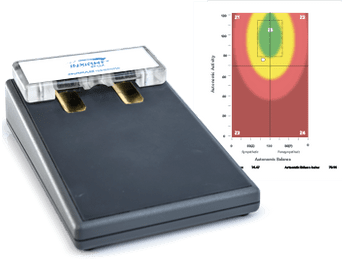
The neuroPULSE analyzes Heart Rate Variability, or HRV. Rather than simply looking at heart rate, HRV examines beat-to-beat variation and provides useful information related to autonomic balance, reserve, adaptability, and recovery.

If we are talking about how the autonomic nervous system responds to demand, then flexibility matters. HRV gives the chiropractor objective information about that broader autonomic picture.

It does not directly analyze neuroception signals. It gives you another window into how the nervous system is performing.

neuroCORE and the Somatic Side of Nervous System Performance

The neuroCORE analyzes surface EMG activity in the paraspinal muscles. It helps chiropractors evaluate postural tension, symmetry, motor tone reactions, energy expenditure, and compensatory patterns.

This adds an important layer because nervous system performance is not purely autonomic. Neural pathways involved in posture, motor control, and compensation also tell us something about how the body is organizing itself under demand.

neuroTHERMAL and Autonomic Patterns Along the Spine

The neuroTHERMAL analyzes paraspinal temperature patterns associated with autonomic regulation. Because autonomic activity influences skin blood flow and temperature regulation, thermal analysis provides another objective view of nervous system status.

With neuroTHERMAL, a chiropractor can complete a full spine nerve system scan in under 30 seconds. Rolling and segmental scan modes help analyze stress patterns along spinal regions and follow how those patterns fluctuate under care.

Again, it is not a neuroception test. It is objective neurological analysis that gives the chiropractor another piece of the physiological picture.

Synapse Turns Complex Neurology Into a Patient Conversation

Put neuroPULSE, neuroCORE, and neuroTHERMAL together, and the story becomes much richer.

INSiGHT neuroTECH and Synapse software organize those findings into scan views and reports that make nervous system performance easier to explain. Instead of asking a patient to understand a lecture on neural circuits, autonomic responses, or Polyvagal Theory, you give them objective information they can see.

  • Baseline scan: Establish where nervous system status begins.
  • Progress analysis: Compare how patterns fluctuate under care.
  • Trajectory: Look beyond one good or difficult day and assess the larger direction of nervous system performance.

The chiropractor combines those findings with the history and examination, applies clinical judgment, and builds the care plan from the complete picture. The technology does not replace the chiropractor. It strengthens the conversation.

Neuroception Gives Chiropractic Better Language for Nervous System Performance

For me, the biggest value of neuroception is not giving chiropractors another complicated term to teach patients. We have plenty of those already. Its real value is reminding us that the nervous system is constantly receiving information and organizing responses before conscious thought catches up.

A patient knows what they consciously feel. They know whether they slept well, whether they feel energetic, whether they can settle, and whether something feels different. That matters. But it does not tell us everything about their underlying physiological responses.

That is why I would resist turning neuroception into the latest chiropractic label. We do not need to tell patients they have a “neuroception problem.” We do not need to diagnose Polyvagal states. And we do not need to move outside our scope into trauma recovery or trauma therapy.

What we can do is ask better questions.

  • Detection: How is the nervous system responding to the information coming in?
  • Adaptation: Can it respond appropriately when demand rises?
  • Recovery: Can it move back toward stability when the demand passes?
  • Flexibility: Can the system recognize both threat and safety rather than remaining stuck in one defensive strategy?
  • Performance: What does objective analysis tell us beyond how the patient feels today?

That is where neuroception matters to chiropractic. It reminds us that conscious experience and physiological state are not always the same thing.

And when so much of nervous system performance happens beneath conscious awareness, objective neurological analysis becomes increasingly valuable. INSiGHT scanning technology gives chiropractors a way to bring part of that invisible story into view without pretending to diagnose what the technology cannot diagnose.

Neuroception reminds us that the nervous system is always listening before the conscious mind has finished the conversation. For chiropractors, that makes neurological scanning more than a technology story. It gives us another way to understand adaptability, communicate the why behind care, and keep the conversation where it belongs: on nervous system performance.

A patient stands up from your consultation chair, pauses for a second, and says, “Give me a minute. My back always feels stiff when I first get up.” You have heard some version of that line a thousand times. Maybe they have lower back stiffness every morning. Maybe sitting for long periods makes it worse. Maybe they keep doing back stretches because they have decided their stiff back is simply the price of getting older.

Back stiffness is common, and sometimes the explanation is straightforward. An unfamiliar workout, lifting something heavy, prolonged inactivity, poor posture, or a minor strain can leave the back muscles guarded and restricted. Back stiffness may occur alongside back pain, or a patient may feel stiffness without much discomfort at all. There are also possible causes involving arthritis, a spinal disc, or other conditions that deserve appropriate evaluation.

But you and I both know the patient’s description is only the beginning. The interesting question is not simply, “How do we loosen this back?” The better question is, “Why is this patient’s body creating or maintaining this tension?” Once you ask that, back stiffness becomes more than a tight muscles conversation. It becomes a conversation about posture, protection, compensation, movement, and neurological control.

Common Causes of Back Stiffness and Why the Back Feels Tight

There is no single cause of back stiffness, and we should not force every patient into the same explanation. The common causes of back stiffness include muscular strain, overuse, prolonged inactivity, postural demands, reduced mobility, arthritis, and other spinal or structural issues. These are some of the same common causes of back pain, which is why a good history and examination matter.

The location can vary too. Some patients experience tension through the upper back. Others describe stiffness in your lower back, particularly when they first stand or begin moving. And while pain and stiffness frequently occur together, one does not necessarily require the other. “My back feels tight” is a description, not a diagnosis. The cause of your back stiffness may be quite different from another patient’s presentation even when both use exactly the same words.

Muscle Strain, Overuse, and Protective Guarding

One of the common causes of stiff back complaints is a strain or sprain. Lifting, bending, twisting, shoveling snow, returning to exercise, or asking the body to do more than it has been accustomed to can overload tissues and lead to stiffness.

The body may respond by protecting the region. Stiff muscles develop, movement becomes guarded, and the patient begins to feel restricted. That protective response makes sense. If the nervous system perceives that movement may cause pain or create further difficulty, restricting movement can be a reasonable short-term strategy.

But here is where I encourage chiropractors to think one layer deeper. Tight muscles are the output we observe. The nervous system is directing that output. That does not mean every strain is a neurological mystery. It means that when back stiffness persists or repeatedly returns, we should be interested in why the system continues asking those muscles to guard.

Sitting for Long Periods and Lack of Movement

You do not have to injure yourself to experience back stiffness. Sometimes you simply stop moving. Patients who spend long periods at a desk, in a car, or in one sustained position may notice stiffness when they finally stand. Gentle physical activity can often help ordinary mechanical stiffness loosen as the body starts moving again.

This is one reason avoiding back movement altogether is rarely a useful long-term strategy for uncomplicated mechanical stiffness. Appropriate movement helps maintain mobility, while regular physical activity can help prevent stiffness associated with prolonged inactivity.

But the chiropractor should still be curious. If relatively ordinary demands repeatedly cause back stiffness, what is the system doing during those hours of sitting? How efficiently is it organizing posture?

Posture, a Tight Hip, and Muscular Compensation

The region that feels stiff is not always the only region worth examining. A tight hip, limited hamstring mobility, reduced shoulder mobility, or poor coordination between the back and hip can contribute to back loading. When mobility is reduced above or below the lower spine, the lumbar region may compensate.

Reduced strength or coordination through the glutes, abdominal muscles, core muscles, and back muscles can also alter how load is managed. That is one reason exercises to strengthen core muscles are commonly included in conservative approaches for certain back complaints. A physical therapist may also use physical therapy, exercises and stretches to address mobility, strength, posture, and body mechanics.

For chiropractors, though, posture deserves more than a mechanical explanation. Posture is not simply where somebody puts their shoulders when told to stand straight. It is an ongoing neurological process. The nervous system continually receives sensory information and coordinates motor output to maintain the body against gravity. Even the curve of your lower back exists within that constantly adapting system.

Arthritis, Disc Changes, and Other Structural Causes

Not every stiff back is primarily muscular. Arthritis can contribute to inflammation, reduced joint mobility, stiffness and pain. Age-related changes involving a disc may also be associated with back pain and restricted movement. Other potential causes described in conventional back pain literature include spinal stenosis, scoliosis, spondylolisthesis, fractures, and inflammatory conditions.

These possibilities are why we should never declare the cause of lower back pain from stiffness alone. A patient saying, “My back is stiff,” does not tell us whether the situation is a temporary muscular response, structural concern, inflammatory process, or something else. History matters. Examination matters. Imaging matters when clinically indicated.

And then there is another question chiropractic is particularly well positioned to ask: What does the functional neurological picture look like?

Looking Beyond Back Pain to Understand a Stiff Back

Back stiffness is what the patient experiences. Postural tension is something we can investigate. That distinction matters because back stiffness and back pain are not interchangeable. Some patients have substantial tightness in your lower back with very little discomfort. Others experience lower back pain accompanied by guarding, spasms, restricted movement, or difficulty standing upright.

Another patient may report that their back pain has improved while compensatory patterns remain evident during the examination. Symptoms matter. They simply do not tell the whole story. If we only ask whether the patient can still feel stiffness today, we are relying on one moment in a much larger physiological story.

Tight Muscles May Be the Response, Not the Whole Problem

Muscles do not independently decide how much work they are going to do. Motor tone is neurologically controlled. The nervous system coordinates posture, movement, stabilization, and protective responses. When the body perceives increased demand or threat, muscular guarding can be part of that response and lead to stiffness.

That gives us a different way to think about the patient whose lower back always feels tight. Instead of stopping at, “Your muscles are tight,” ask a better question:

Why is the nervous system asking this region to work this hard?

That question changes the conversation. The goal is not simply to stretch your back until it temporarily feels looser. The goal is to understand why a spinal region may repeatedly return to guarding, compensation, or excessive postural tension.

Posture, Proprioception, and Compensation

The back and spine are part of a living sensory-motor system. Information about position, movement, load, and balance is continually being fed into the nervous system. Motor output is continually adjusted in response. When that process is efficient, the body can adapt to sitting, standing, walking, lifting, and changing positions without unnecessarily burning through energy.

When compensation enters the picture, certain regions may work harder. Limited hip mobility can contribute to back compensation. Another patient may brace excessively through the lumbar region. Someone else may develop a postural strategy that places different demands on the lower back.

The patient experiences the end result as stiffness. The chiropractor gets to ask what is organizing it.

Why Back Pain Alone Does Not Tell the Whole Story

Imagine asking only one question at every re-exam: “Does your back still feel stiff?” It is useful information, but hardly a complete assessment.

The answer can fluctuate based on yesterday’s workout, how long the patient drove, how they slept, or what they lifted. A subjective report captures experience. It does not necessarily reveal the underlying motor or neurological pattern.

That is why objective findings matter. History tells you what the patient has experienced. Your examination tells you what you find. Neurological scanning can add objective analysis of nervous system performance. Now the conversation is no longer limited to whether the patient feels looser.

Treating a Stiff Back Starts With Finding the Cause

When people search for treating a stiff back, they naturally want something they can do tonight. Back stretches. Heat. Ice. Exercise. Maybe over-the-counter pain relievers. These treatment options appear throughout conventional discussions of back pain relief, and some may be appropriate depending on the individual situation.

But there is no single effective treatment for every stiff back because there is no single cause. The first steps to take depend on the history, duration, severity, associated findings, and cause of your back complaint. The steps to take when treating an uncomplicated muscular strain will not necessarily be appropriate for stiffness associated with arthritis, significant trauma, or another condition.

Movement, Back Stretches, and Exercises to Strengthen the Core

For uncomplicated mechanical stiffness, staying appropriately active is commonly encouraged. Gentle walking, comfortable mobility work, and back stretches may help some people find relief. Prolonged bed rest is generally not the goal.

Strength matters too. Exercises to strengthen the core and surrounding musculature can improve the body’s ability to manage load. Improving hip mobility may reduce unnecessary compensation through the lower back. Depending on the cause of your back symptoms, a physical therapist may use physical therapy to address flexibility, mobility, strength, posture, and body mechanics.

These approaches may help alleviate back pain, prevent back stiffness associated with inactivity, and help prevent stiffness from recurring in some situations. But if a patient has been stretching the same region every morning for years because it always tightens again, the stretch may be addressing the sensation without answering the bigger question.

Why does the body keep returning there?

Heat, Ice, and Conventional Back Pain Relief

Heat is commonly used to relax stiff muscles, while ice may be used in some acute situations involving pain and inflammation. Over-the-counter pain relievers are another common approach people use to reduce pain and inflammation.

Those approaches belong in the larger back pain treatment conversation, but they primarily address symptoms. They do not automatically identify the cause of your pain or explain why recurrent back stiffness keeps returning.

Pain relief can be valuable. Feeling better is worth celebrating. But when evaluating recurrent or chronic back pain, symptom improvement should not automatically end our curiosity about function.

Where Chiropractic Adjustments Fit

Chiropractic adjustments are among the conservative hands-on approaches used for back pain and stiffness. But chiropractors sell ourselves short when we explain the adjustment as nothing more than a way to loosen a stiff joint or relieve pain.

Neurologically-Focused Chiropractic Care asks a bigger question. Where is neurological interference present? How is the patient adapting? What does the examination tell us about function? What findings are asking for attention?

The chiropractor combines the history, examination, neurological findings, and clinical judgment to create the care plan. The goal is not to promise that every adjustment will help alleviate back pain or that every cause back stiffness has a chiropractic answer. It is to understand the patient well enough to make a responsible recommendation.

When Back Stiffness Needs Additional Evaluation

Most chiropractors recognize when the story no longer sounds like routine stiffness. Significant trauma, progressive weakness, numbness, radiating signs, substantial loss of function, or other additional symptoms can require further assessment, imaging, medical evaluation, or referral.

Back and neck pain can have many causes, and appropriate differential assessment matters. The same is true when neck pain or low back pain appears alongside neurological findings or when a presentation is causing pain and stiffness that continues to worsen.

  • History: When did the back stiffness begin, and what was happening around that time?
  • Pattern: Is the stiffness constant, or does movement, sitting, sleep, or activity influence it?
  • Associated findings: Are numbness, weakness, radiating signs, or other neurological findings present?
  • Function: Does the patient have difficulty walking, standing, bending, or completing normal activities?
  • Clinical context: Is there trauma, known arthritis, a disc concern, or another factor requiring further investigation?

Structural imaging and neurological scanning also need to stay in their proper lanes. An X-ray, MRI, or other imaging study may be appropriate when the clinical question involves structure or pathology. Imaging helps evaluate structure. Neurological scanning helps us analyze function.

How INSiGHT Scanning Helps Chiropractors See Beyond Back Stiffness

A patient can tell you, “My back feels stiff.” Your hands may find guarding. Your examination may reveal restricted movement. You may see altered posture or compensation. But what if you could also show the patient objective information about how their nervous system is organizing muscular and neurological responses?

That is where INSiGHT scanning technology fits into the chiropractic examination. Its purpose is not to diagnose the cause of back stiffness, arthritis, a disc condition, or another pathology. INSiGHT provides objective neurological exam data and scan views that support the chiropractor’s interpretation.

Neurological scanning shifts the patient’s attention from simply asking where their back hurts toward nerves and performance. Instead of having another conversation entirely about back pain, the chiropractor can begin showing the patient how their nervous system is performing.

neuroCORE Makes Postural Tension and Energy Use Visible

For the patient with recurring back stiffness, neuroCORE is particularly relevant. INSiGHT neuroCORE uses surface electromyography, or sEMG, to analyze electrical activity in the paraspinal musculature. That provides objective information about postural tension, symmetry, motor tone reactions, energy output, and compensation.

Think about how different that conversation becomes. The patient says, “My lower back always feels tight.” Instead of simply agreeing that they have tight muscles, you can examine what those muscles are actually doing.

Are particular spinal regions producing excessive activity? Is there asymmetry? Does the pattern indicate that the system is expending more energy than expected simply to maintain posture?

A patient may think the answer is another stretch. The more useful chiropractic conversation may be about how much energy their nervous system is spending to hold them upright.

neuroTHERMAL Adds an Autonomic View

The INSiGHT neuroTHERMAL adds another dimension by analyzing paraspinal temperature patterns associated with autonomic regulation. A full spine nerve system scan can be completed in under 30 seconds, making it practical for new patient examinations and progress assessments.

Rolling and segmental scan modes allow chiropractors to analyze neurological distress patterns and see how those patterns fluctuate. Thermal analysis does not diagnose the reason for a patient’s stiff back. It provides another functional view of autonomic patterns along the spinal regions.

A stiff back may be what the patient notices. The neurological picture can be broader than the place where they happen to feel stiffness.

neuroPULSE Looks at Adaptability and Reserve

neuroPULSE analyzes Heart Rate Variability to provide information about autonomic balance, adaptability, recovery, and reserve. That matters because patients do not walk into your office as a collection of isolated body parts. The same nervous system coordinating posture and motor tone is continually responding to demand.

INSiGHT neuroTECH gives the chiropractor a three-dimensional neurological picture:

  • Reserve with neuroPULSE: How much adaptive capacity does the system have?
  • Energy with neuroCORE: How efficiently is the system organizing postural muscular activity?
  • Depth with neuroTHERMAL: What autonomic patterns are showing up along the spinal regions?

Together, INSiGHT neuroTECH and Synapse software help chiropractors look beyond the one region that happens to feel stiff and toward the patient’s broader nervous system performance.

Baseline, Response, and Trajectory

This is where neurological scanning becomes particularly useful for patient communication. Start with a baseline. Assess response. Then follow the trajectory.

Without objective analysis, the progress conversation can easily become, “How’s the back?” “Pretty good.” “Still stiff?” “Sometimes.” That tells you something, but it does not tell you very much about nervous system performance.

With INSiGHT neuroTECH and Synapse software, chiropractors can compare scan views and discuss how nervous system status fluctuates over time. The technology provides objective information. The chiropractor combines those findings with history, examination, clinical judgment, and the patient’s goals to build the care plan.

When patients can see their nervous system in living color, the conversation stops being entirely about today’s back pain. They have a clearer way to understand performance, adaptability, and progress.

A Stiff Back Can Start a Bigger Chiropractic Conversation

“My back is stiff” is useful information. It is simply not enough information.

There are many reasons for a stiff back. Strain, inactivity, posture, tight hips, overuse, arthritis, spinal disc changes, and other structural or inflammatory situations can contribute to back stiffness. Appropriate movement, strengthening, lifestyle changes, physical therapy, chiropractic adjustments, or other options may have a role depending on the cause.

But the chiropractor has an opportunity to go further. When patients repeatedly experience back stiffness or low back pain, we can ask more than where they feel it. We can examine how they move, how they compensate, how posture is being organized, and whether their core and back muscles appear to be working harder than expected.

Most importantly, we can stop asking symptoms to tell us a story they were never capable of telling by themselves.

One patient may experience back stiffness before significant back pain appears. Another may feel better while functional patterns still deserve attention. Another may simply have a short-lived stiff back after doing too much over the weekend. Good chiropractic does not force all three people into the same explanation.

We examine. We analyze. We use objective information where it helps. And we make the recommendation that fits the person standing in front of us.

That is the bigger opportunity neurological scanning gives the chiropractic profession. Back stiffness may be the reason a patient starts the conversation. INSiGHT scanning technology gives chiropractors another way to assess the neurological story underneath it and track how nervous system performance fluctuates over time.

The stiff back gets their attention. The nervous system gives us the bigger story.

Heart Rate Variability (HRV) has become one of the most widely discussed measurements in modern health and performance.

Professional athletes use it to guide recovery.

Researchers use it to study autonomic nervous system function.

Consumer wearables use it to estimate stress and sleep quality.

Increasingly, chiropractors are also incorporating HRV into clinical practice.

But why?

The answer goes far beyond measuring fitness or recovery.

For chiropractors, HRV offers an objective way to evaluate how well the autonomic nervous system is adapting to the physical, chemical, and emotional stresses of everyday life. When combined with a comprehensive clinical examination, HRV provides valuable insight into nervous system function that cannot be observed through symptoms alone.

Chiropractic Has Always Focused on the Nervous System

At its core, chiropractic care has always emphasized the relationship between the spine and the nervous system.

While symptoms often bring patients into the office, chiropractors recognize that symptoms alone don’t always reflect what’s happening beneath the surface.

Some individuals experience significant dysfunction with very few symptoms.

Others report considerable symptoms despite relatively minor objective findings.

This is why chiropractors have long sought objective methods for evaluating nervous system function rather than relying solely on subjective symptom reports.

HRV has become one of those valuable objective measurements.

What HRV Reveals

Heart Rate Variability reflects the ongoing communication between the heart and the autonomic nervous system.

Rather than simply measuring heart rate, HRV provides information about how effectively the body adapts to changing demands.

Higher adaptability generally reflects greater flexibility within the autonomic nervous system.

Reduced variability may suggest the nervous system is operating under increased physiological stress.

Importantly, HRV is not a diagnosis.

Instead, it is one piece of objective information that helps clinicians better understand how the nervous system is functioning.

Stress Is More Than Just an Emotion

When most people hear the word “stress,” they immediately think about work deadlines or financial pressure.

The nervous system, however, responds to many different forms of stress.

These include:

  • Physical stress
  • Emotional stress
  • Chemical stress
  • Sleep disruption
  • Inflammation
  • Illness
  • Injury
  • Environmental demands

Every one of these factors influences autonomic nervous system regulation.

HRV provides one way of observing how effectively the body is adapting to those cumulative demands.

Symptoms Don’t Always Tell the Whole Story

One of the challenges in healthcare is that symptoms often appear late in the process.

Two patients may report similar neck discomfort while demonstrating very different patterns of nervous system function.

Conversely, one patient may report feeling perfectly healthy while objective measurements suggest their nervous system is working much harder than expected to maintain normal function.

This is why objective measurements are becoming increasingly important across healthcare.

Rather than replacing clinical judgment, they add another layer of information to help guide assessment and communication.

HRV Supports More Objective Conversations

One of the greatest advantages of HRV in chiropractic practice is that it provides objective data.

Instead of discussing nervous system function using only descriptive language, chiropractors can incorporate measurable physiological information into patient conversations.

This often helps patients better understand concepts that can otherwise feel abstract.

Rather than simply discussing stress adaptation, clinicians can demonstrate how the autonomic nervous system is responding at the time of the assessment.

Objective data frequently makes complex conversations easier to understand.

Tracking Progress Over Time

HRV is rarely most useful as a single isolated measurement.

Its greatest value often comes from observing changes over time.

By comparing standardized assessments performed under similar conditions, chiropractors can evaluate whether autonomic nervous system function appears to be changing throughout the course of care.

This allows objective progress to become part of the clinical conversation alongside:

  • Patient history
  • Physical examination
  • Functional assessment
  • Clinical findings
  • Patient-reported outcomes

Together, these pieces create a more complete picture of patient progress.

HRV Is Only One Piece of the Puzzle

Although HRV provides valuable insight into autonomic nervous system regulation, it does not measure every aspect of neurological function.

It cannot independently evaluate:

  • Neuromuscular function
  • Surface muscle activity
  • Segmental thermal patterns
  • Biomechanics
  • Joint motion
  • Structural findings

This is why HRV should never be interpreted in isolation.

Like blood pressure or heart rate, HRV contributes important information, but it is only one component of a comprehensive evaluation.

A More Complete Picture of Nervous System Function

Modern chiropractic assessment increasingly combines multiple objective measurements rather than relying on one test alone.

Each measurement contributes different information.

HRV helps evaluate autonomic adaptability.

Surface electromyography (sEMG) provides information about muscle activity.

Thermographic assessment evaluates autonomic patterns associated with spinal temperature distribution.

Viewed together, these objective measurements help create a broader understanding of nervous system performance than any individual measurement alone.

How neuroPULSE Fits Into Neurological Assessment

The neuroPULSE technology within the INSiGHT scanning technology measures Heart Rate Variability under standardized clinical conditions.

Unlike wearable devices that prioritize continuous monitoring throughout everyday life, neuroPULSE is designed to collect reproducible HRV measurements during a controlled assessment.

Patients remain seated while the hand is supported and stabilized throughout the recording, helping reduce unnecessary sources of variability. This standardized approach allows clinicians to compare HRV measurements more confidently over time as part of an objective neurological assessment.

Within the INSiGHT scanning technology, neuroPULSE is combined with:

  • neuroCORE, which evaluates surface electromyography (sEMG) along the spine.
  • neuroTHERMAL, which evaluates thermal patterns associated with autonomic nervous system function.

Together, these technologies provide complementary information about nervous system performance rather than relying on HRV alone.

Objective Data Builds Better Patient Understanding

Many patients have difficulty understanding concepts like autonomic regulation, adaptability, or nervous system function.

Objective measurements can make these conversations much easier.

When patients can see measurable information alongside the clinical examination, discussions often become clearer and more meaningful.

Rather than focusing exclusively on symptoms, conversations can shift toward understanding how the nervous system is functioning and how progress is being monitored over time.

This objective approach often improves patient engagement because it gives both the chiropractor and the patient a common reference point for discussing care.

How Accurate Are HRV Wearables? What the Research Says

Wearables and Chiropractic Assessments Work Together

Some patients already wear devices such as an Oura Ring, WHOOP strap, Apple Watch, or Garmin smartwatch.

These tools provide excellent day-to-day insight into recovery, sleep, and long-term wellness trends.

Clinical HRV assessment serves a different purpose.

Rather than replacing consumer wearables, standardized assessments complement them by providing objective measurements collected under consistent examination conditions.

Many patients benefit from both approaches.

Their wearable helps them monitor lifestyle habits between visits.

Their chiropractor uses standardized HRV assessment as part of a comprehensive neurological evaluation performed within the clinical setting.

The Future of Objective Chiropractic Assessment

Healthcare continues moving toward greater use of objective measurements.

Patients increasingly expect healthcare providers to incorporate technology that helps explain findings, monitor progress, and improve communication.

Heart Rate Variability has become one valuable component of that movement.

When interpreted appropriately and combined with a comprehensive clinical examination, HRV provides meaningful insight into autonomic nervous system function while supporting objective, patient-centered conversations.

As technologies continue to evolve, chiropractors have an opportunity to combine clinical expertise with reproducible physiological measurements that help patients better understand their nervous system and their progress over time.

A patient points to the back of their head and says, “It starts right here.” They may describe a dull headache at the base of the skull, a sudden electric sensation that shoots upward, or a headache that starts in the neck and seems to wrap around the side of the head. Sometimes it reaches behind the eye. Sometimes the scalp itself becomes tender.

It is tempting to put all of that under one familiar label: headache. But for a chiropractor, location is only the beginning of the story. A headache at the base of your skull can involve several different patterns, including tension headaches, a cervicogenic headache, migraine, or occipital neuralgia. The overlap is exactly why the examination matters.

And this is where chiropractic has an important role. Instead of simply asking how to relieve pain, we can ask a better question: What is happening through the skull and neck, particularly the upper cervical region, that may be contributing to what this patient is experiencing?

Why a Headache at the Base of the Skull Deserves a Closer Look

When a headache starts at the base of the skull, the upper neck deserves attention. This region contains a remarkable amount of neurological and mechanical activity in a relatively small space. The cervical spine supports the head, provides substantial neck movement, and sits close to the spinal cord and nerves that carry information between the brain and body.

That does not mean every headache in the back of the head originates in the neck. Migraine is a neurological condition, for example, and there are many medical causes of headache that have little to do with the cervical region. But there are also headache patterns in which structures and nerves around the neck are directly involved.

A useful starting point is distinguishing among a few common presentations:

  • Tension headaches: These headaches often feel like pressure, tightness, or a dull ache and may be associated with postural tension around the neck and shoulders.
  • Cervicogenic headache: This is a type of headache in which the source is in the cervical region, with referred pain experienced in the head.
  • Occipital neuralgia: This involves irritation or injury affecting an occipital nerve and can produce sudden, sharp, shooting, or electric sensations.
  • Migraine: Migraine can involve throbbing pain, nausea, sensitivity to light and sound, and other neurological signs. Some migraine presentations also involve the neck.

The location alone does not make the diagnosis. That distinction is important, especially when a severe headache, a new or unusual presentation, neurological deficits, fever, trauma, or other concerning findings suggest the need for medical evaluation.

Occipital Neuralgia and Pain at the Base of the Skull

Occipital neuralgia is one of the first conditions worth considering when someone describes a sharp headache that starts at the back of the head and travels upward.

The occipital nerves provide sensation across much of the posterior scalp. The greater occipital nerve is primarily associated with C2, while the lesser and third occipital nerves contribute to sensation in nearby areas. When one of these nerves becomes irritated, compressed, or injured, the resulting occipital neuralgia pain can feel dramatically different from an ordinary tension-type headache.

Patients may describe it like a sharp electrical jolt. Others describe burning, piercing, aching, or throbbing pain. The discomfort may start near the base of the skull and radiate toward the top of the head, around the ear, or sometimes behind the eye.

Common signs associated with occipital neuralgia can include:

  • Sharp or shooting sensations: The patient may say it feels like a sharp electrical shock.
  • Scalp tenderness: Even brushing the hair or resting the head against a pillow may trigger discomfort.
  • One-sided or bilateral symptoms: The pattern can affect one or both sides.
  • Sensitivity to movement: Turning the head can aggravate irritated nerves.
  • Radiating sensations: The patient may feel pain traveling from the upper neck through the back of your head and toward the scalp.
  • Tenderness around the nerve: Pressure near the affected occipital region may reproduce the patient’s familiar symptoms.

Occipital neuralgia is considered a headache disorder, but it is different from migraine. That difference matters because a patient who says, “I have migraines,” may actually be describing another type of pain altogether.

What Causes Occipital Neuralgia?

There is not always one identifiable cause. Occipital neuralgia can develop when there is pressure, injury, or irritation affecting an occipital nerve somewhere along its course.

The nerves travel from the upper cervical region through tissues at the back of the head before reaching the scalp. That creates several opportunities for nerve irritation.

Potential causes occipital neuralgia may be associated with include:

  • Trauma involving the head or neck
  • Tight muscles surrounding the nerve
  • Cervical disc problems
  • Osteoarthritis or other forms of arthritis
  • Postural tension and mechanical strain
  • Inflammation or infection
  • Other conditions that may affect the nerve or surrounding tissues

Patients sometimes describe this as a “pinch” in the neck. That language is understandable, but as chiropractors, we want to look deeper than a simple pinched-nerve explanation.

What is happening with cervical motion? Is there significant postural tension? What does the neurological examination reveal? Is the upper cervical region involved? Are there findings that require referral?

Those are better questions than assuming every headache has the same cause.

Cervicogenic Headache and the Cervical Spine

A cervicogenic headache is different because the head symptoms are referred from structures in the neck. In other words, cervicogenic headaches originate from the cervical region even though the patient experiences the discomfort in the head.

That can be confusing for patients. They feel the headache, so naturally they assume the problem must be somewhere inside the head. But referred pain does not always work that way.

A cervicogenic headache often starts in the neck and spreads toward the head. The patient may notice a stiff neck, restricted cervical motion, or pain and stiffness that become more noticeable with certain positions or activities. Symptoms may travel from the neck area into the back or side of the head and sometimes behind the eyes.

A few clues may raise suspicion of cervical involvement:

  • The headache that starts with certain neck positions or movements
  • Restricted or uncomfortable neck movement
  • Tenderness through the upper cervical region
  • Symptoms that originate in the neck and spread toward the head
  • A history of neck injury or sustained postural loading
  • A consistent one-sided pattern involving the head and neck

Cervicogenic headaches can be challenging because their presentation can overlap with other headache categories. That is why treating cervicogenic headaches begins with correctly identifying what is contributing to the presentation rather than simply chasing the location where the patient happens to feel symptoms.

Occipital Neuralgia vs. Migraine

Migraine and occipital neuralgia can look surprisingly similar from across the exam room.

Both can produce significant head pain. Both can involve the back of the head. Both can sometimes produce discomfort behind the eye, and light sensitivity may occur. But when you listen carefully to the patient’s description, differences begin to emerge.

Occipital neuralgia pain is commonly described as sharp, shooting, burning, or electric. Episodes may come suddenly and may be triggered by touching the scalp, turning the head, or other seemingly minor movements.

A migraine more commonly lasts longer and may throb. It may be accompanied by nausea, sensitivity to light, sensitivity to sound, or other neurological signs. A patient may be particularly bothered by sensitivity to light and sound or may be accompanied by nausea during an episode.

The challenge is that real patients do not always read the textbook.

Someone can have migraine and cervical dysfunction at the same time. Migraine may involve neck symptoms. Occipital neuralgia may coexist with another headache condition. Headaches often require more investigation than asking the patient where it hurts.

That is why a comprehensive headache assessment should begin with history and examination rather than assumptions.

How Is Occipital Neuralgia Diagnosed?

Having occipital neuralgia diagnosed requires more than finding tenderness at the base of the head.

A medical provider will typically review the patient’s history, examine the head or neck, evaluate the pattern and triggers, and determine whether other conditions need to be ruled out. Palpation around the occipital nerves may reproduce familiar symptoms.

An occipital nerve block may also be used diagnostically. With a nerve block, a local anesthetic is placed around the suspected nerve. Temporary improvement following an occipital nerve block can provide additional diagnostic information.

Depending on the presentation, imaging or other testing may be appropriate to investigate a suspected underlying cause.

This is where chiropractic responsibility matters. If the presentation does not fit an uncomplicated mechanical or neurological pattern, or if the history raises concern for a secondary cause, the patient deserves the appropriate referral.

A headache may occasionally be associated with a more serious medical situation. A sudden severe headache, significant trauma, fever, new neurological changes, confusion, weakness, visual changes, or a major departure from the patient’s usual headache pattern deserves prompt medical attention.

Why the Upper Cervical Region Matters

For the chiropractor, one of the most interesting parts of this conversation is the relationship between the base of the skull and the upper cervical spine.

C1 and C2 sit immediately below the skull. C2 is especially relevant to the occipital conversation because of its neurological relationship to the greater occipital nerve. This region is also close to the top of the spinal cord and is responsible for a considerable amount of head motion and sensory input.

When you think about the upper cervical region, do not reduce it to a single vertebra being “out.” Look at the whole functional picture.

A patient may have altered motion, postural tension, guarding, compensation, or neurological interference around the upper cervical spine. Those findings can coexist with headaches in the back of the head, but they need to be assessed rather than assumed to be causal.

That is the difference between symptom chasing and Neurologically-Focused Chiropractic Care.

We are not saying every migraine comes from the neck. We are not saying every case of occipital neuralgia is chiropractic in origin. We are saying that when a headache starts at the base of the head and the cervical region is part of the presentation, it makes sense for a chiropractor to carefully evaluate how that region and the nervous system are functioning.

When Postural Tension Becomes Part of the Headache Story

You cannot talk about the back and neck without talking about posture.

Modern life asks people to spend remarkable amounts of time looking downward. Phones, laptops, driving, prolonged sitting, and repetitive work can keep the head forward and place ongoing demand around the neck muscles and cervical spine.

The SEO phrase patients commonly search is muscle tension, but in the office I prefer to think in terms of postural tension. It keeps us from reducing the problem to a tight muscle that simply needs to be rubbed until it relaxes.

The better question is why those muscles are tight.

Are they compensating? Is the head being carried forward? Is cervical movement restricted? Is the nervous system continually recruiting extra motor activity to stabilize the region?

This is also where other approaches may play a role. Depending on the patient’s situation, medical providers may recommend medication such as ibuprofen, heat, massage, physical therapy, or specific physical therapy exercises. There is a place for symptom management and rehabilitation when clinically appropriate.

But for the chiropractor, the examination should still answer the larger functional question: What patterns are present through the cervical region and nervous system, and how are they fluctuating over time?

How Different Headache Patterns Are Commonly Managed

There is no single way to treat every headache because headache is not a single condition.

A tension-type presentation, cervicogenic headache, migraine, and occipital neuralgia can require very different approaches. Even two patients with the same diagnosis may require different clinical management based on their history and findings.

Medical management of occipital neuralgia can include medications, anti-inflammatory approaches, nerve block procedures, injections, and in selected persistent cases, surgical procedures. Physical therapy may be used when mobility, posture, or musculoskeletal function is involved.

For a chiropractor, the goal should not be to compete with those approaches or promise that an adjustment is the one way to treat every presentation.

Our job is to determine whether chiropractic findings are present and whether those findings belong in the patient’s care plan.

When cervical dysfunction and neurological interference are identified, the chiropractor can build recommendations from the full examination. When something outside our lane is suspected, collaboration or referral is the right move.

That is good chiropractic.

How INSiGHT Scanning Adds a Neurological View to Headache Cases

This is where these cases get especially interesting to me.

A patient walks into your office talking about a headache. Their entire attention is fixed on the symptom. Your opportunity is to broaden the conversation from where they feel it to how their nervous system is performing.

INSiGHT scanning technology can help make that shift.

To be clear, INSiGHT scanning technology does not diagnose migraine, cervicogenic headache, occipital neuralgia, or the cause of a patient’s symptoms. It provides objective neurological exam data and reporting that support the chiropractor’s interpretation.

That distinction matters.

With INSiGHT neuroTECH scanning technologies and Synapse software, you can look at the patient from three complementary neurological perspectives:

  • neuroCORE: Surface electromyography analyzes paraspinal muscle activity, symmetry, postural tension, and patterns of energy expenditure. In a patient with significant head and neck complaints, this can add objective information about how the motor system is organizing itself.
  • neuroTHERMAL: A full spine nerve system scan analyzes paraspinal temperature patterns associated with autonomic regulation. Rather than relying solely on where the patient reports symptoms, you gain another objective view of nervous system status.
  • neuroPULSE: Heart Rate Variability analyzes autonomic balance, adaptability, and reserve, broadening the conversation beyond the local neck complaint to how well the nervous system is adapting overall.

The scans do not tell you, “This patient has occipital neuralgia.” They do something different.

They help you see the nervous system story surrounding the presentation.

That is a much more responsible and useful role for neurological scanning.

From a Headache Complaint to Objective Neurological Findings

This is where a good report of findings can change the patient’s understanding of chiropractic.

The patient may have walked in thinking, “I need to get relief from this headache.” After a thorough examination, you can acknowledge that goal without allowing it to become the only metric that matters.

You can show the patient what you found.

If neuroCORE identifies substantial postural tension or inefficient motor patterns, show them. If neuroTHERMAL reveals autonomic patterns needing attention, explain what those scan views mean. If neuroPULSE suggests reduced adaptability, put that information into context.

Now the conversation becomes:

“You came in because of the headache, and of course we care about that. But I also want you to see what we found underneath the symptom. These scans give us a baseline of how your nervous system is performing. As we move through your care plan, we can come back and compare.”

That is a very different conversation from promising to make headaches disappear.

And Doc, patients understand the difference.

When you give them a shared reference point, they can understand why feeling different is important while still recognizing that symptoms alone do not tell the whole story.

Re-Scanning Helps Answer the Question Patients Eventually Ask

Eventually, every patient asks some version of the same question:

“How do we know?”

That is where baseline and progress scanning earn their place.

The initial scan gives you the starting point. A later scan lets you compare nervous system status instead of relying entirely on the patient’s memory of how they felt weeks ago.

Maybe their headache frequency has decreased. Good. Celebrate it.

But now look at the objective findings too. Is postural tension organizing differently? Are thermal patterns fluctuating? What is happening with adaptability and reserve? Is the broader neurological picture moving in a direction that makes sense alongside the patient’s experience?

The technology does not make that judgment for you. The chiropractor does.

INSiGHT scanning technology provides the objective analysis. You bring the examination, clinical reasoning, adjustment, interpretation, and care plan.

That is exactly how technology ought to fit into chiropractic.

Helping Patients See More Than the Headache

A headache at the base of the skull can have several explanations. It may be related to a tension-type pattern, cervicogenic headache, migraine, occipital neuralgia, or another situation entirely. The fact that two people point to the same spot does not mean the same thing is happening underneath.

That is why the examination matters.

When the presentation starts at the base of the head, involves the cervical region, or seems to radiate through the occipital distribution, chiropractors have an opportunity to look beyond the symptom without making promises the findings cannot support.

Listen to the patient’s story. Examine the cervical region. Recognize when medical referral is appropriate. Look at nervous system performance. Establish objective baseline data when neurological scanning is part of your practice. Then build the care plan from what you actually find.

Because the goal is not to convince a patient that every headache comes from the spine.

The goal is to help them understand their own neurological story more clearly.

When patients can see where they started, understand what you found, and compare that with what happens under care, chiropractic becomes about something much bigger than trying to find relief from the next headache.

It becomes a conversation about nerves, function, adaptability, and nervous system performance.

A healthy spine is not a straight spine, which can come as a surprise.

When viewed from the side, the spine of an adult has a series of natural spinal curves that create a gentle S-shaped profile. The cervical and lumbar regions curve inward, while the thoracic and sacral regions curve outward. Those spinal curves are not mistakes that need correcting. They are part of the remarkable engineering that allows the spine to support us, move with us, absorb forces, and adapt to the physical demands of everyday life.

For chiropractors, however, the interesting question is not simply whether a curve in the spine looks normal. It is what that finding means in the context of the person sitting in front of us. Structure matters. So do movement, posture, symptoms, adaptation, and nervous system performance. Understanding the importance of spinal curves gives us a valuable starting point. Knowing when to look beyond spinal curvature gives us a much more complete clinical picture.

1. What Are the Normal Spinal Curves of the Spine?

If you look at a healthy spine from the front or back, the spine appears relatively straight. Turn that same person to the side and the picture changes. The normal spinal curves become visible, creating the characteristic S-shaped profile of the spinal column.

The spine has four primary curves. These four curves allow the spine to be both stable and mobile. Rather than thinking about a healthy spine as a rigid stack of bones, it is more useful to picture a dynamic structure built to carry load while still allowing the body to move.

Cervical, thoracic, lumbar, and sacral curves

Each region contributes a different curve to the overall shape of the spine:

  • Cervical: The cervical spine in the neck normally forms an inward curve. This is called lordosis, or more specifically cervical lordosis.
  • Thoracic: The thoracic spine through the middle and upper back forms an outward curve. This normal kyphotic shape is known as thoracic kyphosis.
  • Lumbar: The lumbar spine in the lower back curves inward again. This lordotic curve is known as lumbar lordosis.
  • Sacral: The sacral spine curves outward, completing the series of distinct curves seen through the spine.

Put these regions together and you get the natural S curve of the adult spine. The curves alternate between lordotic and kyphotic orientations as you move from the neck toward the pelvis.

Kyphosis and lordosis are not automatically disorders

This is an important distinction because patients frequently encounter the words lordosis and kyphosis online and assume they describe a problem.

They do not. Kyphosis and lordosis describe the direction of normal spinal curves. A lordotic curve bends inward, while a kyphotic curve bends outward. Problems may arise when the degree of curvature becomes unusually increased or reduced, when there is a loss of curvature, or when a curve reverses from its expected orientation.

The exact size and appearance of spinal curves can also vary from person to person. There is no reason to assume that every individual must conform to one visual template for the formation of an ideal spine. For the chiropractor, a structural measurement becomes more meaningful when it is considered alongside the patient’s history, examination, symptoms, function, and other objective findings.

2. The Importance of Spinal Curves in Chiropractic Biomechanics

Why is the ideal spine not shaped like a perfectly straight column?

Because human beings are not statues. We walk, bend, rotate, lift, reach, sit, run, and carry loads. Even standing quietly requires the body to continually manage gravity. The natural curvature of the spine helps the body handle these demands while maintaining a useful balance between mobility and stability.

Think about the difference between a rigid pole and a spring. A rigid pole can carry load, but it has relatively little ability to adapt when forces arrive from different directions. The curved architecture of the spine gives the body more options. Spinal curves contribute to shock absorption and help distribute mechanical forces through the vertebra, disc, muscles, ligaments, and surrounding tissues.

That is one reason curves are important for more than appearance. A healthy spine naturally has curves because the spine needs to manage movement and load rather than simply remain straight.

Each spine curve has a different job

The cervical region supports the head while maintaining enough mobility to look up, down, and around us. The cervical lordosis portion of the spine exists in an area where mobility and stability have to coexist throughout the day.

The thoracic region has a different job. It works closely with the rib cage and provides considerable stability through the trunk. The normal kyphosis curve through the upper back is therefore part of healthy spinal architecture, not something that should simply be straightened.

The lumbar region faces yet another set of demands. The lumbar curve sits beneath the spinal regions above it and handles substantial mechanical forces during standing, walking, bending, lifting, and twisting. The vertebrae and discs of the lower back are built differently from those in the neck because their jobs are different.

When one curve changes, the rest of the body may adapt

The spine functions as one connected structure. When the position or shape of one region changes meaningfully, mechanical demands elsewhere may change as well. The body is remarkably good at maintaining balance, keeping the eyes level, and finding ways to continue moving. Sometimes that involves compensatory positioning elsewhere in the spine.

This does not mean that every altered spine curve creates a predictable chain reaction or inevitably causes back pain. Human biomechanics are far more complex than that. It does explain why chiropractors often examine the entire spine rather than focusing exclusively on one painful area or one vertebra.

Posture can offer useful clues too. Forward head carriage, rounded shoulders, or altered pelvic positioning may accompany changes in spinal mechanics. But posture is information, not a diagnosis. A photograph cannot tell us everything about health, just as looking at the back of the spine cannot tell us everything about how the nervous system is performing.

This is where good chiropractic reasoning matters. We want to understand spinal curves without becoming trapped by the structure alone.

3. When Spinal Curvature Changes: Lordosis, Kyphosis, and Scoliosis

Patients often hear lordosis, kyphosis, and scoliosis mentioned together and assume all three are spine disorders. We have already established why that is not quite right.

Lordotic and kyphotic spinal curves are normal when they occur in their expected regions. The clinical conversation changes when there is excessive curvature, a meaningful reduction or reversal of a normal curve, or an abnormal lateral curving of the spine.

For chiropractors, identifying the curvature is only the beginning. We also want to know why it is present, whether it is structural or functional, whether it is progressing, how the person moves, and how the finding relates to the rest of the examination.

Changes in lordosis

The cervical and lumbar regions normally have lordotic curves. In some people, the inward curvature becomes more pronounced. In others, the normal curve may become reduced or reversed.

Excessive lumbar lordosis is sometimes called hyperlordosis or “swayback.” The appearance may include a more pronounced inward lumbar curve along with changes in overall posture. Some people experience lower back discomfort, stiffness, or muscular fatigue, while others may have a noticeable curvature with few symptoms.

The same principle applies to cervical lordosis. Changes in the cervical curve may be seen alongside altered head and neck positioning, including forward head posture. But the appearance of a curve does not automatically tell us how a person feels or functions.

When thoracic kyphosis becomes excessive

The thoracic region is normally kyphotic. When the outward curve becomes unusually pronounced, it may be described as hyperkyphosis.

Patients may encounter the older term “hunchback,” but that label tells us very little about why the curvature in the upper spine exists. There are different forms of kyphosis, including postural, Scheuermann-type structural, and congenital presentations. Their causes and clinical implications are not interchangeable.

That is why identifying an abnormal curve is not the same as understanding it. Age, history, mobility, symptoms, progression, and physical examination findings all matter. Significant structural findings may also require collaborative assessment with another healthcare professional or spine surgeon.

How scoliosis differs from normal spinal curves

Scoliosis deserves its own distinction because it is not simply an exaggerated lordotic or kyphotic curve.

When viewed from the front or back, scoliosis involves an abnormal lateral curvature. Structural scoliosis also involves vertebral rotation, making it a three-dimensional condition rather than simply a spine shaped like a C when viewed from one direction.

An X-ray may be used when clinically appropriate to assess structural curvature and calculate a Cobb angle. A lateral spinal curvature of approximately 10 degrees or greater is generally used as the radiographic threshold for scoliosis. Curves less than 20 degrees may still require appropriate clinical monitoring depending on factors such as age, skeletal maturity, progression, and individual presentation.

For people with scoliosis, the existence of the curve alone does not determine the appropriate management. Curve magnitude, location, age, progression, skeletal maturity, symptoms, and function can all matter.

How chiropractors evaluate changes in spinal curvature

No single examination finding tells the whole story. Depending on the patient’s presentation, a chiropractic evaluation may include:

  • Health history: Understanding symptoms, previous injuries, development, lifestyle, and relevant health history.
  • Postural and movement assessment: Looking at how the patient stands, moves, bends, and adapts to load.
  • Physical examination: Range of motion, palpation, orthopedic testing, neurological testing, and other clinically appropriate procedures.
  • Structural assessment: Imaging may provide additional information about spinal alignment and the curvature of the spine when clinically indicated.
  • Functional assessment: Objective neurological information can add another dimension that cannot be determined from spinal structure alone.

Chiropractic care may be part of managing patients with altered spinal mechanics, but chiropractic adjustments should not be presented as a guaranteed way to produce one ideal spine curve. Depending on the presentation, management could involve chiropractic care, exercise or rehabilitation, observation, bracing, referral, or specialist assessment.

The same caution applies when discussing long-term chiropractic care. The purpose should not be to promise that every structural curve will be permanently changed. Good care begins by understanding what is clinically relevant for the individual patient.

4. Beyond the Spine Curve: Adding INSiGHT Neurological Scanning to Chiropractic Care

Here is where the conversation about spinal curves gets especially interesting for me.

Chiropractors have always been interested in structure. We look at the spine, how it moves, how its segments relate to one another, and how the person organizes themselves around it. Techniques such as Chiropractic BioPhysics have also contributed to a profession-wide conversation about spinal alignment and structural measurement.

But if we stop at structure, we risk making an assumption I have never been comfortable with: that what the spine looks like automatically tells us how the nervous system is functioning.

It doesn’t.

A structural image and a neurological scan answer different questions

Structural imaging can provide useful information about vertebral relationships, alignment, healthy curvature, and changes in spinal curves. What it cannot directly measure is nervous system performance.

Likewise, someone can have a substantial structural finding without experiencing a corresponding level of symptoms. Another person may have significant functional concerns even though the shape of the spine does not immediately look dramatic.

So rather than asking only, “What does this curve look like?” I believe a neurologically focused chiropractor should also ask, “What else can we objectively measure about this person?”

That is why neurological scanning can be such a valuable addition to the chiropractic examination.

How INSiGHT Scanning adds another layer

INSiGHT Scanning is designed to give chiropractors objective information about nervous system performance. It does not diagnose scoliosis, hyperlordosis, hyperkyphosis, or structural pathology, and it does not replace structural imaging when imaging is clinically appropriate.

It measures something different.

The INSiGHT neuroTECH brings together three scanning technologies:

  • neuroPULSE™: Uses heart rate variability, or HRV, to provide information about neurological reserve and adaptability.
  • neuroCORE™: Uses surface electromyography, or sEMG, to measure patterns of paraspinal muscle activity, symmetry, and energy use.
  • neuroTHERMAL™: Measures paraspinal temperature patterns to add information about patterns of neurological regulation along the spine.

That distinction becomes particularly valuable when discussing spinal curves. An image can show us architecture. INSiGHT Scanning lets us ask another question about the person living inside that architecture: How is their nervous system performing?

From a spinal snapshot to an objective neurological baseline

A first examination gives us a starting point. With INSiGHT Scanning, chiropractors can establish objective baseline neurological findings and compare those findings during later progress examinations.

The INSiGHT software (powered by Synapse) transforms scan findings into visual reports that can support the Report of Findings and subsequent re-examinations. The CORESCORE brings the three neuroTECH perspectives together into a clear reporting framework, helping the doctor communicate where the patient started and compare neurological findings over time.

The technology does not decide what care a patient needs. INSiGHT provides objective examination data and reports. The chiropractor builds the care plan by bringing those findings together with history, physical examination, structural information, clinical judgment, and the individual patient’s needs.

Technology should strengthen clinical reasoning, not replace it.

Helping patients see more than the curve

Images of spinal curvature can be powerful because patients immediately see them. Show someone an unusual curve and their eyes naturally go straight to it. But the conversation can become misleading if we jump from “Your spine looks like this” to “Therefore, this is exactly how your body must be functioning.”

Patients are more complex than their pictures.

Objective neurological scanning gives chiropractors another way to build the clinical story. We can discuss what the patient is experiencing, what we found during the examination, which structural findings matter, what neurological scans are showing, and what objective findings we will follow over time.

At the progress examination, we can come back to those measurements: Where did we start? What has changed? What has remained? How does that compare with what the patient is experiencing now?

That is a richer conversation than simply pointing to the curves in your spine and declaring them good or bad. Structure and function are not competing ideas. They are different windows into the same patient.

5. A Healthy Spine Is More Than the Shape of Its Curves

There is a simple idea worth remembering: a healthy spine is supposed to have spinal curves.

The cervical and lumbar regions normally curve inward, while the thoracic and sacral regions curve outward. Those spinal curves help the spine manage weight, movement, balance, and mechanical forces. A perfectly straight spine is not the definition of normal anatomy.

At the same time, changes in spinal curvature deserve thoughtful evaluation. Increased lordosis, excessive kyphosis, loss of curvature, or scoliosis can provide meaningful structural information. But a structural finding does not automatically tell us whether someone will have pain, how well their nervous system is functioning, or what care they need.

That is why maintaining a healthy spine cannot simply mean chasing the appearance of one theoretical ideal. We want to ensure the spine and the person attached to it are assessed in context.

Good chiropractic assessment looks wider. It considers history, structure, mechanics, movement, symptoms, neurological findings, function, and change over time. For the neurologically focused chiropractor, INSiGHT Scanning adds objective information that helps make nervous system performance visible and measurable alongside the rest of the examination.

A spinal curve is something we can see. In many cases, it is something we can measure. But the person attached to that spine is where the real clinical conversation begins.

The better question is not simply, “What shape is the spine?”

It is, “What does this finding mean for this person, how are they functioning, and what can we objectively measure as we move forward?”

That is where chiropractic examination becomes more than a picture of the spine. And it is where INSiGHT neurological scanning can help chiropractors see a much bigger story.

+