
That is where functional neurology enters the conversation. Rather than asking only what the brain looks like, functional neurology asks how well the brain is communicating with the spinal cord, sensory systems, vestibular pathways, muscles, joints, and autonomic nervous system. It is concerned with brain function, neurological performance, and whether a person can respond to stimulation without quickly losing accuracy or becoming fatigued.
For chiropractors, this is familiar territory. We have always understood that structure and function are related, but they are not interchangeable. Imaging can show anatomy. A neurological examination can show performance. Objective neurological scanning adds another layer by helping the chiropractor analyze autonomic balance, postural tension, energy use, and adaptability beyond symptoms alone.
What Functional Neurology Means in Chiropractic
Functional neurology is a specialized approach to evaluating and supporting the way neurological pathways perform. It studies how the brain receives information, processes that information, and creates an appropriate response. Functional neurology focuses on communication among different regions of the brain rather than looking only for visible damage or disease.
Within the chiropractic profession, this field is also commonly called chiropractic neurology. Many practitioners begin as a doctor of chiropractic and complete postgraduate education in neurological examination, vestibular function, movement, cognitive performance, sensory integration, and rehabilitation. A chiropractic neurologist may combine adjustments with visual exercises, balance tasks, motor activities, or other forms of therapy when those procedures are appropriate for the patient.
Communication, Integration, and Neurological Performance
The brain does not operate as a collection of isolated parts. Different areas of the brain are continuously exchanging information with the spinal cord, vestibular system, visual pathways, muscles, joints, organs, and sensory receptors. A functional brain must receive those signals, organize them, and produce a response that matches the situation.
A functional neurologist may therefore ask questions such as:
- Visual control: Can the patient accurately follow a moving target or shift their eyes between two points?
- Balance: Can they remain stable when visual or surface information changes?
- Vestibular function: Does head movement reproduce dizziness, nausea, or disorientation?
- Motor output: Can the patient coordinate the eyes, head, spine, and limbs during movement?
- Cognitive function: Can the patient maintain focus as a task becomes more demanding?
- Fatigability: Does an initially accurate response deteriorate after repetition?
These questions help identify functional patterns without assuming that one finding proves a specific neurological condition. A balance error does not diagnose a brain injury. An unusual eye movement does not identify one exact region of the brain. Findings must be considered alongside the history, examination, and any appropriate medical testing.
Neuroplasticity and the Functional Brain
The neurological foundation of this approach is neuroplasticity. Nerve cells and neurological networks can adapt in response to movement, repetition, learning, sensory stimulation, and experience. This principle is well established in neuroscience and forms part of many recognized rehabilitation methods.
That does not mean every damaged pathway can be restored or that every neurological condition will respond to the same therapy. It means the brain has some capacity to reorganize the way it processes and responds to information. Functional neurology treatments may use eye movements, vestibular rehabilitation, balance exercises, cognitive tasks, motor activities, or carefully selected brain stimulation to encourage a more organized response.
The goal is not to overwhelm the system. The goal is to provide enough useful stimulation to create a response, then reassess what happened. That is the heart of an evidence-informed approach to functional neurology.
Education and Credentials Matter
The title functional neurologist should not be treated as a casual marketing label. A practitioner should be able to explain their professional license, postgraduate education, and scope. The American Chiropractic Neurology Board is one organization associated with certification in chiropractic neurology, but patients and referring health professionals should still verify a provider’s qualifications.
Functional neurology is also not medical neurology under another name. A medical neurologist is trained in the diagnosis and treatment of neurological disorders, including medication, imaging, laboratory testing, and specialist referral. The chiropractor’s role remains grounded in their own license, examination findings, clinical judgment, and responsibility to refer when necessary.
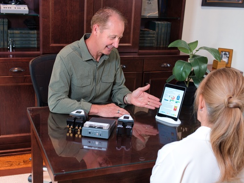
How Functional Neurology Assesses Neurological Function
Functional neurology is not one machine, one adjustment, or one exercise. It begins with a detailed history and neurological examination. The practitioner needs to understand when the presentation began, what makes it better or worse, how the patient responds to activity, and whether red flags require urgent referral or medical evaluation.
The examination may include eye movements, balance, gait, reflexes, posture, coordination, proprioception, sensory discrimination, cognitive tasks, and vestibular system performance. The practitioner may also observe whether a response is accurate, repeatable, symmetrical, and sustainable.
Sensory, Cognitive, Motor, and Vestibular Testing
The brain is constantly collecting information from the environment and the body. Vision provides information about movement and position. Proprioception tells the brain where the joints and body are located. The vestibular system contributes to balance, spatial orientation, and stabilization of the eyes during head movement.
These streams must be integrated before the body can create an appropriate motor or autonomic response. When that process becomes inefficient, the patient may notice dizziness, motion sensitivity, poor coordination, brain fog, visual difficulty, or reduced cognitive function.
A functional neurological examination may include:
- Eye movements: Visual tracking, rapid eye shifts, and gaze stability
- Balance and gait: Static balance, walking patterns, and postural transitions
- Coordination: Fine motor control, timing, and accuracy
- Sensory integration: The ability to combine visual, vestibular, and proprioceptive input
- Cognitive testing: Attention, processing speed, and task tolerance
- Autonomic response: Heart rate or postural responses to demand
No single test should carry the entire interpretation. Functional neurology looks for patterns across multiple systems and asks whether those patterns make sense in the context of the patient’s presentation.
From Examination to Individualized Therapy
Once a functional pattern is identified, the practitioner may select a specific activity and reassess the response. A visual exercise may be followed by another balance test. A vestibular task may be followed by an eye movement assessment. A chiropractic adjustment may be followed by a postural or neurological scan.
A practical treatment approach may follow this sequence:
- Gather the history and identify referral concerns.
- Complete the neurological examination.
- Establish functional and objective baselines.
- Select an appropriate sensory, motor, cognitive, or vestibular activity.
- Reassess the response.
- Adjust the intensity or direction of the activity.
- Track functional recovery over time.
This process is often individualized because two people with similar neurological symptoms may respond differently to the same activity. One person with dizziness may struggle with visual motion, while another is more affected by head movement or postural change.
The phrase treatment plan is often used in broader healthcare, but within chiropractic we would call this a care plan. The chiropractor develops that care plan from the full clinical picture rather than from the symptom name alone.
Functional Neuroimaging and Structural Imaging
Functional neuroimaging, structural imaging, and office-based examination answer different questions. MRI and CT can identify bleeding, tumors, fractures, stroke, and other structural concerns. Functional neuroimaging may examine activation or blood flow to the brain. An office examination evaluates observable neurological performance.
Chiropractic neurological scanning adds another category of information. It does not image specific areas of the brain or map different regions of the brain. Instead, it helps analyze autonomic balance, postural tension, motor activity, and adaptability.
A responsible approach never treats normal imaging as proof that nothing is wrong. It also never treats a functional finding as proof of structural disease. Comprehensive care depends on knowing which question each form of testing can answer.
Learn more about INSiGHT scanning?
Fill this out and we’ll get in touch!
"*" indicates required fields
Functional Neurology for Concussion, Brain Injuries, and Neurological Symptoms
Functional neurology and traditional neurology should not be framed as opposing treatment options. Traditional neurology is essential when a person needs medical diagnosis, medication, imaging, laboratory testing, or specialist management for a serious neurological condition.
A neurologist is the appropriate provider for concerns such as stroke, epilepsy, brain tumors, progressive weakness, multiple sclerosis, Parkinson’s disease, or other complex neurological disorders. Functional neurology may become part of the conversation when the clinical question is how the patient is performing after urgent or structural concerns have been addressed.
Concussion and Mild Traumatic Brain Injuries
Concussion is one of the most common reasons people explore functional neurology. A concussion is a mild traumatic brain injury, yet structural imaging may appear normal even when the patient continues to experience meaningful changes in function.
Common post-concussion traits may include:
- Dizziness: Feeling unsteady, lightheaded, or disoriented
- Brain fog: Difficulty concentrating or processing information
- Visual sensitivity: Discomfort with light, screens, or moving environments
- Cognitive fatigue: Reduced tolerance for reading, work, or conversation
- Balance changes: Difficulty with gait, coordination, or head movement
- Exercise intolerance: Symptoms increasing with physical demand
Visual processing, autonomic regulation, vestibular function, and cognitive workload may all contribute to the presentation. Functional neurology may use visual, motor, cognitive, and vestibular rehabilitation as part of a broader neurological rehabilitation strategy.
No chiropractor should claim that an adjustment or exercise cures concussion or traumatic brain injuries. Acute head trauma, worsening signs, unusual behavior, loss of consciousness, progressive weakness, or severe symptoms require prompt medical attention. Persistent cases may also need collaboration with physicians, neuropsychologists, occupational therapists, and other health professionals experienced with acquired brain injuries.
Migraine, Dizziness, Vertigo, and Vestibular Challenges
Migraine, dizziness, vertigo, motion sensitivity, and balance difficulty are also commonly associated with functional neurology. An examination may include visual tracking, gait, head-position responses, vestibular testing, and the patient’s ability to remain stable when sensory conditions change.
The vestibular system has connections with many areas of the brain. It contributes to balance, posture, spatial awareness, and the ability to keep vision stable while the head moves. When vestibular input does not integrate efficiently with visual and proprioceptive information, the patient may feel unsteady or unusually sensitive to motion.
Some clinics use equipment such as GyroStim as part of motion-based or vestibular activities. Any statement about GyroStim should remain consistent with its current regulatory status, intended use, and supporting evidence. No device should become more important than the reasoning behind its use.
Dysautonomia, Movement Disorders, and Complex Presentations
Functional neurology is also discussed in relation to dysautonomia, chronic pain, functional movement disorders, post-traumatic stress disorder, and functional neurological symptoms. These presentations require careful language because similar signs can arise from very different causes.
A formal functional neurological disorder or movement disorder should be diagnosed by an appropriately qualified provider. Chiropractic scanning does not diagnose dysautonomia, functional neurological conditions, or neurological disease from a color pattern.
Chiropractors can still contribute meaningful information about posture, proprioception, autonomic balance, neurological imbalances, and nervous system performance. The key is knowing when to address neurological function within chiropractic scope and when collaboration or referral is the responsible next step.
The evidence supporting individual therapies varies. Neuroplasticity is established, and many visual, motor, cognitive, and vestibular methods have recognized uses. Evidence for the broader functional neurology model is less uniform, especially when studies use small samples, mixed procedures, or highly individualized protocols.
That does not mean the approach has no value. It means the strength of the claim should match the strength of the evidence. The aim should be improving brain performance and quality of life without promising a guaranteed outcome.

INSiGHT Scanning for Brain Health and Functional Neurology Treatments
A neurological examination can reveal meaningful patterns, but many findings are difficult for patients to see or remember. They may hear an explanation about autonomic regulation, motor output, or vestibular function and still wonder what it means for them.
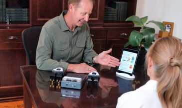
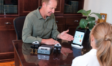
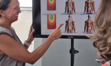
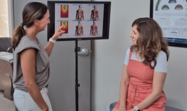
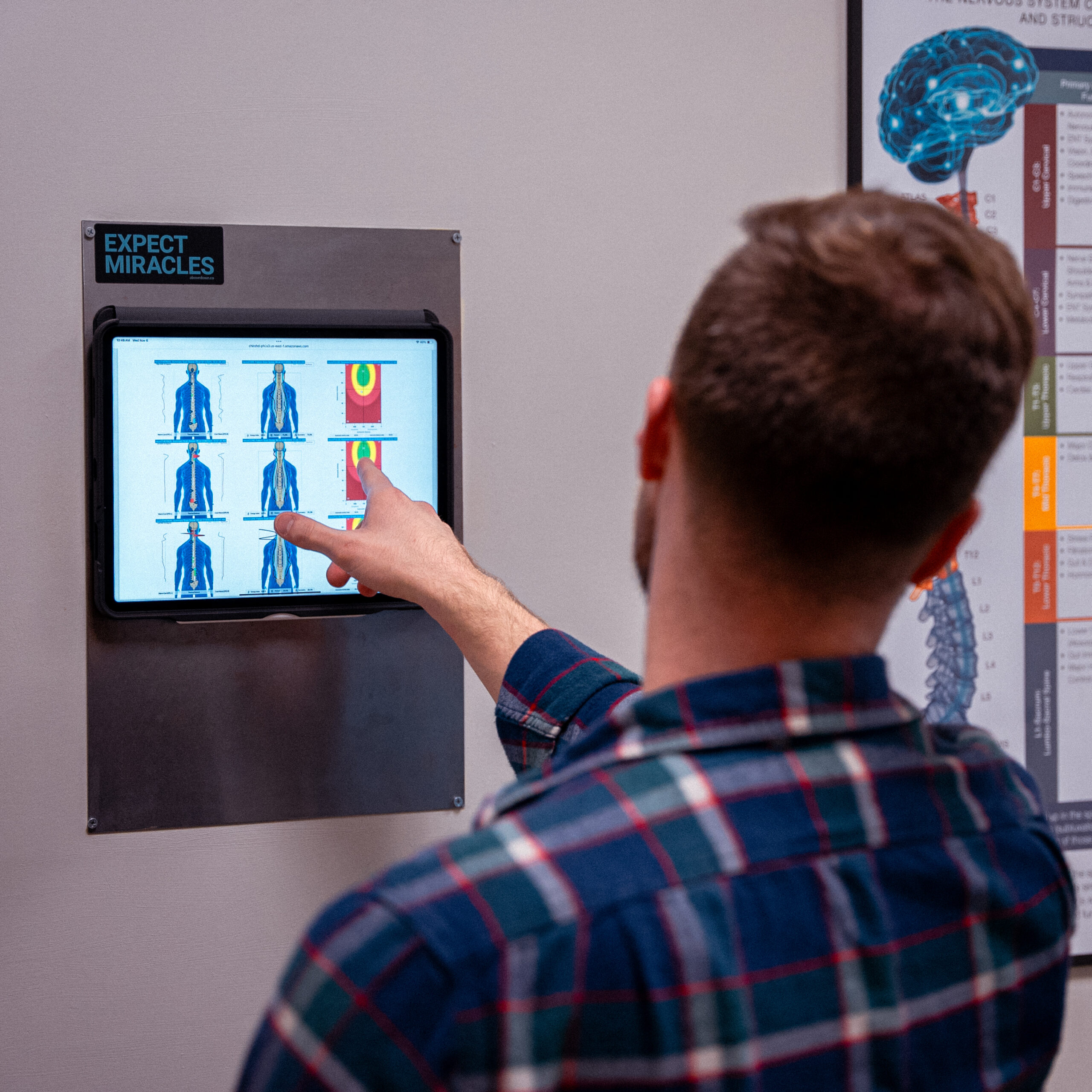
This is where INSiGHT scanning technology gives the chiropractor a clearer way to communicate. It provides objective exam data that can be reviewed and compared over time. When patients see their nervous system status in living color, the conversation moves from abstract explanation to a shared reference point.
INSiGHT scanning is not functional neuroimaging. It does not show brain structure, diagnose concussion, or identify specific regions of the brain. It provides non-invasive analysis of autonomic and somatic patterns that support the chiropractor’s examination, interpretation, and care plan.
neuroPULSE and Autonomic Adaptability
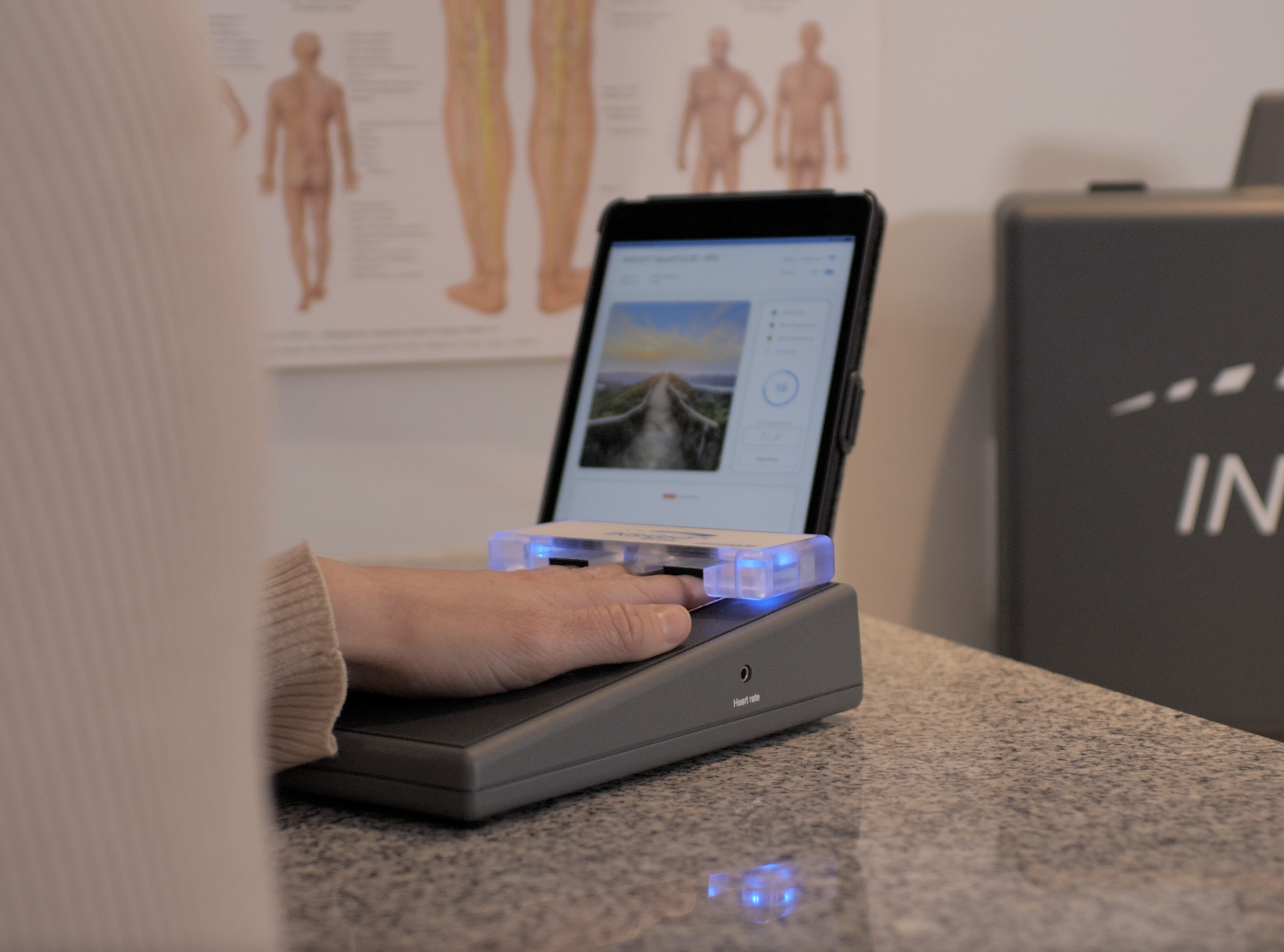
The neuroPULSE analyzes Heart Rate Variability, the beat-to-beat variation in heart pacing. HRV provides information about autonomic activity, balance, adaptability, and reserve.
A resilient system should be able to increase output when demand rises and recover when the demand passes. The goal is not to remain calm every moment. The goal is flexibility.
This becomes relevant when patients with concussion history, migraine, dysautonomia, dizziness, or poor activity tolerance also show signs of altered autonomic regulation. HRV does not establish a diagnosis, but it can help the chiropractor determine whether the system appears adaptable or is operating with limited reserve.
neuroCORE and Motor Output
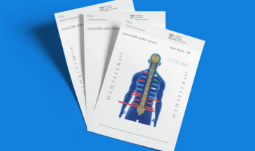
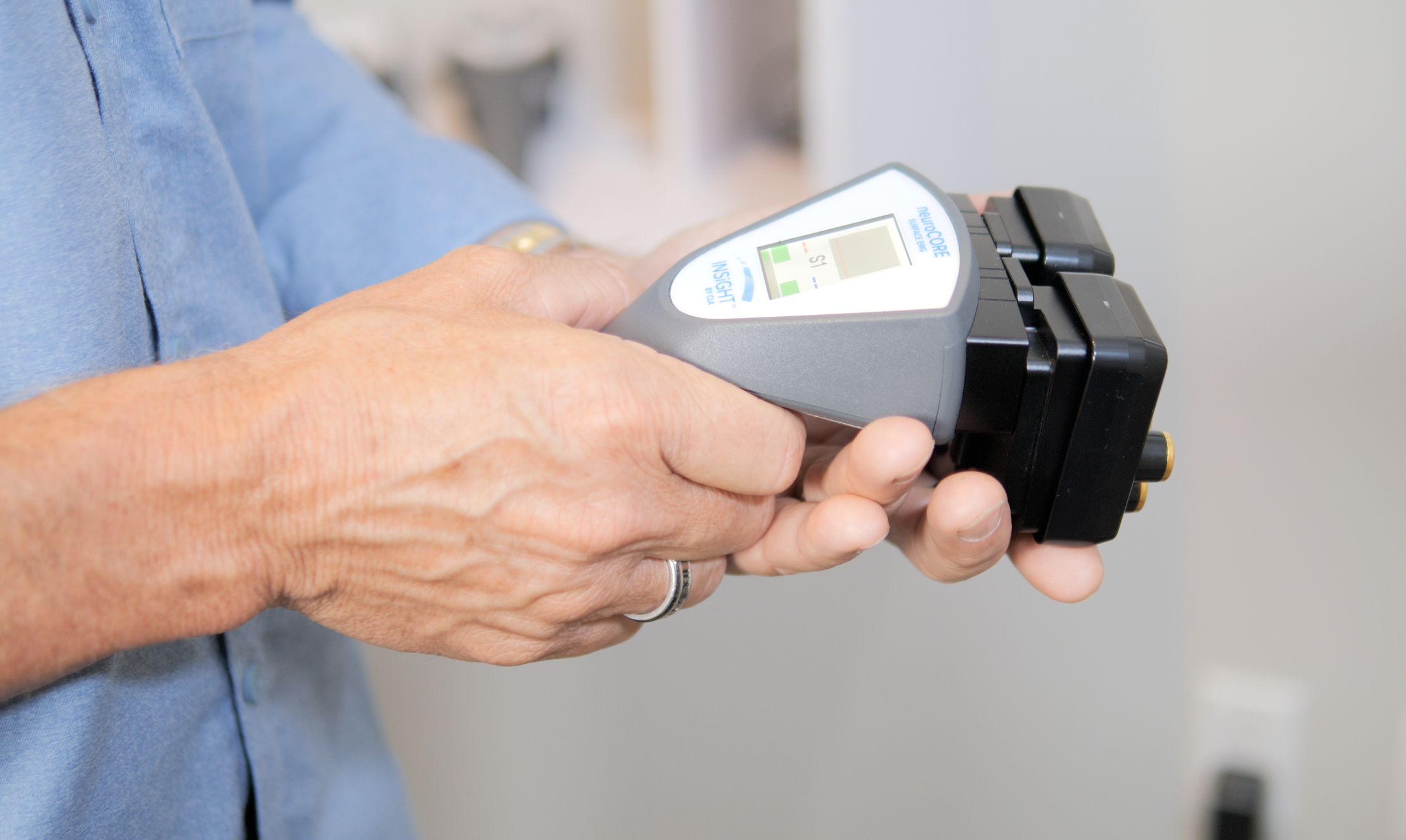
The neuroCORE uses surface electromyography to analyze electrical activity in the paraspinal muscles. Its scan views help the chiropractor evaluate postural tension, symmetry, motor tone, compensation, and energy expenditure.
Functional neurology is deeply interested in how sensory input influences motor output. The brain receives information from the body and uses it to organize posture and movement. When neurological interference is present, the body may spend more energy stabilizing or compensating.
The neuroCORE does not identify a movement disorder or tell the doctor which part of the brain produced the pattern. It shows how motor output is being expressed along the spinal region.
neuroTHERMAL and Autonomic Patterns
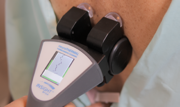
The neuroTHERMAL performs a full spine nerve system scan in under 30 seconds. It analyzes bilateral paraspinal temperature patterns connected to autonomic regulation and blood flow.
Temperature regulation is influenced by the autonomic nervous system. Persistent asymmetry or pattern breaks can give the chiropractor another functional view of how neurological distress is being expressed along the spine.
A thermal scan does not diagnose inflammation, organ disease, or brain pathology. It provides objective information about autonomic patterns and spinal regions needing attention.
The INSiGHT Software, Powered by Synapse
The INSiGHT software, powered by Synapse, organizes neuroPULSE, neuroCORE, and neuroTHERMAL findings into patient-friendly scan views and reports. The chiropractor can compare initial findings with progress scans and communicate whether patterns are improving, fluctuating, or remaining under demand.
The CORESCORE brings the three forms of analysis together into one neurological efficiency score. It is not a diagnostic grade. It is a report-card-style metric that simplifies information about reserve, energy, and depth.
A scan-led process may include:
- Baseline scan: Establish the patient’s starting nervous system status.
- Report of findings: Connect the scan views to the examination and patient goals.
- Care plan: Let the chiropractor build recommendations from the complete clinical picture.
- Response scan: Assess how the patient is adapting under care.
- Progress scan: Compare findings and establish a longer-term trajectory.
- Referral: Involve other providers when findings require additional evaluation.
The technology does not create the care plan. The chiropractor does. INSiGHT neuroTECH and Synapse software provide objective analysis that strengthens clinical interpretation and patient communication.
A More Measurable Future for Optimizing Brain and Nervous System Performance
Functional neurology asks chiropractors to look beyond whether a structure appears damaged. It asks how the patient receives information, how the function of the brain organizes it, and whether the resulting response is efficient, adaptable, and sustainable.
That perspective brings sensory input, motor output, cognitive performance, vestibular function, autonomic regulation, and neuroplasticity into one larger neurological story. It does not remove the spine from chiropractic. It helps explain why spinal motion and the constant flow of sensory information are so important to brain function.
Some people describe this as a holistic approach to brain health. Within chiropractic, I would call it a vitalistic, drug-free approach that respects neurological health while remaining clear about scope. Responsible functional neurology requires appropriate education, careful examination, evidence-informed therapy, and collaboration when a patient needs medical care.
The profession does not move forward through larger claims. It moves forward through better questions, stronger objective analysis, and clearer communication. The goal is not simply to enhance cognitive function for a few minutes or promise that every brain will recover the same way. The goal is to promote brain health, support functional recovery, and help patients understand what their nervous system is expressing.
That is why neurological scanning belongs in the approach to functional neurology. The adjustment remains central, but the scan provides a visible baseline, a response, and a trajectory. It helps the chiropractor see how well the patient is adapting beyond what they feel on one particular day.
When patients can see where neurological distress is building, how much energy their system is using, and whether their adaptability is improving, they begin to understand the why behind chiropractic care. Once that happens, they stop seeing care as something reserved only for symptoms and begin valuing it as part of a broader strategy for optimizing brain and nervous system performance.
That is the real opportunity ahead for functional neurology in chiropractic: not bigger promises, but a more complete examination and a clearer way to make the invisible visible.