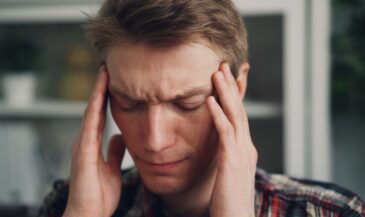
That is why the paraspinal region matters so much in chiropractic. This is not just a strip of tissue beside the spine. It is a highly active system of musculature, connective support, and neural relationships that helps the body stay upright, rotate, extend, and protect itself under stress. When the paraspinal muscle system loses coordination, the body often buys stability with stiffness. The result may look like muscle pain, severe pain, or chronic low back pain, but the deeper issue is often function.
For a Neurologically-Focused Chiropractor, the paraspinal region is not simply where symptoms show up. It is where strategy shows up. It is where you can begin to see whether the body is moving with options or moving with protection. And when that strategy becomes measurable through neurological scanning, the whole care conversation gets clearer.
What the paraspinal region is and why it matters in chiropractic
The paraspinal region refers to the tissues running vertically along both sides of the spine, from the cervical region through the thoracic spine and lumbar spine down toward the sacrum. This area sits location relative to each vertebra and spinous process and includes muscles around the spinal column that help guide motion and maintain stability. In chiropractic, that makes the paraspinal region one of the most clinically useful regions in the body.
The paraspinal muscles are a group, not a single structure. In fact, paraspinal muscles are a group of layered support systems that must work together if the spine is going to stay stable and still move well. These paraspinal muscle groups support posture, help control extension and rotation, and protect each intervertebral segment during movement. When this group of muscles works efficiently, the patient moves with confidence. When the system is overworked or poorly coordinated, patients become prone to pain, fatigue, and guarding.
That is why chiropractors pay close attention to the paraspinal region. A tight or tender paraspinal muscle is rarely just a local issue. It may be associated with paraspinal compensation, poor load sharing, or deeper neurological interference. Patients with similar symptoms can present with completely different motor strategies. One patient may hinge well through the lower back. Another may brace through the entire spine. Their symptoms may sound alike, but their nervous system strategy is not.
In day-to-day practice, this is one of the most valuable ideas to remember: the paraspinal region often shows how the body is protecting itself. It gives chiropractors insight into posture, movement tolerance, and the way the spine is being managed under load. That is what makes the assessment of paraspinal function so important.
Anatomy of the paraspinal region chiropractors need to know
The anatomy of the paraspinal is best understood as a layered system. The paraspinal region contains a broad muscle group, deeper segmental stabilizers, connective support, and neural relationships that all contribute to spinal control. The musculature in this area is not there simply to move the body. It helps coordinate the body’s response to gravity, load, and motion.
The best-known layer is the erector spinae. The erector spinae muscle system includes the iliocostalis, longissimus, and spinalis muscle groups. These extensor muscles help extend the spine, maintain upright posture, and manage longer-range trunk support. The erector spinae and lumbar erector spinae are especially important in the lumbar spine, where prolonged loading, poor endurance, or compensation patterns often show up first. In some back pain patients, you may even find a dominant left erector spinae pattern that reflects asymmetrical stabilization.
Below that larger system sits the transversospinalis layer, including the multifidus muscle. The lumbar multifidus and related segmental muscles are essential for fine control around each vertebra and spinous process. These muscles include deep stabilizers that help manage motion between levels rather than simply creating large movement. When the multifidus muscle or right multifidus underperforms, the body often shifts extra demand to the larger lumbar paraspinal muscle system. That can contribute to muscle fatigue, poor muscle strength, and inefficient movement in the lumbar spinal region.
The anatomy of the paraspinal also includes important relationships with nearby structures. The paravertebral and paravertebral muscles work beside the spine to support control and posture. The psoas muscle, abdominal muscles, and other back muscles influence how the paraspinal and psoas muscle systems share load. Muscle fibers in these regions must coordinate with the thoracic spine and upper back just as well as they do with the lower back. When one system loses efficiency, another muscles may try to compensate.
- Erector spinae: The long extensor system that supports posture and extension.
- Longissimus and iliocostalis: Key parts of the erector spinae that help manage trunk support and movement.
- Multifidus muscle: A deep stabilizer that helps control motion at the segmental level.
- Lumbar multifidus: Especially important in protecting the lumbar spine during load and transition.
- Paravertebral muscles: Support structures that work with the broader paraspinal musculature.
That is the practical takeaway. The paraspinal muscle is not just a mover. The paraspinal musculature is a control system. It supports the spine, manages force, and helps determine whether the body moves with freedom or with stiffness.
Learn more about INSiGHT scanning?
Fill this out and we’ll get in touch!
"*" indicates required fields
Common paraspinal problems chiropractors see in practice
In practice, the paraspinal region is involved in both acute and chronic presentations. An acute complaint may begin with a muscle strain, an awkward lift, or a sudden disc irritation. The patient may describe severe pain, muscle spasms, or the sense that the lower back “locked up.” In those moments, the paraspinal muscle system is often acting protectively. The body is trying to reduce motion and create safety around the spine.
Over time, those patterns can become more complicated. Acute and chronic presentations often blend together when the original protection strategy never fully resolves. A patient with chronic low back pain may no longer present with obvious spasm, but the changes in the paraspinal region can still be significant. The lumbar paraspinal may remain overactive, the lumbar muscle system may lose endurance, and the body may keep relying on compensation rather than recovery. That is one reason chronic low back pain is so often associated with low back pain patterns that persist even when symptoms fluctuate.
Research has paid more attention to this in recent years. Chiropractors will see discussions of paraspinal muscle morphology, paraspinal muscle atrophy, muscle mass loss, and atrophy in the lumbar paraspinal region. These changes in the paraspinal system may be associated with low back pain, altered control, and long-term movement limitations. In some studies, paraspinal abnormalities and abnormalities in the paraspinal system are described alongside degenerative findings, soft tissue conditions, or intervertebral disc changes. That does not mean every patient follows the same path, but it does mean the paraspinal story is not always a simple one.
You may also see patients with an imbalanced lumbar paraspinal muscle pattern, asymmetry through the thoracic spine, or protective stiffness extending into the cervical paraspinal region. Some presentations are clearly associated with paraspinal overload. Others are associated with low back pain even when imaging does not look dramatic. In both cases, the chiropractor’s job is to ask a more useful question: can this patient stabilize the spine well, or is the body using protection as its only strategy?
- Acute presentation: Muscle strain, guarding, disc irritation, or a sudden lower back flare-up.
- Chronic presentation: Endurance loss, compensation, and chronic low back pain patterns.
- Structural change: Atrophy, paraspinal muscle atrophy, or changes in the paraspinal seen over time.
- Functional change: Reduced control, altered movement, poor muscle strength, and ongoing protection.
That is why these findings matter in chiropractic. A sore paraspinal muscle may reflect much more than local irritation. It may reveal how the nervous system is managing load, and whether the body still has enough reserve to move well.
How chiropractors assess the paraspinal region with more precision
The best assessment of paraspinal function is layered. It starts with observation. A chiropractor watches how the patient stands, hinges, rotates, extends, and transfers load through the spine. That movement story matters because posture, asymmetry, and guarding often show up before the patient has the language to describe them. This early assessment of paraspinal function is one of the most honest parts of the exam.
Hands-on evaluation still plays an essential role. Palpation, tenderness, tissue tone, and motion findings around each spinous process help build the clinical picture. So does medical history. The history of prior trauma, recurrent flare-ups, training habits, sedentary behavior, and previous disc episodes all influence how the chiropractor interprets the spine and surrounding musculature. This is where clinical reasoning begins to separate local irritation from deeper adaptation.
Still, structure is not the same as function. That is a critical point. MRI can describe tissue. CT scan can describe architecture. Radiology can add detail about a vertebra, spinous alignment, or an intervertebral disc. But an MRI scan does not tell you in real time how the nervous system is organizing the paraspinal musculature under load. It does not tell you whether the body is using the lumbar spine efficiently or bracing through the whole spinal system to reduce pain.
That is why chiropractors must move beyond static description. The assessment of paraspinal function should include what the patient can do, what the tissues reveal, and how the spine behaves under movement demand. When combined with objective neurological scanning, that process becomes far more precise.
Why neurological scanning changes the paraspinal conversation
If chiropractors want the paraspinal region to be more than a conversation about tightness or tenderness, they need objective analysis. This is where INSiGHT scanning technology becomes so useful. It helps make the paraspinal region measurable rather than mysterious. Instead of relying only on what you feel or what the patient says, you can support the analysis of paraspinal function with objective exam data.
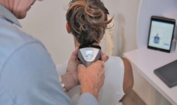
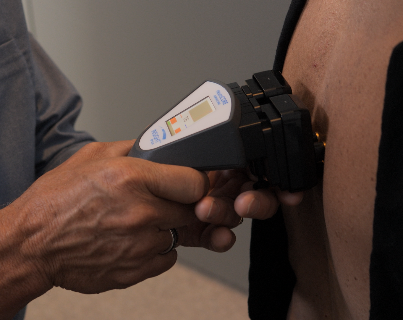
The most direct technology here is neuroCORE. neuroCORE uses surface electromyography to evaluate paraspinal muscle activity and reveal patterns of asymmetry, overuse, exhaustion, and inefficient recruitment. This matters because the paraspinal muscle system is one of the clearest expressions of how the motor nervous system is organizing itself. When the erector spinae muscle is carrying too much of the load, when lumbar multifidus support is poor, or when the lumbar paraspinal is compensating for deeper dysfunction, neuroCORE helps make those patterns visible. That improves the assessment of paraspinal activity in a way palpation alone cannot.
Then there is neuroTHERMAL, which adds a segmental stress picture across the spinal column. The paraspinal region is not only a motor story. It is also an autonomic story. neuroTHERMAL helps visualize stress patterns that may be associated with segmental dysfunction and broader neurological distress. neuroPULSE then broadens the picture again by evaluating adaptability and reserve through heart rate variability. This matters when back pain patients are not just dealing with local tension, but with a nervous system that is stuck in protection and has less capacity to recover.
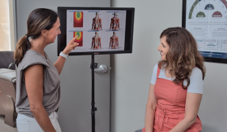
All three technologies work together through INSiGHT neuroTECH and Synapse software. That combination turns complex findings into scan reports the chiropractor can interpret and communicate more clearly. It is important to say this accurately: INSiGHT does not create the care plan. It provides objective exam data and reports. The chiropractor interprets those findings and then designs the care plan. That keeps the clinical decision-making exactly where it belongs, while giving the doctor stronger information to work from.
This is where the paraspinal conversation becomes much more useful in practice. A chiropractor can compare baseline and progress scans, explain why certain treatment options are being used, and support the treatment of paraspinal dysfunction with greater precision. That may include adjustments, progressive exercise, and other treatment options for paraspinal muscle coordination. It may also help explain why over-the-counter pain or pain relievers do not address the deeper functional issue. The goal is not just to reduce pain. The goal is to improve how the spine is being stabilized and how the nervous system is adapting over time.
- neuroCORE: Helps document paraspinal muscle activity, asymmetry, and motor output patterns.
- neuroTHERMAL: Helps visualize segmental stress patterns across the spine.
- neuroPULSE: Helps explain adaptability and resilience when protection patterns persist.
- Synapse software: Helps translate complex neurological data into clear reports.
In the end, that is why INSiGHT scanning belongs in the conversation. It brings objective data to a region that chiropractors already know matters deeply. It helps connect the paraspinal region back to nervous system performance, and it gives both doctor and patient a clearer way to see progress.
From sore tissue to smarter strategy
The paraspinal region deserves more than a quick note about tightness in the chart. It is one of the clearest places a chiropractor can observe stability, movement strategy, adaptation, and nervous system performance all at once. When the paraspinal muscle system is coordinated, the spine can move with more options. When it is not, the body often turns to stiffness, guarding, and compensation.
That is why this subject matters so much in chiropractic. The paraspinal region sits at the center of posture, load management, and movement quality. It is influenced by the erector spinae, multifidus muscle function, the lumbar paraspinal, the thoracic spine, and even the relationship between the paraspinal and psoas muscle systems. When these systems work well together, the patient has more adaptability. When they do not, the body often substitutes protection for performance.
For the chiropractor, the opportunity is simple but powerful. Evaluate the region carefully. Interpret it functionally. Understand when symptoms are associated with paraspinal compensation rather than just local irritation. And when possible, use neurological scanning to make those patterns visible. The neuroCORE helps document motor tone. The neuroTHERMAL helps visualize stress patterns. The neuroPULSE helps explain reserve and resilience. Together, they bring more clarity to the exam and more certainty to the report.
That is how the paraspinal region becomes more than a site of discomfort. It becomes a measurable part of the bigger neurological story. And when patients can see that story clearly, they are far more likely to understand the value of care and stay engaged in the process.