
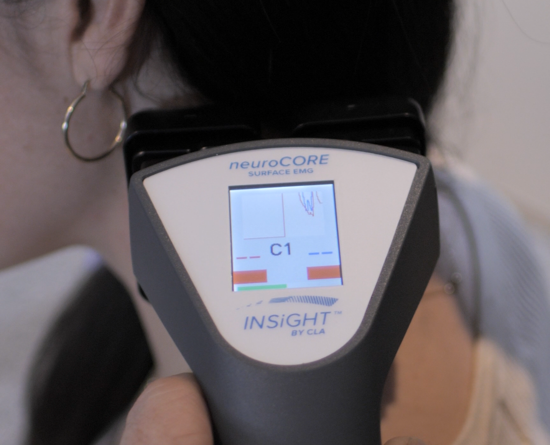
That is why so many doctors ask how they can best explain sEMG to patients in a way that feels clear, simple, and valuable?” The answer is to stop leading with muscle language and start leading with nerve language. sEMG is not just about muscle activity. It is about how the nervous system is organizing posture, tone, compensation, and energy around the spine.
Once patients understand that their muscles are responding to stress, gravity, posture, and nerve signaling, the scan begins to make sense. They stop seeing chiropractic care as something focused only on symptoms or joints. They begin to understand that chiropractic is about nervous system performance, and that objective neurological scanning helps make that story visible.
Start With a Simple Patient Explanation of sEMG
Explaining sEMG to patients starts with a comparison patients already understand. Most people have heard of an EKG, even if they do not know all the details. That gives you a simple bridge.
You might say, “An sEMG scan is a little like an EKG for your spine. An EKG looks at electrical activity around the heart. An sEMG looks at electrical activity in the muscles around your spine so we can better understand how your nervous system is organizing posture, tension, and compensation.”
That explanation works because it is familiar and accurate. Surface electromyography reads signals from the body. It helps show how hard the postural muscles are working, whether one side is working harder than the other, and whether the body is using energy efficiently.
Patients do not need to become experts in electromyographic language. They need to understand the purpose of the scan. sEMG measures the electrical activity of the paraspinal muscles through a sensor or surface electrode placed on the skin. It does not send electricity into the body. It is non-invasive, quick, and easy to understand once the scan view is explained well.
This is especially important because some patients hear EMG and think of a hospital-style EMG study. A traditional procedure called an EMG study may involve a needle electrode placed into muscle. Surface EMG is different. It uses the placement of small sensors on the surface of the body to read electrical signals from specific muscle groups.
- Simple explanation: “This scan helps us see how your nervous system is communicating with the muscles that support your spine.”
- Purpose: “We are not measuring strength. We are looking at how efficiently your body is stabilizing posture.”
- Safety: “The sensor reads your body’s signals. It does not send current into your body.”
- Baseline: “We use this scan to establish a baseline, then compare future scan views as care continues.”
Keep it visual. Tell them the scan helps make the invisible visible. That phrase may sound simple, but it is exactly what patients need. They need to see what their body has been trying to tell them.
What sEMG Measures in Chiropractic
In chiropractic, sEMG provides a way to analyze the electrical activity of muscles that support and stabilize the spinal region. These muscles are constantly responding to nerve signaling, posture, movement, gravity, and compensation. They are not acting randomly. They are following instructions from the nervous system.
Surface electromyography does not simply tell you whether a muscle is tight. It helps show levels of electrical activity, amplitude, asymmetry, muscle activity, and whether the muscles are responding efficiently. When you explain this clearly, patients begin to understand that the scan is not looking for a “bad muscle.” It is looking at how the nervous system is organizing function.
Christopher Kent has written on the use of surface electromyography in the assessment of changes in paraspinal muscle activity associated with vertebral subluxation. That matters because it frames sEMG as part of a neurological chiropractic analysis, not a stand-alone test that replaces the doctor’s judgment.
One helpful way to explain sEMG is to break it down into what the scan is showing.
- Motor tone: The scan helps show how nerves are instructing muscles to contract, relax, stabilize, or compensate.
- Amplitude: This reflects the intensity of the electrical activity in the muscle and can show where the body is using more energy than expected.
- Asymmetry: This shows whether one side is working harder than the other, which may point to imbalance or compensation.
- Overactivity and underactivity: Some areas may show abnormal muscle firing or firing too much, while others may appear underactive.
- Energy use: The scan helps reveal whether the body is using energy in an efficient or inefficient way to manage posture.
This is where the phrase “muscle differentials around the spine” becomes useful. sEMG measures muscle differentials around spinal regions, helping you show whether the right and left sides are working in a balanced way. In plain language, sEMG provides a map of how the muscles are responding to neurological demand.
You can tell a patient, “When one side is working much harder than the other, it gives us a clue that your body is trying to compensate. The scan helps us see whether that compensation is happening along the spine and whether certain areas are using more energy than they should.”
That kind of explanation keeps the conversation connected to chiropractic care. It also helps patients understand why palpation alone does not tell the whole story. Palpation is valuable, but sEMG scanning adds measurable information about nerve firing in the muscles and the electrical activity of muscles that cannot always be felt by hand.
Learn more about INSiGHT scanning?
Fill this out and we’ll get in touch!
"*" indicates required fields
Why sEMG Matters in a Chiropractic Conversation
Most patients naturally focus on how they feel. They want to know why they have symptoms, why their posture feels off, or why certain health concerns keep coming back.
sEMG gives you a better conversation. Instead of saying, “This area feels tight,” you can say, “This scan helps us see how your nervous system is asking these muscles to work.” That one shift moves the patient from symptom thinking to function thinking.
In chiropractic, subluxation is not just a simple structural issue. A vertebral subluxation can involve neurological interference, altered movement, and changes in how the body coordinates itself. When that interference affects the spinal nerve and motor control, the muscles around the spine may show abnormal patterns, spasm, postural tension, or inefficient energy use.
This is where sEMG technology measures the nerve response in a way patients can see. You can explain, “When a spinal region is not moving or functioning well, nearby muscles may start working harder to protect, stabilize, or compensate. The scan helps us identify where that may be happening.”
That does not mean sEMG can pinpoint every root cause by itself. It means sEMG provides information that helps you interpret the patient’s nervous system status alongside their history, examination, posture, movement, and scan views. It can provide valuable insight, but the chiropractor still brings the clinical interpretation.
This is also how you explain why care continues even after a patient starts feeling better. You might say, “Your symptoms tell us how you feel. Your scan helps us see how your nervous system is organizing function. We want both to improve, but they do not always change at the same speed.”
How to Explain sEMG Scan Results Without Overwhelming Patients
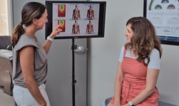
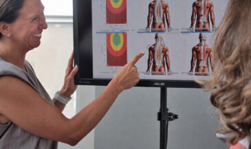
The scan view is often the moment when patients start to understand. They may not remember every detail about surface electromyography, but they will remember seeing their own nervous system patterns in color.
The key is to explain the colors without creating fear. Red does not mean the patient has a bad muscle. Blue does not mean something is broken. Yellow does not mean failure. The colors are a way to show activity associated with how the muscles are firing and how the nervous system is organizing postural control.
A clear explanation might be, “Think of this like a weather map for your spine. The colors help us see where your nervous system is organizing muscle activity efficiently and where your body may be using more energy than expected.”
Then explain the scan view in simple terms.
- White or green: These areas generally suggest more efficient or balanced muscle firing.
- Blue: These areas may show mild to moderate postural tension, asymmetry, or increased electrical activity.
- Red: These areas may show higher levels of electrical activity or increased neuromuscular demand.
- Yellow: These areas may show lower output or underactive patterns.
- Side-to-side differences: These may show that one side is working harder than the other.
You can add, “A red area does not mean you have a bad muscle. It means that part of your spine may be working harder than expected, and your nervous system may be using extra energy there.”
This is a better way to explain tight or contracted muscles as well. Instead of making the conversation only about muscle tension, you can explain that the muscles are responding to nerve signaling, posture, and compensation. When muscles are firing abnormally, it may be the result of neurological interference, not simply a local muscle problem.
Patients often ask whether sEMG measures symptoms. The answer is no, not directly. sEMG measures the electrical activity related to muscle firing. Those patterns may relate to compensation, neurological stress patterns, or subluxations, but the scan is not a symptom meter.
Patients may also ask if this scan replaces the chiropractor’s examination. It does not. Chiropractors can use sEMG as part of a broader clinical picture. It supports the exam by adding instrumentation that helps make function measurable and easier to explain.
If the patient asks why the scan is repeated, the answer is simple: “We repeat it so we can compare your baseline with future scans. That helps us see whether your body is adapting and whether your nervous system is organizing muscle activity more efficiently.”
How INSiGHT neuroCORE Makes sEMG Easier to Explain
This is where INSiGHT scanning technology becomes especially helpful. INSiGHT neuroCORE uses sEMG technology to analyze the motor side of nervous system performance. It helps show how the muscles around your spine are responding to neurological demand, postural load, and compensation.
INSiGHT sEMG helps turn electrical signals into scan views patients can understand. That matters because patients do not need a lecture on electromyographic theory. They need to see the pattern, understand what it means, and connect that information to their chiropractic care.
A clear patient explanation would be, “We use INSiGHT neuroCORE to help us see how your nervous system is communicating with the muscles around your spine. Then we use that information, along with your exam and history, to better understand what needs attention.”
This language is important because INSiGHT technology does not create the care plan. INSiGHT™ scanning provides objective exam data and reports. The chiropractor interprets that information with clinical expertise and uses it to support personalized care.
neuroCORE becomes even more valuable when it is used with the other INSiGHT scanning technologies.
- neuroCORE: Uses surface EMG to analyze postural energy, motor tone, amplitude, electrical activity, and muscle differentials around the spine.
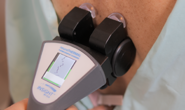
- neuroTHERMAL: Helps analyze autonomic regulation patterns, including temperature differences related to blood flow and nervous system control.
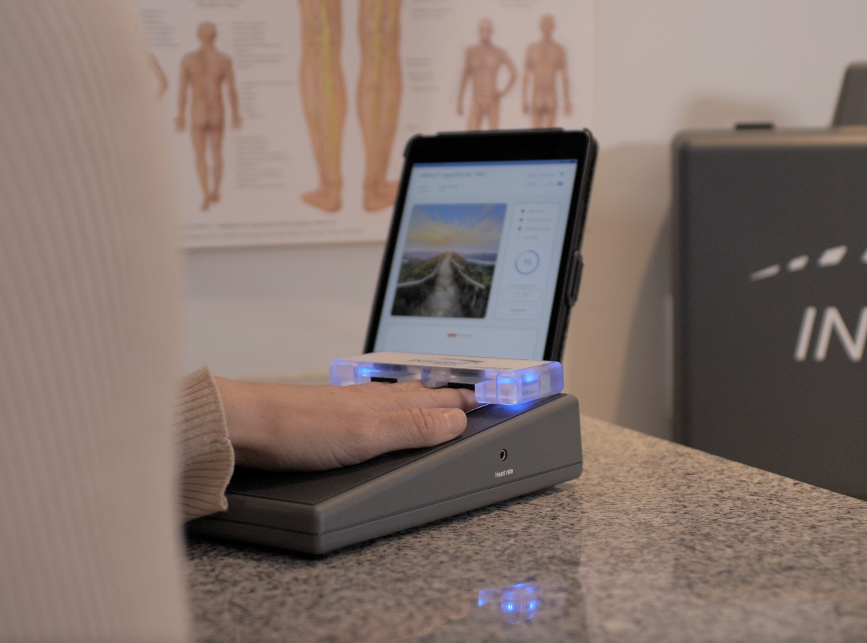
- neuroPULSE: Uses Heart Rate Variability to assess adaptability, autonomic balance, and reserve.
- Synapse software: Helps organize scan data into reports and scan views that make complex neurological information easier to understand.
This matters because sEMG and thermography do not tell the same story. sEMG looks at motor activity and postural organization. neuroTHERMAL looks at autonomic regulation. neuroPULSE adds the adaptability picture. Together, INSiGHT neuroTECH and Synapse software help chiropractors create a more complete neurological profile.
The scan helps identify patterns in how the muscles are responding, while the full INSiGHT scanning process gives a broader view of nervous system performance.
When patients can see their scan views, compare progress, and understand why chiropractic adjustments are being recommended, the conversation changes. They are not being asked to believe blindly. They are being shown objective information that helps explain why care continues and how the chiropractor is tracking change.
That is the role of better neurological scanning. It helps doctors explain more clearly. It helps patients participate more confidently. It helps the profession move from vague explanations to measurable communication.
Give Patients a Clearer Way to Understand Their Nervous System
Patients do not need to understand every technical detail of sEMG, EMG, amplitude, and signal processing. They need to understand that their nervous system controls the muscles that support their spine, and that sEMG provides a measurable way to see how those muscles are being organized.
When you explain sEMG as an EKG for the spine, the patient has a reference point. When you explain that the scan uses a surface electrode placed on the skin, the patient feels safe. When you explain that the scan helps show compensation, imbalance, abnormal muscle firing, and inefficient energy use, the patient begins to understand the why behind care.
This is how you can include even difficult SEO language like treatment plans or misalignment without drifting from the CLA message. Patients may use those words, but you can gently reframe them. You might say, “Some people think this is about a misalignment or a standard treatment plan. What we are really looking at is neurological interference, compensation, and the data that helps us shape your care plan.”
That kind of explanation respects the patient’s language while elevating the conversation. It keeps chiropractic grounded in objective findings and nervous system performance.