
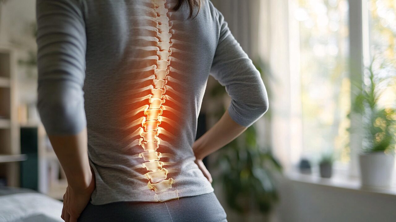
As chiropractors, we have an opportunity to give that conversation more depth. What the patient feels matters. So do their strength, coordination, examination findings, and physiological responses. Understanding spinal nerve function helps us connect those pieces without reducing the person to one complaint or one location on a chart.
Good anatomy gives our explanations a foundation. Thoughtful examination gives them clinical meaning. Neurological scanning adds objective findings we can interpret, explain, and compare as care progresses.
Spinal Nerve Anatomy and Connections With the Spinal Cord
Spinal nerve function begins with communication. The brain and spinal cord form the central nervous system. The peripheral nervous system includes spinal and cranial nerves, ganglia, and associated pathways connecting central structures with the body.
Nerves contain bundles of nerve fibers, primarily axons, supported by connective tissue and glial cells. These axons belong to nerve cells called neurons. Their electrical activity carries information toward central processing areas and outward toward responding tissues. Understanding that organization helps distinguish the pathway itself from the physiological response recorded during scanning.
How Spinal Nerve Roots Form Mixed Pathways
Spinal nerve roots have different roles. The posterior, or dorsal, root carries sensory information toward the spinal cord. The associated cell bodies lie in the dorsal root ganglia. The anterior, or ventral, root carries outgoing motor fibers, including preganglionic autonomic fibers at particular levels.
The roots join to form a short mixed nerve, which then divides into branches. Both dorsal and ventral branches contain incoming and outgoing fibers. A sensory nerve pathway and a motor nerve pathway therefore contribute to the same mixed structure, although their messages travel in different directions.
The Regional Nerve Groups
There are 31 pairs of spinal nerves. Their regional organization provides a useful starting point for understanding the distribution of nerve signals:
- Cervical: Eight pairs of cervical nerves contribute to pathways serving the neck, diaphragm, shoulders, and upper limbs.
- Thoracic: Twelve pairs of thoracic nerves supply much of the chest and abdominal wall.
- Lumbar: Five pairs of lumbar nerves contribute to lower back and lower-limb supply.
- Sacral: Five pairs of sacral nerves contribute to pathways serving the pelvis, lower limbs, and perineum.
- Coccygeal: One coccygeal nerve pair contributes to a small territory near the tailbone.
There are eight nerve pairs despite 7 cervical vertebrae. C1 through C7 leave above their corresponding vertebra; C8 leaves between C7 and T1. The first thoracic pair leaves below T1, and subsequent nerves exit below their correspondingly numbered vertebrae.
The adult spinal cord usually ends near L1–L2 within the spinal column. Lower roots descend as the cauda equina before reaching their exits. Consequently, levels of the spinal cord do not consistently align with similarly numbered vertebral levels.
A spinal cord segment is a section of the spinal cord associated with one pair of roots. The gray matter of the spinal cord contains neuronal cell bodies and local circuits; surrounding white matter contains ascending and descending pathways. The enlargements of the spinal cord accommodate additional circuitry serving the limbs. These distinctions help us explain why a regional scan finding requires interpretation rather than a direct leap to a diagnosis.
How Spinal Nerves Function in Sensation, Movement, and Regulation
Spinal nerve function extends well beyond what a patient consciously notices. Incoming information helps the body monitor its position and surroundings. Outgoing activity coordinates muscle responses and contributes to regulation.
For chiropractors, that gives everyday observations greater meaning. How a person stands, moves, and responds during examination reflects multiple interacting processes. Neurological scanning samples particular physiological expressions within that larger picture.
Sensory Input and Motor Function
Sensory pathways carry information about touch, temperature, tissue-threatening stimuli, and body position. Some information reaches conscious awareness as sensation. Other input contributes to coordination without requiring deliberate attention.
Proprioceptive receptors in muscles, tendons, and joints supply information about position and movement. Motor output helps organize the muscular response. The upper cervical region, trunk, and limbs all participate in this continuing exchange.
Clinical maps help organize findings:
- Dermatomes: Skin territories associated with individual roots, with overlapping borders.
- Myotomes: Muscle actions associated predominantly with particular spinal levels.
- Named nerve distributions: Territories supplied by pathways that frequently contain fibers from several roots.
Many ventral branches combine into plexuses, creating a network of nerves before reaching the limbs. These nerves branching and regrouping explain why one muscle or skin region rarely provides the whole answer. Surface EMG adds information about recorded muscular activity; it does not replace examination of sensation or strength.
Spinal Reflex Coordination
A spinal reflex links incoming information with an outgoing response through spinal circuitry. Muscle spindle input, for example, participates in the stretch reflex examined with a tendon hammer. More complex responses involve additional connections.
The spinal cord can organize a rapid response while information also travels toward the brain. Descending pathways influence these circuits, too. Reflex findings therefore deserve interpretation alongside other examination results, independently of what a scan shows.
Autonomic Pathways and Involuntary Functions
Autonomic pathways contribute to circulation, gland activity, digestion, and other involuntary functions. Sympathetic preganglionic neurons arise primarily from T1–L2. Parasympathetic outflow has cranial origins and sacral spinal origins at S2–S4.
The vagus belongs to the cranial nerves and carries substantial parasympathetic supply to thoracic and abdominal organs. It is not a cervical spinal pathway. Similarly, breathing involves several forms of control, including motor supply to respiratory muscles.
These distinctions keep our explanations of spinal nerve function accurate. HRV and regional temperature analysis contribute information relevant to regulation, but neither establishes how every organ is performing.
Learn more about INSiGHT scanning?
Fill this out and we’ll get in touch!
"*" indicates required fields
Assessing Spinal Nerve Function in Chiropractic Practice
A useful assessment begins with the patient’s story. What feels different? When did it begin? Is the distribution consistent? Has strength, coordination, or the ability to complete everyday tasks changed?
Spinal nerve function belongs within that complete clinical picture. A complaint in the hand may involve the cervical spine, a pathway farther along the arm, or another process. Good examination helps establish which possibilities deserve attention before scanning findings are interpreted.
Begin With History and Examination
Ask about onset, progression, medical history, and activities that influence the presentation. Then select examination procedures that address the clinical question:
- Sensory testing: Compare appropriate modalities and distributions.
- Strength testing: Look for patterns across relevant muscle actions.
- Response testing: Evaluate tendon responses, symmetry, and associated findings.
- Coordination and gait: Observe function that may suggest involvement beyond one spinal segment.
- Orthopedic assessment: Use relevant procedures to inform the working clinical assessment.
Dermatomal findings may suggest root involvement, while another distribution may raise questions about a peripheral nerve. Neither pattern should be interpreted in isolation. Objective scanning can complement this process without replacing localization or differential assessment.
Recognize When Further Investigation Is Needed
Progressive weakness, worsening coordination, or findings suggesting spinal cord damage require appropriate investigation. New bladder or bowel disturbance or perineal numbness accompanying severe radiating lower back complaints requires immediate assessment through local pathways.
Spinal cord injuries and other serious conditions need appropriate medical evaluation. Neurological scanning should never delay referral or be used to exclude those conditions.
Combine Structural and Functional Findings
Imaging, when indicated, investigates anatomical questions involving discs, the vertebral canal, and surrounding tissues. INSiGHT Scanning contributes physiological observations. These categories become useful together when their findings are connected with the examination.
Within chiropractic, vertebral subluxation provides a clinical framework for considering altered spinal function and associated neurological findings. It should be distinguished from a confirmed compressive lesion. Neither a postural observation nor an isolated scan pattern establishes nerve compression.
Document baseline findings and the conditions under which they were collected. Reassessment can then examine spinal nerve function through the patient’s experience, clinical tests, and complementary scan comparisons.
How INSiGHT Scanning Supports Spinal Nerve Function Assessment
INSiGHT Scanning gives the chiropractor objective findings that support interpretation and patient communication. Its value comes from combining complementary physiological assessments with the history, examination, and clinical judgment you already bring.
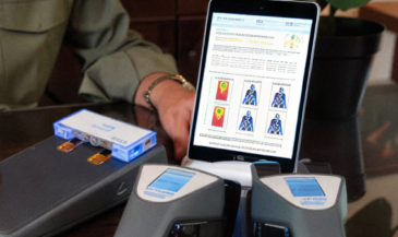
CLA’s neuroPULSE, neuroCORE, and neuroTHERMAL examine different responses. Together, they help broaden the discussion of spinal nerve function while preserving an important distinction: these assessments do not directly test conduction through every spinal pathway.
neuroPULSE and Adaptive Reserve
The neuroPULSE analyzes heart rate variability, the variation in timing between consecutive beats. HRV reflects influences on cardiac rhythm, including autonomic modulation. CLA uses these findings within its adaptive reserve framework to support conversations about regulation, recovery, and resilience.
Explain the distinction simply: heart rate describes how often the heart beats; HRV examines variation between beats. Age, breathing, posture, recent activity, medications, and recording quality influence interpretation. One lower reading does not establish sympathetic overdrive or localize a root lesion.
neuroCORE and Paraspinal Activity
The neuroCORE uses surface EMG to analyze electrical activity recorded over paraspinal muscles. Activity levels and symmetry provide information relevant to postural tension and muscular activation.
Placement, electrode contact, posture, movement, and protocol influence the recording. Higher and lower values need context. This assessment differs from diagnostic needle EMG and conduction studies; it does not independently diagnose root compression.
neuroTHERMAL and Temperature Patterns
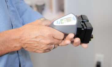
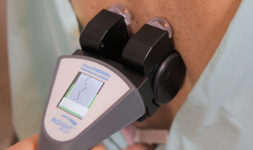
The neuroTHERMAL uses infrared technology to analyze bilateral paraspinal skin temperature differences. Blood flow and vasomotor regulation influence those temperatures, making the findings relevant to a broader functional assessment.
A full spine nerve system scan can be completed in less than 30 seconds. Consistent technique, room conditions, and acclimatization support useful comparisons. An asymmetry remains a finding for interpretation rather than proof of organ dysfunction or a specific structural condition.
Reporting With the INSiGHT Software
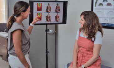
The INSiGHT software (powered by Synapse) organizes findings into scan views and comparative reports. CORESCORE combines information from the three assessments into a patient-friendly composite metric. Its underlying findings remain essential to interpretation.
CLA’s RED framework provides a consistent sequence:
- Reserve: Begin with neuroPULSE and HRV findings.
- Energy: Review neuroCORE activity and distribution patterns.
- Depth: Consider neuroTHERMAL findings within the regional and overall presentation.
The Rule of 3 Scans organizes baseline, response, and trajectory. CLA’s Rule of 12 provides a reporting cadence, while clinical circumstances guide appropriate timing. Reassessments should answer meaningful questions about progress.
When findings fluctuate, review collection conditions, recent events, examination results, and the patient’s experience. INSiGHT provides objective analysis and reporting. The chiropractor interprets that information and builds the care plan.
Making Spinal Nerve Function Meaningful to Patients
Patients do not need to memorize the anatomy of the spinal cord to understand their care. They need a clear explanation of what you examined, what you found, and how your recommendations relate to their goals.
Spinal nerve function gives that explanation a practical foundation. Movement, posture, sensation, and regulation are familiar experiences. Connecting them with appropriate findings helps patients understand why your examination extends beyond their immediate complaint.
- Explain: Describe the relevant function in everyday language.
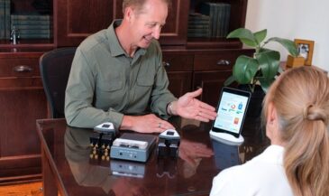
- Show: Review examination findings and appropriate scan views.
- Connect: Explain how the information fits together and where uncertainty remains.
- Plan: Discuss recommendations, goals, and the purpose of reassessment.
At the re-exam, return to the same questions. Is daily function improving? What does the clinical examination show? Do scan comparisons support the broader picture? When findings disagree, investigate rather than forcing every result into the same story.
That is the opportunity INSiGHT Scanning brings to the spinal nerve function conversation. Objective findings become something you and the patient can review together. With neuroTECH and the INSiGHT software, chiropractors can strengthen examinations, explain recommendations clearly, and make progress easier to understand.