

As a chiropractor, you recognize that structure deserves attention. You also want to understand how the person moves, recruits supporting muscles, and manages daily demands. A picture provides one perspective. Examination and objective neurological scanning contribute additional information that helps you explain the bigger picture.
The opportunity is to give patients clarity about their cervical curve while keeping the conversation grounded in their findings, functional goals, and nervous system performance.
The Cervical Curve and Its Clinical Significance
The cervical spine supports the head, accommodates movement, and surrounds the spinal cord. Its seven vertebrae work with joints, discs, muscles, and connective tissues to provide strength and flexibility. This portion of the spine participates in a coordinated relationship with the thoracic spine and other spinal regions.
A cervical curve describes one aspect of that structure. Its significance depends on the patient’s anatomy, presentation, and examination. The appearance alone cannot tell you how comfortably someone moves or how well they tolerate an ordinary workday.
Cervical Lordosis, Straightening, and Cervical Kyphosis
Cervical lordosis describes the inward, lordotic curve commonly seen when viewing the neck from the side. Patients may recognize the natural c-shaped curve shown in educational illustrations. Different types of cervical alignment include:
- Lordotic alignment: The neck retains its inward curvature.
- Reduced lordosis: The neck appears flatter, sometimes described as a straight neck or military neck.
- Cervical kyphosis: The neck develops an opposite curve rather than the usual lordotic configuration.
Loss of cervical lordosis and a reversed configuration deserve careful distinction. Neither description establishes the cause, severity, or need for a particular intervention. A straighter neck may occur without symptoms, and an apparently normal neck curve does not rule out a functional complaint.
What Does Normal Curvature Mean?
There is variation among people without symptoms. Research involving more than 15,000 asymptomatic individuals documented substantial differences in cervical alignment. That finding encourages thoughtful interpretation rather than treating a single angle as a universal target.
When patients ask about a healthy cervical curve, explain its relevance alongside their examination. The term “healthy curve” should never become a promise that one shape guarantees better function. Similarly, loss of curvature does not automatically mean nerve compression.
Connecting Structure With Function
The curve of the cervical spine contributes to the region’s mechanical arrangement, while muscles actively help stabilize the neck. Through neurological scanning, you can assess aspects of paraspinal electrical activity alongside those structural observations. Scanning adds functional information; it does not determine the shape of the neck.
Why Cervical Curvature Can Change
Patients often blame a screen when they learn about an altered cervical curve. Sustained positioning belongs in the history, but the discussion deserves more depth. Ask about work, driving, previous injuries, exercise, sleep, and how often the patient varies their position.
Understanding the context helps you distinguish everyday mechanical demands from situations requiring additional investigation. The alignment of the cervical spine should be interpreted within that history rather than attributed to one habit.
Forward Head Posture and Daily Demands
Forward head posture describes the position of the head relative to the body. It is different from the curvature assessed on an appropriate structural image. These findings can occur together, but one does not establish the other.
From a biomechanics perspective, head position influences muscular demand and tissue loading. The relationship between center of gravity and balance also involves adjacent regions. Calling this “poor posture” offers little guidance unless you identify the specific task, its duration, and the patient’s tolerance.
Imaging position matters, too. Research shows that acquisition posture can influence apparent alignment. Before describing a neck straighter than it was previously, consider whether the images provide a valid comparison.
Injury, Degeneration, and Supporting Tissues
Trauma, degenerative processes, congenital differences, certain underlying conditions, and previous surgery can contribute to altered alignment. Muscles and ligaments in the neck provide support, but an image showing straightening cannot by itself establish ligament damage.
If the curve is lost or reduced, the next question concerns its clinical significance. Is the presentation flexible or fixed? Is there an injury history? Are there neurological findings? The answers guide evaluation and recommendations.
Where Neurological Scanning Contributes
Surface EMG can document paraspinal electrical activity when collected using a consistent protocol. This may help you discuss muscular recruitment and postural compensation alongside the cervical curve finding. A scan cannot identify the cause of structural alteration or establish instability. Those questions require the appropriate examination and investigations.
Learn more about INSiGHT scanning?
Fill this out and we’ll get in touch!
"*" indicates required fields
Assessing Neck Pain and Planning Individualized Chiropractic Care
Patients usually want to drive comfortably, sleep better, or finish a workday without persistent neck pain. Start with those priorities. Ask what they notice, when it began, what aggravates it, and which activities they want to regain.
A cervical curve finding becomes useful when connected to that individual story. Symptoms, examination findings, structural observations, and functional scan data each contribute something different to your judgment.
History, Examination, and Imaging
Your assessment should address onset, trauma, previous surgery, medical conditions, and progression. Examination may include strength, reflexes, sensation, coordination, balance, and range of motion. Cervical radiculopathy and suspected cord involvement require appropriate evaluation.
Imaging should answer a clinical question. An x-ray can characterize structural alignment, while other imaging may be indicated for different concerns.
Progressive weakness, walking difficulty, loss of hand coordination, or new bowel or bladder dysfunction require timely medical assessment.
Care Goals and Appropriate Options
When findings support conservative care, recommendations should reflect the presentation, preferences, and goals. Chiropractic adjustments may be appropriate for selected patients after consideration of contraindications, risks, and informed consent. Exercise, education, and collaboration with physical therapy or medical specialists may also contribute.
Patients researching “treatment options” need practical explanations of what each approach is intended to accomplish. Relevant components may include:
- Adjusting workstation setup to improve comfort during familiar tasks.
- Introducing movement breaks during prolonged activities.
- Selecting strengthening exercises that match current capacity.
- Progressing home activities according to tolerance.
- Reassessing goals and examination findings at appropriate intervals.
Exercise and manual approaches can support symptom and functional improvement in suitable presentations. That evidence should not be stretched into a guarantee of structural correction.
Setting Expectations for Curve Restoration
When discussing restoring the natural curve of the neck, distinguish symptom improvement, functional improvement, and documented structural change. These are separate outcomes. A patient may become more comfortable without a demonstrated change in alignment.
Traction or extension exercises require appropriate selection and supervision. A generic recommendation to force the neck backward is unsuitable, particularly when instability or neurological compromise is a concern.
At reassessment, revisit daily function, repeat relevant examination procedures, and compare scans when indicated. Improved scan findings cannot prove correction of the cervical curve. Persistent complaints deserve attention even when a report looks more favorable.
How INSiGHT Scanning Supports Cervical Curve Assessment
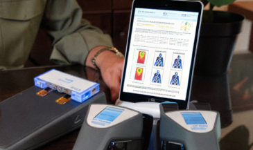
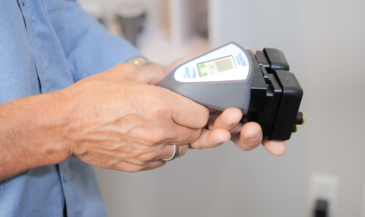
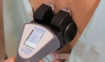
INSiGHT Scanning brings objective physiological information into this conversation. CLA’s neuroPULSE, neuroCORE, and neuroTHERMAL provide complementary analyses that help you discuss adaptability, muscular recruitment, and regulation within the broader examination.
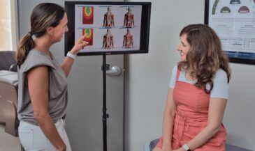
For a patient concerned about their cervical curve, those findings can make the functional conversation easier to understand. The chiropractor interprets the data alongside history, examination, and structural findings, then develops the care plan.
neuroPULSE: Autonomic Patterns and Adaptive Reserve
neuroPULSE analyzes heart rate variability, or HRV. This describes variation in the intervals between successive heartbeats rather than pulse rate alone. It contributes information relevant to autonomic regulation and physiological adaptability.
Recent activity, breathing, sleep, illness, medications, and recording conditions can influence HRV. One reading should not become a verdict about resilience or an explanation for the curve of your neck. Document context and interpret trends carefully.
For the chiropractor, this creates an opportunity to discuss recovery and daily demands without claiming that cervical alignment caused an autonomic pattern.
neuroCORE: Paraspinal Activity and Compensation
neuroCORE uses surface EMG to analyze paraspinal electrical activity, including amplitude and symmetry. These findings add information about muscular recruitment that you can consider alongside movement, palpation, and activity tolerance.
Explain the output plainly. A higher reading does not automatically mean greater strength, and a lower reading does not by itself establish weakness. Surface EMG differs from needle EMG and nerve conduction testing used in medical investigations.
This distinction helps patients understand the purpose of their scan. You are documenting functional data relevant to your assessment, rather than claiming to locate a compressed nerve or measure mechanical tension in the cervical tissues directly.
neuroTHERMAL: Temperature Regulation Patterns
neuroTHERMAL analyzes bilateral skin temperature patterns along the spine. Skin temperature reflects several influences, including blood flow and physiological regulation, making preparation and acquisition conditions important.
Interpret differences within the complete examination. They do not independently prove inflammation, a specific lesion, or an organ condition. They provide another set of objective findings for clinical interpretation and comparison.
Reports That Make Re-Exams More Meaningful
The INSiGHT software organizes scan views, reports, and comparisons. CORESCORE summarizes combined scan data in a patient-friendly format. It is not a score for cervical curve quality or evidence of structural restoration.
The INSiGHT software supports a useful baseline, response, and trajectory framework, with reassessment timing guided by clinical need. A clear reporting sequence helps:
- Begin with the patient’s reported experience and functional goals.
- Review relevant examination and structural findings.
- Explain what each scan assessed.
- Compare current results with the baseline under documented conditions.
- Describe how the complete findings inform your recommendation.
If paraspinal activity fluctuates, explain that observation accurately. If HRV differs, consider relevant influences. Neither finding alone establishes the effect of an adjustment. Careful interpretation strengthens the credibility of neurological scanning.
Give the Cervical Curve Conversation a Bigger Purpose
When patients ask about the curve in your neck, they want to understand what it means for everyday life. A useful answer connects anatomy with their examination, concerns, and goals. It also explains what remains uncertain and how you will assess progress.
A healthy neck conversation makes room for several outcomes: greater activity tolerance, improved comfort, relevant examination findings, and meaningful scan comparisons. Structural improvement requires its own appropriate assessment.
For the Neurologically-Focused Chiropractor, the cervical curve offers a starting point for explaining nervous system performance. INSiGHT Scanning supplies objective data and understandable reports that help patients follow the functional side of that conversation.
Bring those findings into your exams and re-exams with clear explanations and sound judgment. Patients gain a shared reference point, and you gain a more grounded way to explain your recommendations. That is how a question about a curve becomes a clearer understanding of care.