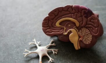
Unlike conventional Magnetic Resonance Imaging (MRI), which discloses only anatomy and pathology, functional MRI is a non-invasive neuroimaging technique which demonstrates alterations in brain physiology. The technique relies on the relationship between neuronal synaptic activity, energy metabolism, and blood circulation. During neuronal stimulation, there is a subtle change in signal intensity which is attributed to a local change in blood oxygenation and blood flow. Changes in the oxygenation state of hemoglobin are induced by task activation. In response to activation, the MRI signal intensity increases as a result of an increase in blood flow and oxygen. (1) Brain activation patterns have been recorded using visual, sensorimotor, and auditory stimuli, as well as higher-order cognitive processes. (2-6)
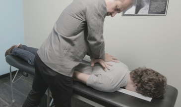
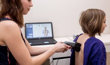
It has been suggested that chiropractic care may improve brain function. Stevens and Gorman (7) proposed that “spinal derangement” may adversely affect cerebral function. Terrett notes that “manipulation” can result in increased cerebral blood flow, resulting in normal cerebral function. Gorman (9,10) has described alterations in visual function which resolved following “manipulation,” and hypothesized that the favorable clinical responses were due to increased blood flow to the retina and brain.
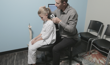
Furthermore, chiropractic care has been associated with favorable changes in children with learning disorders and attention deficit- hyperactivity disorder (11,12,13,14,15). Other authors have reported an association between chiropractic adjustments and improved mental function (16.17). Objective assessment of brain function using EEG spectral analysis was performed on five children. Analysis revealed more normalized brainwaves after chiropractic adjustments (18).
Functional MRI may be a useful technology for studying the effects of chiropractic adjustment on brain function. In one case example, the task of voluntary unilateral ankle motion was selected for evaluation before and after a chiropractic adjustment. Before the adjustment, generalized areas of activation were seen scattered bilaterally throughout the brain. Immediately following the adjustment, the regions of activation were smaller, and were unilateral (19). It has been conjectured that chiropractic adjustments lead to improved neural efficiency, evidenced by fewer and more specific foci of altered brain activity (20).
References
1. LeBihan D, Jezzard P, Haxby J, et al: “Functional magnetic resonance imaging of the brain.” Ann Intern Med 1995;122:296.
2. LeBihan D, Turner R, Zeffiro TA, et al: “Activation of human primary visual cortex during visual recall: a magnetic resonance imaging study.” Proc Natl Acad Sci USA 1993;90:11802.
3. Hinke RM, Hu S, Stillman AE, et al: “Functional magnetic resonance imaging of Broca’s area during internal speech.” Neuroreport 1993;4:675.
4. Rueckert L. Appollonio I, Grafman J, et al: “Magnetic resonance imaging functional activation of left frontal cortex during covert word production.” J Neuroimaging 1994;4:67.
5. Rao SM, Binder JR, Bandettini PA, et al: “Functional magnetic resonance imaging of complex human movements.” Neurology 1993;43:2311.
6. Belliveau JW, Kennedy DN Jr, McKinstry RC, et al: “Functional mapping of the human visual cortex by magnetic resonance imaging.” Science 1991;254:716.
7. Stephens D, Gorman RF: “The association between visual incompetence and spinal derangement: an instructive case history.” J Manipulative Physiol Ther 1997;20:343.
8. Terrett AGJ: “The cerebral dysfunction theory.” In: Gatterman MI (ed): “Foundations of Chiropractic: Subluxation.” Mosby-Year Book, Inc. St. Louis, MO. 1995. P. 340.
9. Gorman RF: “The treatment of presumptive optic nerve ischemia by manipulation.” J Manipulative Physiol Ther 1995;18:172.
10. Gorman RF: “Monocular vision loss after closed head trauma: immediate resolution associated with spinal manipulation.” J Manipulative Physiol Ther 1993;16:138.
11. Giesen J, Center D, Leach R: “An evaluation of chiropractic manipulation as a treatment of hyperactivity in children.” J Manipulative Physiol Ther 1989;12:353.
12. Phillips C: “Case study: the effect of using spinal manipulation and craniosacral therapy as the treatment approach for attention deficit-hyperactivity disorder.” Proceedings of the National Conference on Chiropractic and Pediatrics 1991, P. 57.
13. Anderson C, Partridge J: “Seizures plus attention deficit hyperactivity disorder.” International Review of Chiropractic Jun 1993; P. 35.
14. Barnes T: “A multi-faceted approach to attention deficit hyperactivity disorder: a case report.” International Review of Chiropractic Jan/Feb 1995; P. 41.
15. Barnes T: “Attention deficit hyperactivity disorder and the triad of health.” Journal of Clinical Chiropractic Pediatrics 1996;1(2):59.
16. Thomas M, Wood J: “Upper cervical adjustments may improve mental function.” Manual Medicine 1992;6(6):215.
17. Walton EV: “The effects of chiropractic treatment on students with learning and behavioral impairments due to neurological dysfunction.” International Review of Chiropractic 1975;29(4-5):24.
18. Hospers LA: “EEG and CEEG studies before and after upper cervical or SOT Category II adjustment and children after head trauma, in epilepsy, and in ‘hyperactivity.’” Proceedings of the National Conference on Chiropractic and Pediatrics 1992:84.
19. Epstein D: “Network Spinal Analysis: a system of health care delivery within the subluxation-based chiropractic model.” Journal of Vertebral Subluxation Research 1996;1(1):51.
20. Kent C, Vernon L: “Case Studies In Chiropractic MRI.” Chapter 2, page 23. International Chiropractors Association. Arlington, VA. 1998.


