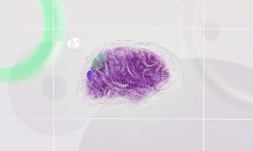
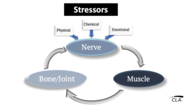
Every blink, taste, smile, or breath starts with the powerful network of nerves in your head. These cranial nerves carry signals that keep you sensing, moving, and adapting every day — a point strongly supported by Kent’s review. Yet they’re often overshadowed when chiropractors think only of vertebrae.
In today’s Neurologically-Focused Chiropractic Care, understanding the nerves in the head is non-negotiable. These pathways bridge the brain to the senses, muscles, and organs — and they reflect how well your patients handle stress and adapt to life’s demands.
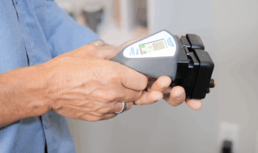
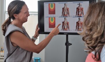
This guide will help you revisit the cranial nerves, see how nerve tension and interference show up, and use the INSiGHT neuroTECH to prove your care is making a difference that your patients can see and trust.
What Are the Cranial Nerves?
Your 12 pairs of cranial nerves branch out directly from your brain — not from the spinal cord. They reach your eyes, ears, nose, mouth, face, neck, and shoulders, delivering sensory and motor signals that run everything from blinking to digesting.
Some cranial nerves carry pure sensory input, like sight or smell. Others control muscle movement, or they do both — coordinating complex functions like chewing, swallowing, or making facial expressions.
For Neurologically-Focused Chiropractors, these nerves are a window into your patient’s full nervous system status. When they face tension or interference, you often see subtle signs first — reminders that your scans and assessments are about more than just vertebrae.
How Many Cranial Nerves Do You Have?
Twelve pairs, left and right. They’re symmetrical highways connecting the brain with tissues and organs on both sides of the body. Understanding where each pair travels helps you interpret neurological function and connect the dots when stress begins to overwhelm their control.
Key Functions of the Cranial Nerves
Every cranial nerve has a job, and together they form an elegant system that keeps your patients’ senses sharp and their bodies in balance. Here’s a quick look:
- Olfactory (CN I): Sense of smell.
- Optic (CN II): Vision.
- Oculomotor (CN III), Trochlear (CN IV), Abducens (CN VI): Eye movement and focus.
- Trigeminal (CN V): Sensation across the face; chewing.
- Facial (CN VII): Facial expressions; taste.
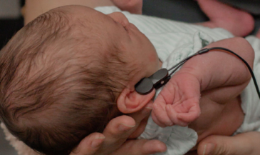
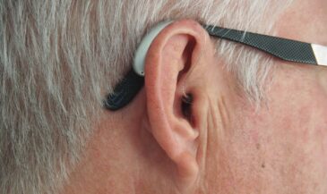
- Vestibulocochlear (CN VIII): Hearing and balance.
- Glossopharyngeal (CN IX) & Hypoglossal (CN XII): Taste, swallowing, tongue movement.
- Vagus (CN X): Heart rate, digestion, mood — your “rest-and-digest” powerhouse. The great “Wandering Nerve” setting the tone for adaptability.
- Accessory (CN XI): Neck and shoulder movement.
When you think function-first, these connections come alive. They reveal how a well-adjusted nervous system supports every aspect of daily life.
Olfactory Nerve Anatomy (CN I)
The olfactory nerve is the first cranial nerve and your direct line to smell. Its nerve fibers pass through the nasal cavity and the cribriform plate of the ethmoid bone to reach the olfactory bulb — an essential structure in human anatomy.
If there’s tension along the upper neck or cervical spine, signals here can be affected. This is why baseline scans and clear nervous system assessments help you catch subtle sensory changes.
Optic Nerve Anatomy (CN II)
The optic nerve, known as cranial nerve II, transmits visual information from the retina through the optic canal and back to the visual cortex. A healthy optic nerve is crucial for interpreting the world around you.
As you review patients’ cases, remember that optic nerve pathways can be influenced by cervical nerve tension or upper neck misalignments, highlighting the importance of clear anatomy knowledge combined with objective scans.
Oculomotor, Trochlear, and Abducens Nerves: Eye Movement Anatomy
The oculomotor nerve (CN III), trochlear nerve (CN IV), and abducens nerve (CN VI) control eye muscle movement. They pass through the superior orbital fissure, innervating muscles that keep vision steady.
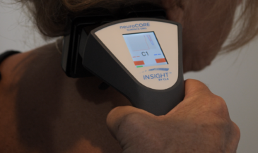
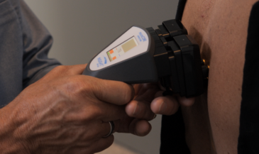
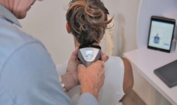
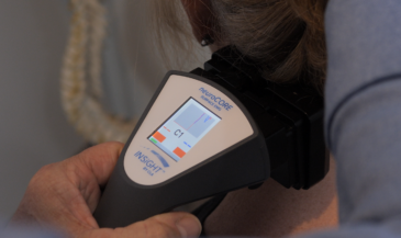
Disruption here can lead to double vision or eye tracking problems. Checking eye reflexes alongside your neuroCORE surface EMG scans can reveal muscle tension patterns in the head and neck that might otherwise go unnoticed.
Trigeminal Nerve Anatomy: CN V
The trigeminal nerve, also called cranial nerve V or CN V, is the largest cranial nerve and a key mixed nerve for the head and neck. Its three divisions — ophthalmic, maxillary, and mandibular — branch from the trigeminal ganglion, providing sensation to the skin of the face and controlling muscles of mastication.
This nerve does double duty: it carries sensory information and controls chewing muscles. When tension builds here, patients may experience sharp discomfort, jaw stiffness, or heightened sensitivity — all signs discussed in Kent’s paper.
Conditions like trigeminal neuralgia arise when a blood vessel puts pressure on this nerve, leading to episodes of intense facial pain. Using surface EMG scans helps identify muscle exhaustion patterns related to the mandibular division of the trigeminal, while thermal scans highlight autonomic shifts.
Facial Nerve Anatomy (CN VII)
The facial nerve innervates the muscles of facial expression, the lacrimal glands, and part of the tongue. It exits the temporal bone through the stylomastoid foramen. Tension along the upper neck can affect this nerve, showing up as drooping on one side of the face — a sign seen in Bell’s palsy.
Vestibulocochlear Nerve Anatomy (CN VIII)
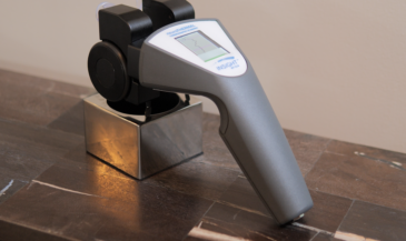
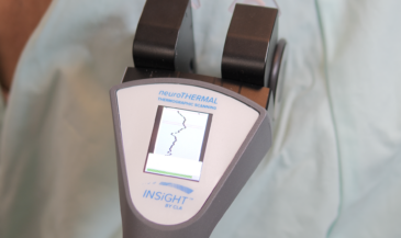
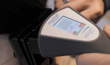
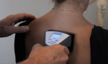
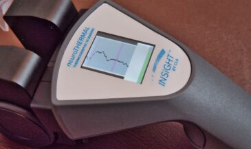
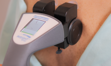
This cranial nerve, also known as the vestibulocochlear nerve, controls hearing and balance. Its nerve fibers pass through the internal acoustic meatus in the temporal bone. Balance issues or hearing disruptions can sometimes connect back to autonomic pathways, making your neuroTHERMAL scans invaluable for full analysis.
Glossopharyngeal and Hypoglossal Nerve Anatomy (CN IX & XII)
The glossopharyngeal nerve assists with taste and swallowing, while the hypoglossal nerve moves the tongue. Both rely on clear signals through the cervical region. Subtle weakness here can show up during muscle tests or when a patient reports slurred speech. The INSiGHT scans make these patterns clear.
Vagus Nerve Anatomy: The Longest Cranial Nerve
Known as the longest cranial nerve, the vagus nerve (CN X) extends from the brainstem through the neck and torso, connecting to organs like the heart and gut. The ganglion of the vagus nerve modulates these pathways.
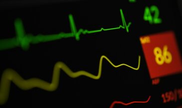
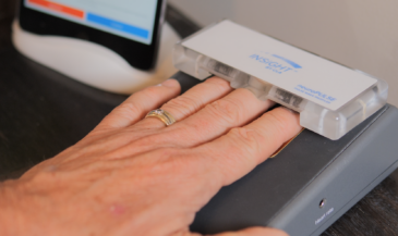
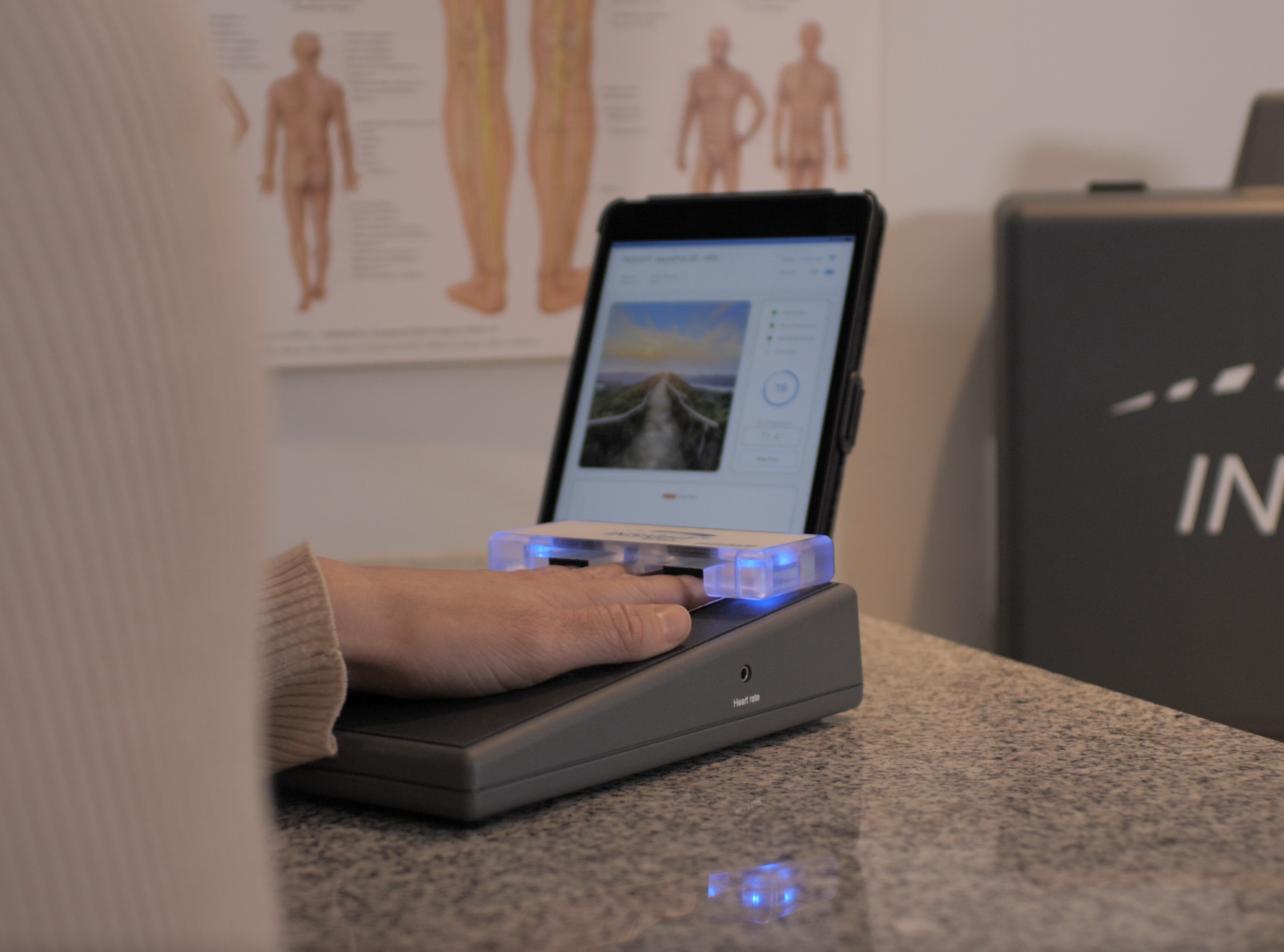
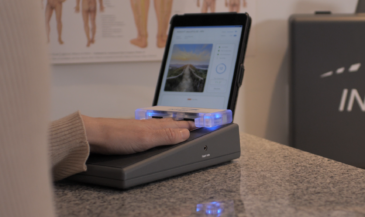
Heart Rate Variability (HRV) is one of the best ways to track vagus nerve function. Research shows that a well balanced HRV means your nervous system can switch smoothly between “fight-or-flight” and “rest-and-digest” states. The neuroPULSE gives you this insight in seconds.
Accessory Nerve Anatomy (CN XI)
The accessory nerve, sometimes called the spinal accessory nerve, innervates the sternocleidomastoid and trapezius muscles, helping move the head and shoulders. Tension in the upper neck and cervical plexus can disrupt this nerve, so it’s essential to include it in your scans.
Signs of Cranial Nerve Interference
How does tension here show up? Often through subtle but telling signs:
- Numbness, tingling, or pain along the face (think trigeminal neuralgia).
- Sudden facial drooping on one side, as seen in Bell’s palsy.
- Changes in taste or smell.
- Difficulty swallowing or speaking clearly.
- Eye tracking problems or double vision.
- Jaw fatigue while chewing.
These signs aren’t random — they reflect how vertebral tension can disrupt cranial nerve pathways, as noted in Kent’s recent review. Your role is to see these patterns early and use scans to make the invisible visible.
Sympathetic and Parasympathetic Pathways in the Head
Your cranial nerves don’t just handle senses and muscles. They’re deeply tied to the autonomic system that keeps you alive behind the scenes — a connection well detailed in Kent’s research.
The sympathetic system, your “fight-or-flight” wiring, starts in the spinal cord and travels through cervical ganglia to the brain. It controls pupil dilation, blood vessel tone, and sweat response when stress hits — but chronic neurological distress can push this into sympathetic overdrive.
The parasympathetic system does the opposite: it helps you “rest and digest.” These pathways run through cranial nerves like the vagus and glossopharyngeal, with key relay ganglia that keep your body’s internal processes balanced.
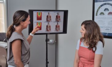
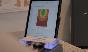
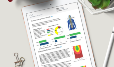
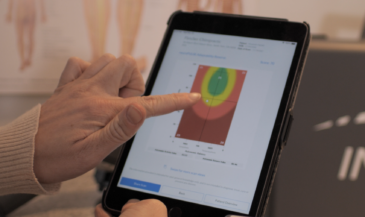
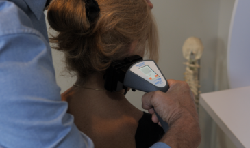
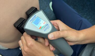
INSiGHT’s neuroTHERMAL scans show how well these pathways adapt to chronic and acute stress. Paired with HRV analysis, you get a clear view of stress resilience and balance.
The Vagus Nerve: The HRV Connection
The vagus nerve (CN X) is the longest cranial nerve and the backbone of your parasympathetic system. It helps regulate your heart rate, digestion, and mood. One of the best ways to check its status is Heart Rate Variability (HRV).
HRV is widely used to gauge autonomic adaptability — as shown in Kent’s HRV study. A balanced HRV means the nervous system can switch smoothly between “fight-or-flight” and “rest-and-digest.”
The neuroPULSE scan shows your patients their HRV in living color — proof their care plan is helping them handle stress and thrive.
Conditions Linked to Cranial Nerve Tension
Cranial nerve tension shows up in real, disruptive ways:
- Bell’s palsy: Sudden facial drooping.
- Trigeminal neuralgia: Intense facial discomfort.
- Cranial nerve palsies: Eye misalignment, swallowing problems.
These conditions highlight how upper cervical and brainstem tension can affect cranial pathways — as noted in Kent’s review. Objective scans connect these visible traits to deeper patterns of stress.
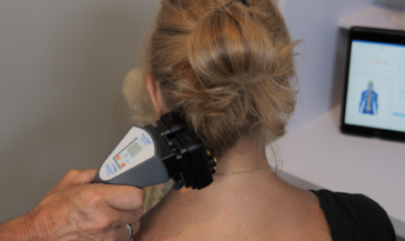
How Chiropractors Assess Cranial Nerve Health
You don’t need complex tools to check cranial nerves. Start with simple tests — reflexes, sensory checks, muscle strength — then back it up with data.
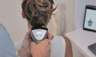
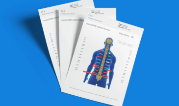
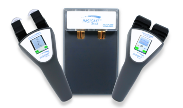
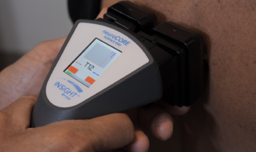
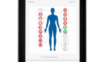
Full spine nerve system scans help you see what you might otherwise miss.INSiGHT research confirms that neuroTHERMAL, neuroCORE, and neuroPULSE work together to show somatic and autonomic balance. The CORESCORE™ brings it all together as your patient’s “neurological report card.”
Bringing It All Together with INSiGHT neuroTECH
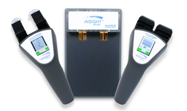
Your INSiGHT neuroTECH is your greatest ally.
- The neuroPULSE measures HRV and vagal tone —Kent’s HRV paper shows how valuable this is.
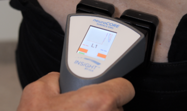
- The neuroCORE reveals neuromuscular tension patterns that can tie back to cranial nerve distress.
- The neuroTHERMAL shows subtle autonomic shifts at the spinal nerve level.
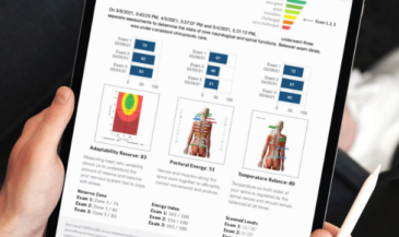
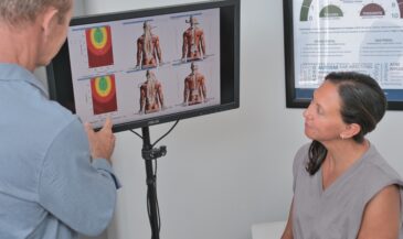
Together, these scans build your CORESCORE™ — the trusted report card that makes your care plan measurable and transparent. CLA’s Adaptability Project shows that patients who see these visuals stop counting visits and start valuing the why behind their care.
Bringing Certainty to the Nerves in the Head
When you see the bigger picture — from the spine to the cranial nerves — you stand out as a chiropractor who looks beyond structure. You prove your care is making a difference patients can see, trust, and share.
Stay curious. Keep scanning. Use your INSiGHT neuroTECH every day to transform invisible pathways into visible progress. That’s how you build certainty, trust, and a truly adaptable community.
If you want to bring this kind of transformational technology into your practice,book a call with an INSiGHT Advisor. We’ll show you how to implement scanning, reporting, and care planning tools that boost your retention and help your patients reach their full potential.
Sources:
Kent, C. (2024). Assessment of Somatic and Autonomic Nervous System Changes Associated with Vertebral Subluxation: A Review and Commentary. Vertebral Subluxation Research
Kent, C. (2017). Heart Rate Variability to Assess the Changes in Autonomic Nervous System Function Associated with Vertebral Subluxation. Vertebral Subluxation Research
Kent, C. (2017). Surface Electromyography in the Assessment of Changes in Paraspinal Muscle Activity Associated with Vertebral Subluxation: A Review. Vertebral Subluxation Research
INSiGHT CLA. (2024). Publications Supporting the Use of INSiGHT Scanning. Vertebral Subluxation Research
Kent, C., Haas, A., Chung, J., Mills, B., McCoy, M. (2024). Vertebral Subluxation and Systems Biology: An Integrative Review Exploring the Salutogenic Influence of Chiropractic Care. PubMed Central
Hartenburg, M. (2018). Heart Rate Variability as an Objective Outcome Measure for Subluxation Based Chiropractic Care for Athletes. Vertebral Subluxation Research
McCoy, M. (2017). Paraspinal Thermography in the Analysis and Management of Vertebral Subluxation: A Review of the Literature. Vertebral Subluxation Research
Zhang, J., Dean, D., Nosco, D., Strathopulos, D., Floros, M. (2006). Effect of Chiropractic Care on Heart Rate Variability and Pain in a Multisite Clinical Study. PubMed
McCoy, M., Campbell, I., Stone, P., Fedorchuk, C., Wijayawardana, S., Easley, K. (2011). Intra-Examiner and Inter-Examiner Reproducibility of Paraspinal Thermography. PubMed Central