If you’ve ever checked your pulse and noticed it’s not always perfectly steady, you’ve observed a natural phenomenon known as heart rate variability (HRV). But what is a heart rate variability, and why does it matter for your health, your nervous system, and your ability to adapt?

Heart rate variability is the measure of the small but significant differences in time between each heart beat. Unlike your average heart rate—which tells you how many times your heart beats per minute—HRV looks at the beat-to-beat variability, the subtle shift from one beat to the next. Scientists and doctors have learned that these variations in heart rate reveal how well your autonomic nervous system is working to keep you resilient and adaptable, even as life’s stresses, joys, and demands shift from moment to moment.

With new technology and deeper research, we now understand that heart rate variability is much more than a fitness trend or a feature in your smartwatch. HRV may be one of the most meaningful indicators of your overall heart health, stress, and nervous system function—making it a key tool for planning care, measuring progress, and living well.

What Is Heart Rate Variability?

Heart rate variability (HRV) refers to the tiny, natural changes in the timing between each heart beat. When your heart rate is measured, you may be told that your pulse is, for example, 70 beats per minute. However, your heart doesn’t beat every 0.857 seconds with machine-like precision. Instead, each beat is separated by slightly different intervals—sometimes shorter, sometimes longer—based on the body’s ongoing internal regulation.

This “instantaneous heart rate” and interval variability are not signs of a problem; in fact, a healthy heart and a responsive nervous system produce variations in heart rate as the body adapts to its environment. The concept of “heart rate variability” is the foundation for understanding the body’s adaptability, emotional regulation, and physical recovery. Higher heart rate variability generally reflects better adaptability and greater nervous system balance, while lower heart rate variability can indicate stress or limited recovery capacity.

The study and analysis of heart rate variability has grown to include advanced clinical tools such as EKGs and spectral analysis of heart rate, helping doctors, researchers, and chiropractors better understand and support nervous system health in their patients.

How the Autonomic Nervous System Controls Heart Rate Variability

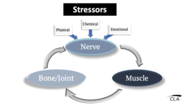
The autonomic nervous system (ANS) is the body’s automatic control system, overseeing everything from heart rate and blood pressure to breathing, digestion, and your ability to adapt to stress. It has two primary branches:

  • Sympathetic Nervous System: Activates the “fight or flight” response, increasing heart rates, redirecting blood flow, and boosting alertness when needed.
  • Parasympathetic Nervous System: Drives the “rest and digest” functions, promoting calm, recovery, and restoration.

Heart rate variability is the real-time record of how these two systems are interacting. When your body is in a calm, adaptive state, your parasympathetic system and vagal control of the heart predominate, leading to a wider range of intervals between heart beats (high HRV). When the sympathetic system is overactive—such as during chronic stress, pain, or illness—your HRV typically narrows (decreased heart rate variability).

The ANS communicates with the heart via the sinoatrial (SA) node, regulating the mean heart rate, and continually adjusting it based on needs and external demands. Researchers use frequency domain measures of heart and spectral analysis of heart rate to study these patterns and understand the deeper workings of the nervous system.

Why Heart Rate Variability Matters for Health and Adaptability

The more your heart rates are able to adapt—moment to moment—the healthier and more resilient your body tends to be. HRV may predict recovery from exercise, stress, and even major life events. In clinical research, decreased heart rate variability has been linked to an increased risk of conditions such as congestive heart failure, arrhythmias, and even sudden cardiac death.

reduced heart rate variability is often observed in patients with chronic stress, sleep disorders, or inflammatory illnesses. On the other hand, higher heart rate variability has been associated with improved recovery, emotional resilience, and lower overall mortality.

For patients recovering from cardiac procedures, like heart transplant or rate variability after acute myocardial infarction, HRV is used to track recovery progress. Some studies show that heart rate variability in patients after a heart attack or transplant can be a reliable rate variability as a predictor for future complications.

HRV may also play a crucial role in neurological care, where chiropractors and other providers use it as an index of adaptability, nervous system regulation, and progress over time.

What Affects Your Heart Rate Variability

Many factors can affect your heart rate variability, some of which are within your control and some that are not. The most important influences include:

Factors that Lower HRV

  • Chronic Stress: Emotional strain, overwork, and persistent anxiety
  • Sleep Disturbances: Poor quality or inconsistent sleep
  • Sedentary Lifestyle: Lack of regular movement
  • Poor Nutrition: Diets high in processed foods, sugar, and low in key nutrients
  • Alcohol or Drug Use: Excessive consumption reduces HRV
  • Illnesses: Inflammation, chronic pain, diabetes, and cardiovascular issues like chronic congestive heart failure
  • Certain Medications: Beta-blockers, antidepressants, and others
  • Aging: Natural declines in variability in healthy individuals over time

Factors that Improve HRV

  • Restorative Sleep: Regular, high-quality rest
  • Consistent Exercise: Aerobic activity and strength training
  • Healthy Nutrition: Emphasis on whole foods, healthy fats, lean proteins, and vegetables
  • Breathing Exercises: Slow, controlled breathing that regulates respiratory rate and the hf component
  • Mindfulness and Relaxation: Practices such as meditation, yoga, or relaxation techniques
  • Chiropractic Care: Evidence suggests adjustments can improve autonomic inputs to the heart, especially in patients with dysautonomia or nervous system imbalance

Over time, positive lifestyle habits can increase your average heart rate adaptability and strengthen your nervous system’s response to daily life.

How Is Heart Rate Variability Measured?

Measuring heart rate variability requires capturing the precise timing between each heart beat. The gold standard is an electrocardiogram (EKG), which tracks R–R intervals—the peaks between each beat on the heart’s electrical signal. EKGs are used in clinical and research settings to provide highly accurate, reliable HRV data.

However, technological advances have made heart rate variability measurement more accessible for everyday monitoring. Popular tools include:

  • Smartwatches and Fitness Trackers: Devices like Apple Watch, Oura Ring, and WHOOP use photoplethysmography sensors to estimate HRV
  • Chest-Strap Heart Rate Monitors: Used by athletes for more detailed heart rate monitoring and data
  • HRV Biofeedback Devices: Tools and apps designed to provide real-time feedback and stress reduction

These devices often use domain measures of heart period and calculate measures of heart period variability to provide useful information for both individuals and clinicians. Some even allow for power spectral analysis of heart rate data, making advanced HRV analysis available to a wider audience.

It’s important to note that changes in HRV are most meaningful when tracked over time, rather than relying on a single measurement.

Heart Rate Variability to Plan Care and Recovery

One of the most promising uses for heart rate variability is its role in guiding personal health, athletic performance, and clinical care. Coaches, trainers, and clinicians use HRV to decide how hard to train or when to rest. Variability to plan your workouts is becoming a standard for elite athletes, but is just as valuable for everyday individuals looking to maximize their well-being.

By monitoring 24-h heart rate variability and tracking patterns like resting heart, individuals can make smarter decisions about exercise, stress management, and recovery. In patients recovering from patients with a recent heart event, HRV trends can help doctors and care teams make evidence-based decisions.

Because HRV reflects the delicate balance between stress and recovery, it is a measure known as heart rate that supports regulation of heart rate and helps personalize plans for both patients and practitioners.

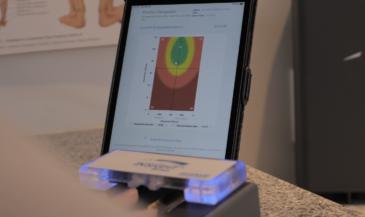
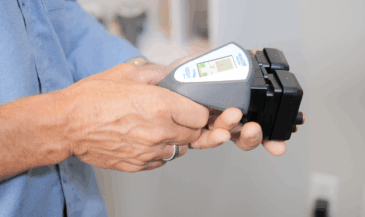
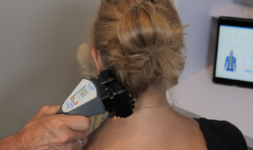
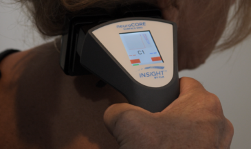
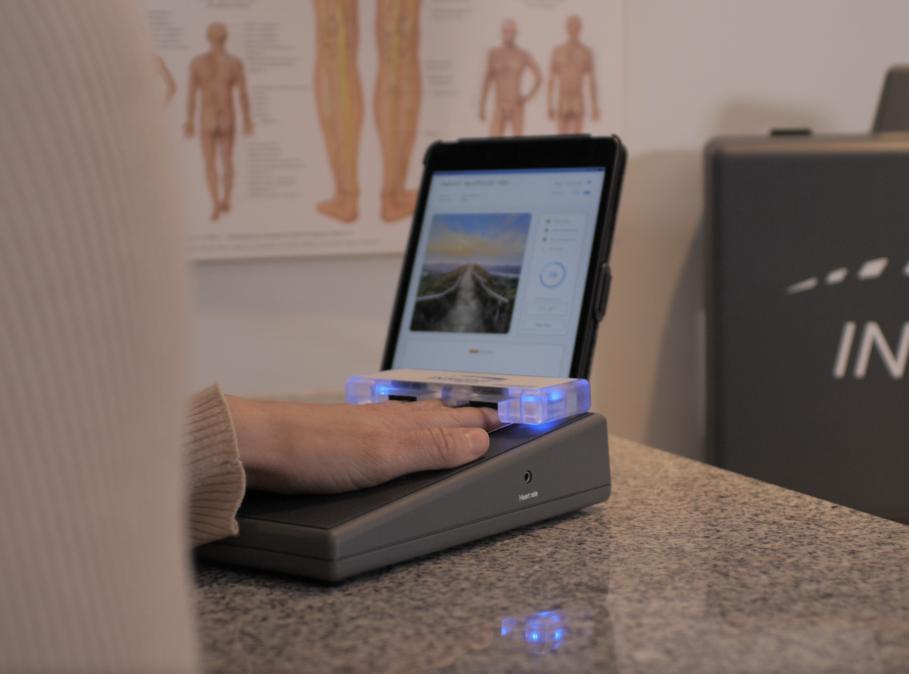
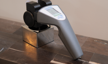
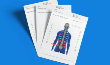
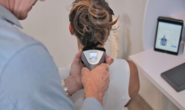
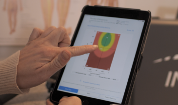
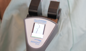
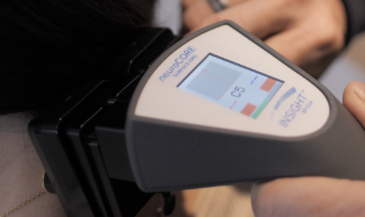
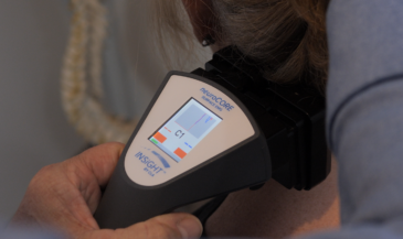
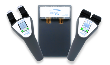
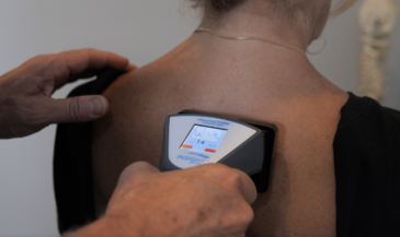
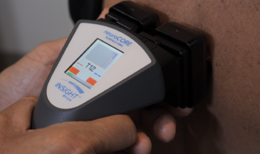
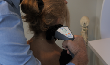
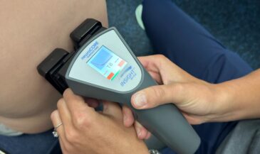
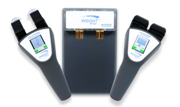
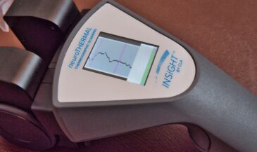
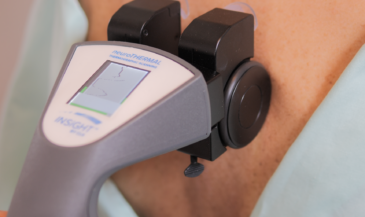
INSiGHT CLA’s Role: Advanced Heart Rate Variability Analysis in Practice

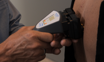
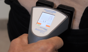
INSiGHT CLA’s NeuroPulse HRV scanning technology allows chiropractors to perform comprehensive heart rate variability analysis right in the clinic. Using non-invasive HRV scanning modules, providers can:

  • Obtain an objective analysis of heart rate variability and identify alterations of heart rate in patients over time
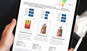
  • Track the effects of adjustments, lifestyle changes, and care plans using HRV as a rate variability as an index
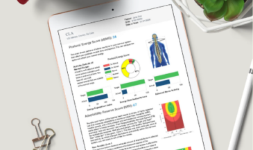
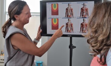
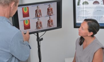
  • Visualize key metrics including component of heart rate variability, inputs to the heart rather than the mean, and variability in the power spectrum analysis of heart
  • Provide patients with actionable data to guide care, improve resilience, and support better outcomes

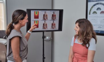
INSiGHT CLA’s scans look beyond the mean, focusing on variations of heart intervals and overall nervous system adaptability. This not only guides chiropractic care, but also empowers patients to become more active participants in their wellness journey.

Chiropractic research leaders have shown that regular chiropractic adjustments can support autonomic regulation and healthy HRV.

Special Applications: Heart Rate Variability in Special Populations

Heart Rate Variability in Patients

HRV isn’t just for athletes or adults—it’s relevant across the lifespan. From fetal heart rate patterns preceding birth, to rate patterns preceding fetal death in critical care, to heart rate variability in chronic illness management, HRV is now used as an early warning and progress tool in many fields.

It is used to monitor variability on the basis of health status, indices of heart rate during rate and blood pressure variability, and even for assessment in those with a heart transplant. Research shows that heart rate variability after acute myocardial events and heart rate variability after myocardial infarction can predict both short- and long-term outcomes.

In healthy populations, heart rate variability in healthy individuals is often higher, but certain conditions—like chronic pain, depression, or cardiovascular issues—can reduce HRV.

Frequently Asked Questions About Heart Rate Variability

Q: How often should I check heart rate variability?
A: For most people, measuring HRV once daily—ideally in the morning—provides the most consistent results. Monitoring trends, rather than one-off readings, gives the best insight into nervous system adaptability.

Q: Can I improve low HRV?
A: Yes. By focusing on better sleep, more physical activity, healthy nutrition, stress reduction, and—when needed—neurologically focused chiropractic care, you can support higher HRV over time.

Q: What does a high HRV mean?
A: Generally, a high HRV reflects a robust, adaptable nervous system, but the ideal range varies from person to person. Always interpret HRV alongside other health factors and in consultation with a knowledgeable provider.

Q: Is HRV the same as heart rate?
A: No. Heart rate is how many times your heart beats per minute; heart rate variability is the subtle difference in timing between each beat. Both are important for assessing health.

Your Next Step: Using Heart Rate Variability for Lifelong Adaptability

Understanding and leveraging what is a heart rate variability can be transformative. HRV is more than a metric—it’s a reflection of your body’s ability to handle stress, recover from challenges, and maintain balance.

From daily life to clinical care, heart rate variability empowers you, your provider, and your care team to make data-driven decisions that promote well-being, resilience, and adaptability at any age. By regularly tracking HRV, applying science-backed lifestyle habits, and working with providers who use advanced tools like INSiGHT CLA, you can ensure your heart rates and nervous system stay strong for the road ahead.


SOURCES

Shaffer, F., & Ginsberg, J. P. (2017). An Overview of Heart Rate Variability Metrics and Norms. https://www.ncbi.nlm.nih.gov/pmc/articles/PMC5624990/ 

McCraty, R., & Shaffer, F. (2015). Heart Rate Variability: New Perspectives on Physiological Mechanisms, Assessment of Self-regulatory Capacity, and Health Risk. https://www.ncbi.nlm.nih.gov/pmc/articles/PMC4599622/ 

Task Force of the European Society of Cardiology and the North American Society of Pacing and Electrophysiology. (1996). Heart rate variability: Standards of measurement, physiological interpretation, and clinical use. https://pubmed.ncbi.nlm.nih.gov/8737210/ 

Jandackova, V. K., et al. (2016). Heart rate variability and sudden cardiac death. https://pubmed.ncbi.nlm.nih.gov/8822992/ 

Umetani, K., et al. (1998). Twenty-Four Hour Time Domain Heart Rate Variability and Heart Rate: Relations to Age and Gender Over Nine Decades. https://www.ncbi.nlm.nih.gov/pmc/articles/PMC5624990/ 

Goldberger, J.J., et al. (2006). Relationship of Heart Rate Variability to Parasympathetic Effect. https://pubmed.ncbi.nlm.nih.gov/23994477/ 

Camm, A.J., et al. (1996). Guidelines: Heart Rate Variability. https://www.ncbi.nlm.nih.gov/pmc/articles/PMC5624990/ 

American College of Cardiology. (2013). Heart Rate Variability in Health and Disease. https://pubmed.ncbi.nlm.nih.gov/19776734/ 

P. Ginsberg. (2017). Biofeedback in the Treatment of Heart Rate Variability. https://pubmed.ncbi.nlm.nih.gov/31506233/

    Kleiger, R. E., et al. (1991). Decreased heart rate variability and its association with increased mortality after acute myocardial infarction. https://pubmed.ncbi.nlm.nih.gov/8822992/ 

    King, R., et al. (2016). Wearable Devices for Monitoring of Heart Rate and Heart Rate Variability: Validation Studies. https://www.ncbi.nlm.nih.gov/pmc/articles/PMC6213340/ 

    Kent, C. (2017). Heart Rate Variability and Chiropractic Care: Clinical Applications. The Journal of Vertebral Subluxation Research. https://www.chiroindex.org/?search_page=article&article_id=13685 

    Kent, C. (2019). NeuroPulse HRV Module: A New Paradigm in Chiropractic Assessment. The Journal of Vertebral Subluxation Research. https://www.chiroindex.org/?search_page=article&article_id=15036 

    Get Started with INSiGHT Scanning

    Take our Free Practice Strategy Assessment. A Personalized Guide and Expert Strategy Call to Help Determine How Scanning will Help you Grow
    ABOUT THE AUTHOR

    Dr. David Fletcher is actively involved in all aspects of innovation teaching and research connected to the INSiGHT™ scanning technologies. He is widely recognized for his ability to share his expertise in compelling and easy to understand ways.

    Dr David is a renowned chiropractor who practiced for many years with his associates in a scan-centric thriving principled family-based practice in Toronto. He is a sought-after teacher mentor and keynote speaker who takes every opportunity to share the wisdom and the power of chiropractic as it is meant to be.

    https://insightcla.com/wp-content/uploads/2022/07/david-1.png
    Dr. David Fletcher
    DC FRCCSS(C) – Founder & CEO CLA Inc.
    LIKE THIS ARTICLE? HELP US SPREAD THE WORD

    Be Seen. Be Heard. Be Certain

    In today’s saturated healthcare market, chiropractors face an ongoing challenge: being visible. Discover key marketing tactics to ensure you can BE SEEN, BE HEARD, and BE CERTAIN in your practice!

    Read these Next…

    Get Informed

    Join 23,121 other Chiropractors and receive topics covering day to day challenges of running your practice.

    If you’ve ever dealt with a herniated disc, you know the question that quickly rises to the top: how long does a herniated disc take to heal? It’s not just about easing the pain—it’s about understanding what’s going on beneath the surface, and what it takes to truly recover. At INSiGHT CLA, we see this all the time. People show up with stories of lingering pain, numbness in a leg or arm, and a timeline that just doesn’t add up. The disc may be the source, but healing doesn’t happen on the calendar—it happens when the nervous system can adapt. Let’s break this down from the ground up.

    What’s Really Going on When a Disc Herniates?

    Between each vertebra in your spine is a disc—think of it like a jelly-filled cushion. The outer ring, called the annulus fibrosus, keeps the gel-like center (nucleus pulposus) in place. This structure helps absorb shock and maintain flexibility in the spine, according to researchers at NCBI. But when that outer ring weakens or tears, the nucleus pushes out. That’s what we call a herniated disc—and when that disc material presses on nearby nerves, it can set off a chain reaction. Depending on whether the issue is in the cervical spine or lumbar disc, you might feel tingling in your fingers or pain down your leg. This has been well-documented in clinical literature. Interestingly, the size of the herniation doesn’t always dictate the severity of your symptoms. As shown in a landmark study from the New England Journal of Medicine, many people with visible disc issues on MRI have no symptoms at all. Why? Because the nervous system—not the disc itself—dictates how pain is interpreted and amplified.

    So… How Long Does a Herniated Disc Take to Heal?

    Most herniated discs heal in about 6 to 12 weeks. Some resolve faster, and others—especially if severe or ignored—can take up to 6 months. Healing happens in phases:

    • Inflammation (1–2 weeks): Your body responds with swelling and muscle guarding. Pain is usually at its peak.
    • Tissue Repair (2–12 weeks): The herniated material may shrink or dry out. Pressure on the nerve lessens.
    • Remodeling (6+ weeks): The spine re-coordinates. Muscles stabilize. The nervous system learns how to move without overreacting. JAMA confirms this sequence.

    What matters more than the clock is your nervous system’s ability to shift out of stress and into recovery. If your system is stuck in “defense mode,” healing can stall—even if the disc looks better on a scan.

    What Affects How Fast You Heal?

    Your healing timeline isn’t random. It’s influenced by:

    • Severity: A small bulge recovers faster than a ruptured disc pressing on a nerve.
    • Location: A herniated lumbar disc often causes leg pain, while cervical issues affect the arms.
    • Overall health: Smokers, those with high stress, or systemic inflammation often heal more slowly.
    • Movement: Gentle motion helps; too much rest or too much exertion can backfire.
    • Neurological adaptability: A well-regulated nervous system can support faster recovery.

    We’ve seen patients with similar MRIs heal at wildly different rates. It often comes down to how adaptable their nervous system is—not just what’s on the film.

    How Do You Know You’re Healing?

    Improvement often looks like this:

    • Radiating pain starts to ease.
    • Muscle tightness softens.
    • Sleep and focus improve.
    • You need less pain medication.

    But the best signs of improvement come from within. Your nervous system begins to regulate, adapt, and respond more effectively—even if the pain hasn’t completely vanished. That’s why INSiGHT scanning is so vital: it allows us to see changes in neural function long before symptoms disappear.

    The INSiGHT Difference: Measuring More Than Symptoms

    INSiGHT scanning gives chiropractors powerful tools to measure how your nervous system is adapting.

    • HRV shows how well you’re shifting between stress and rest. HRV is widely used in chiropractic to assess adaptability and is central to neurological care strategies.
    • sEMG tracks how muscles are firing around the spine.
    • Thermography shows heat patterns that may indicate nerve stress or autonomic imbalance.

    While traditional care may rely on how you “feel,” INSiGHT scans reveal how you’re truly functioning—offering a smarter, more holistic way to track progress.

    Give Your Body a Clear Path to Heal

    So, how long does a herniated disc take to heal? For many, it’s a matter of weeks. For others, it takes a bit more time. But no matter the timeline, what determines your recovery is your nervous system’s ability to adapt. You were built to get better. The disc may be the starting point, but the real healing happens when your brain and body reconnect—and chiropractic care helps facilitate that connection. Choose care that honors your body’s design. Work with a chiropractor who uses INSiGHT to guide decisions, track progress, and truly listen to what your nervous system is telling you. Because healing doesn’t just happen. It’s something your body already knows how to do—once the interference is cleared.


    Frequently Asked Questions

    How Long Does a Herniated Disc Take to Heal?

    The healing time for a herniated disc can vary significantly depending on various factors, including the severity of the herniation, the location of the herniation, and the individual’s overall health. In many cases, a herniated disc can heal on its own within a few weeks to a few months with appropriate conservative treatment.

    What Are the Symptoms of a Herniated Disc?

    Common symptoms of a herniated disc include back pain, leg pain, numbness, and weakness in the affected area. Individuals may also experience symptoms like severe pain that radiates down the leg, often referred to as sciatica. The symptoms can vary based on the location of the herniation and the extent of nerve involvement.

    What Conservative Treatments Are Recommended for Herniated Discs?

    Conservative treatments for a herniated disc typically include physical therapy, pain management strategies such as medications, and lifestyle modifications. Cold therapy and regular physical activity can also help alleviate symptoms and support the healing process.

    Can a Herniated Disc Heal on Its Own?

    Yes, many individuals with a herniated disc can heal on their own within a few weeks to a few months. The body’s ability to heal depends on various factors, including the severity of the herniation and adherence to a proper treatment plan.

    How Does Physical Therapy Aid in Recovery from a Herniated Disc?

    Physical therapy plays a crucial role in the recovery process by helping individuals strengthen their back muscles, improve flexibility, and restore normal movement patterns. A tailored physical therapy program can help you heal faster and reduce the risk of future injuries.

    When Should You Consider Surgery for a Herniated Disc?

    Surgery for a herniated disc is usually recommended if conservative treatments fail to relieve symptoms after several months, or if there is significant weakness or loss of function. Your healthcare provider will assess your condition and determine if surgery is the best option for you.

    What Factors Influence the Time for a Herniated Disc to Heal?

    The healing of a herniated disc can be influenced by several factors, including the severity of the herniation, the individual’s age, overall health, and adherence to treatment recommendations. Individuals with chronic back pain may experience a longer recovery journey.

    Is There a Timeline for Herniated Disc Recovery?

    The timeline for herniated disc recovery varies from person to person. Generally, individuals may start noticing improvement within a few weeks, with significant recovery occurring within 6 to 12 weeks. However, full healing of the disc may take longer, especially in cases of severe pain or complicated conditions.

    SOURCES

    Jensen, M. C., Brant-Zawadzki, M. N., Obuchowski, N., Modic, M. T., Malkasian, D., & Ross, J. S. (1994). Magnetic resonance imaging of the lumbar spine in people without back pain. New England Journal of Medicine, 331(2), 69–73. https://pubmed.ncbi.nlm.nih.gov/8208267/ 

    Urban, J. P., & Roberts, S. (2003). Degeneration of the intervertebral disc. Arthritis Research & Therapy, 5(3), 120–130. https://www.ncbi.nlm.nih.gov/pmc/articles/PMC165042/ 

    Spine-health. (n.d.). Herniated Disc – Causes and Treatments. Spine-health. https://www.spine-health.com/conditions/herniated-disc 

    MedlinePlus. (2023). Herniated disk. U.S. National Library of Medicine. https://medlineplus.gov/ency/article/000442.htm 

    Chiu, C. C., Chuang, T. Y., Chang, K. H., Wu, C. H., Lin, P. W., & Hsu, W. Y. (2014). Management of herniated lumbar disc with radiculopathy: an updated clinical practice guideline. The Spine Journal, 14(8), 1770–1774. https://pubmed.ncbi.nlm.nih.gov/24768732/ 

    Ropper, A. E., & Zafonte, R. D. (2017). Lumbar disk herniation: A review. JAMA, 318(17), 1743–1754. https://jamanetwork.com/journals/jama/fullarticle/2662437 

    Get Started with INSiGHT Scanning

    Take our Free Practice Strategy Assessment. A Personalized Guide and Expert Strategy Call to Help Determine How Scanning will Help you Grow
    ABOUT THE AUTHOR

    Dr. David Fletcher is actively involved in all aspects of innovation teaching and research connected to the INSiGHT™ scanning technologies. He is widely recognized for his ability to share his expertise in compelling and easy to understand ways.

    Dr David is a renowned chiropractor who practiced for many years with his associates in a scan-centric thriving principled family-based practice in Toronto. He is a sought-after teacher mentor and keynote speaker who takes every opportunity to share the wisdom and the power of chiropractic as it is meant to be.

    https://insightcla.com/wp-content/uploads/2022/07/david-1.png
    Dr. David Fletcher
    DC FRCCSS(C) – Founder & CEO CLA Inc.
    LIKE THIS ARTICLE? HELP US SPREAD THE WORD

    Be Seen. Be Heard. Be Certain

    In today’s saturated healthcare market, chiropractors face an ongoing challenge: being visible. Discover key marketing tactics to ensure you can BE SEEN, BE HEARD, and BE CERTAIN in your practice!

    Read these Next…

    Get Informed

    Join 23,121 other Chiropractors and receive topics covering day to day challenges of running your practice.

    If you’ve been in practice for even a short while, you’ve seen it: a patient comes in, clutching their MRI and wearing the weight of the world on their face. “Doc, I’ve got a herniated disc. Does this ever heal?” It’s the kind of question that cuts to the heart of what we do—not just because it’s about symptoms, but because it’s about hope, function, and getting folks back to living their lives.

    I’ve sat across from patients with stories just like this more times than I can count. Some got their disc injury wrestling with a grandkid, some by simply tying their shoes. The names change—bulging disc, slipped disc, ruptured disc—but the worries are always the same: “Will my disc heal? Will I need surgery? Can I get back to being me?”

    The good news, confirmed by peer-reviewed research, is that most herniated discs do heal—and there’s a lot we can do to make that recovery smoother, more predictable, and truly trackable. Even better, modern tools like INSiGHT scanning technology let us see and measure real progress, not just hope for it.

    We’ll walk through how the healing process unfolds, what conservative care (especially neurologically-focused care) looks like, and how INSiGHT scanning lets us see and celebrate true disc healing.

    What Is a Herniated Disc? (And Why Do They Happen?)

    Let’s take it from the top: your spine is a sturdy stack of vertebrae separated by round, cushiony spinal discs—think of them as the shock absorbers and “jelly donuts” of your back. Each disc has a tough exterior (the annulus) and a gel-like center (the nucleus) according to the NCBI.

    A herniated disc, sometimes called a bulging disc, slipped disc, or ruptured disc, is what happens when that jelly center pushes through a crack in the outer ring—often the result of wear, trauma, or a simple twist the wrong way as described by the NCBI.

    But the real trouble isn’t just the bulge—it’s when the herniated disc puts pressure on nearby nerves, causing neurological interference and sometimes radiating symptoms. If you’ve seen this, you know how quickly it can impact daily life.

    More than 3 million people in the U.S. each year are affected by herniated discs, usually in the lower back (lumbar disc) or neck (cervical spine).

    Common causes and risk factors:

    • Disc degeneration: Natural aging and wear
    • Trauma: Accidents or injuries, sometimes sports-related
    • Improper lifting: Bending at the waist instead of the knees
    • Repetitive motions: Certain jobs or hobbies
    • Genetics: Some families are more prone than others

    Risk factors also include being between 30 and 50, carrying extra weight, sitting for long periods, and smoking—which weakens discs.

    Signs, Symptoms, and When to Worry

    A herniated disc can show up in a hundred different ways, but most patients come in with a story that sounds a lot like this: “My lower back hurts and there’s a shooting sensation down my leg,” or “My neck feels stiff and my arm keeps going numb.” These are the classic symptoms—and they’re all about the nervous system getting irritated.

    Typical signs by location:

    • Herniated Lumbar Disc (Lower Back):
      • Persistent lower back discomfort or stiffness
      • Sharp, shooting traits down the leg (classic sciatica)
      • Numbness or tingling in the legs or feet
      • Weakness in the legs
    • Herniated Cervical Disc (Neck):
      • Neck discomfort radiating to the shoulders or between the shoulder blades
      • Numbness or tingling in the arms or hands
      • Weakness in the arms, hands, or fingers

    These aren’t just about pain—they’re signals that the nerve is under tension and the nervous system’s adaptability is being tested.

    Red flags that require urgent attention:

    • Loss of bladder or bowel control
    • Sudden or progressive muscle weakness
    • Numbness in the saddle area (inner thighs, buttocks)

    If you see these, don’t wait—get that patient evaluated right away. These are standard clinical warning signs.

    Diagnosing a herniated disc:

    • Physical exam: Checking reflexes, strength, sensation, and movement
    • Imaging: MRI or CT to see the disc bulge and nerve involvement
    • Nerve tests: EMG and nerve conduction studies to pinpoint areas of nerve tension

    But a good diagnosis is more than confirming a spinal disc bulge—it’s about understanding nervous system status and adaptability.

    Can a Herniated Disc Heal? (And What Does “Healing” Mean?)

    So, do herniated discs heal? Here’s the honest answer, grounded in current research: Yes, most herniated discs heal, and most patients recover without surgery.

    Healing isn’t just about “feeling better.” In the neurologically-focused chiropractic world, healing is about restoring function, adaptability, and reducing nerve tension.

    What’s happening under the surface?

    • The body’s immune system goes to work, reabsorbing some of the disc material—a process described in disc literature.
    • Inflammation decreases, the bulge shrinks, and the pressure on the nerve lessens.
    • Scar tissue forms around the damaged disc, providing stability.

    Most people see significant progress in a few weeks to a few months, but disc healing can take up to a year, especially if the herniation is more severe or the patient is older.

    And remember, it’s not just about symptom relief—it’s about the nervous system’s ability to adapt, as outlined in the INSiGHT Knowledge Base.

    How Herniated Discs Heal: The Science Behind Natural Recovery

    Healing a herniated disc isn’t magic; it’s a biological process. The body is wired to recover—sometimes with a little help from us.

    The healing process looks like this:

    • Inflammation signals the body’s “cleanup crew” to get to work on the affected disc.
    • The herniated disc material dries up or shrinks—documented in disc healing studies.
    • Scar tissue forms, patching up the outer ring of the disc.
    • Nerves regain function as the pressure and tension ease.

    Healing isn’t a straight line. Some days, patients feel almost normal; other days, there are flare-ups. That’s just the nervous system adapting and recovering.

    Factors that can affect healing:

    • Age: Younger patients tend to heal faster, but recovery is possible at any age.
    • Overall health and activity: Movement, strength, flexibility, and nutrition all matter.
    • Nerve tension and adaptability: The more we can reduce interference, the better the disc healing.
    • Lifestyle: Smoking, poor posture, chronic stress, and inactivity can slow progress.

    Most herniated discs heal on their own within a few months, especially with a supportive, neurologically-focused approach.

    Conservative Care Plans: What Really Works?

    When it comes to disc healing, conservative treatment is the name of the game. Only a small percentage of patients ever need spine surgery.

    What’s included in a solid conservative treatment plan?

    • Short-term rest (if symptoms are severe), but keeping folks moving is key.
    • Physical therapy: Strengthening and stretching are critical.
    • Chiropractic adjustments: Gentle, neurologically-focused chiropractic care can improve adaptability and reduce nerve tension.
    • Over-the-counter medications: Sometimes needed for symptoms, but not the main event.
    • Steroid injections: Reserved for stubborn cases where inflammation is a big player.
    • Lifestyle upgrades: Posture, workplace ergonomics, healthy weight, and avoiding smoking.

    Progress is monitored regularly. If the herniated disc is healing, we keep going. If there’s a setback or new neurological symptoms, we re-evaluate and adjust the care plan.

    When Is Surgery Needed? (And Why It’s Rare)

    A lot of folks walk into the office worried their herniated disc will require surgery. But about 90% of herniated discs heal without surgery.

    Surgery may be recommended if:

    • There’s severe or worsening neurological loss.
    • Symptoms are disabling after months of conservative care.
    • There’s clear nerve compression not improving otherwise.

    Minimally invasive surgery, such as microdiscectomy, removes just the herniated portion of the disc. Other surgical options are considered only in rare or complex cases.

    But surgery is generally a last resort. Most people don’t require surgery, especially with a neurologically-focused, objective approach.

    Neurologically-Focused Chiropractic Care: Beyond Symptom Relief

    Here’s what sets Neurologically-Focused Chiropractic Care apart: We’re not just chasing symptoms or treating the “bad disc”—we’re looking at the whole nervous system.

    Our approach:

    The INSiGHT Advantage: Making Nerve Healing Visible

    This is where it all comes together. The INSiGHT technology suiteneuroTHERMAL, neuroCORE, neuroPULSE, and the Synapse software—gives us a window into the healing process that was invisible before.

    With INSiGHT, we can:

    All this data rolls up into the CORESCORE—an easy-to-understand number reflecting overall neural efficiency. It’s a report card for the nervous system, showing real, measurable progress.

    With regular INSiGHT scans, practitioners and patients alike can see the healing journey unfold, building trust, compliance, and real hope.

    Preventing Disc Issues: Strategies for Lifelong Spinal and Nervous System Health

    The best way to handle a herniated disc? Prevent it in the first place. While you can’t avoid every accident, you can stack the deck in your favor.

    Prevention strategies:

    • Use proper lifting techniques.
    • Maintain a healthy weight.
    • Stay active, stretch, and strengthen the core.
    • Take movement breaks if you sit a lot.
    • Improve ergonomics.
    • Don’t smoke.
    • Get regular neurologically-focused chiropractic assessments to keep the nervous system adaptable.


    Prevention is always better than repair—and it’s never too early or too late to start.

    Frequently Asked Questions (FAQ)

    Do herniated discs heal on their own?
    Yes, many herniated discs can heal on their own over time. The body has natural healing mechanisms that can reduce the inflammation and pain associated with a herniated disc. However, the healing process may vary depending on the location of the herniated disc and the severity of symptoms. While some patients may experience relief within weeks, others may take several months to heal completely.

    What are the symptoms of a herniated disc?
    The symptoms of a herniated disc may include back pain, leg pain, numbness, tingling, and weakness in the affected area. In cases of lumbar disc herniation, pain may radiate down the leg, often described as sciatica. It’s important to consult a healthcare professional for a proper diagnosis and treatment plan if you experience these symptoms.

    What treatment options are available for a herniated disc?
    Treatment for a herniated disc typically includes conservative methods such as chiropractic, physical therapy, medication, and lifestyle changes. In cases where pain persists, a doctor may recommend surgical treatment options such as minimally invasive spine surgery or artificial disc replacement. The choice of treatment depends on the severity of the herniation and the patient’s overall health.

    What causes a herniated disc to heal?
    The healing of a herniated disc is influenced by various factors, including the body’s natural repair processes and the individual’s response to treatment. Rest, physical therapy, and proper nutrition can support disc healing. Additionally, avoiding activities that cause pain can help the disc heal more effectively.

    How long does it take for a herniated disc to heal?
    The time it may take to heal from a herniated disc varies from person to person. While some individuals may find relief within a few weeks, others may require several months for complete recovery. Factors such as age, overall health, and adherence to treatment can impact healing time.

    What is the role of surgery for a herniated disc?
    Surgery for a herniated disc is generally considered when conservative treatments do not provide relief. Surgical options may include discectomy, where the herniated portion of the disc is removed, or artificial disc replacement. The goal of surgery is to alleviate pain, restore function, and improve the patient’s quality of life.

    Can natural healing help with a herniated disc?
    Natural healing can indeed play a significant role in the recovery from a herniated disc. Engaging in low-impact activities, maintaining a healthy weight, and practicing good posture can promote healing. Additionally, therapies such as acupuncture and chiropractic care may provide relief and enhance the body’s natural healing processes.

    What are the common causes of herniated discs?
    Herniated discs can occur due to a variety of reasons, including age-related degeneration, injury, and repetitive strain on the spine. Activities that put excessive pressure on the discs, such as heavy lifting or poor posture, can also contribute to disc herniation. Understanding these causes can help in taking preventive measures.

    Do herniated discs heal without surgery?
    Yes. The majority of herniated discs heal naturally, especially with a conservative, neurologically-focused care plan.

    How long does disc healing take?
    Most patients feel better in weeks to months. For some, disc healing can take up to a year, depending on the herniation’s location and severity.

    Is it safe to move with a herniated disc?
    Absolutely. Gentle movement and guided physical therapy are essential for the healing process.

    What symptoms mean I need help now?
    If you lose bladder or bowel control, develop sudden weakness, or have numbness in the saddle area—get medical attention right away.

    Can I prevent another herniated disc?
    With healthy habits and regular neurologically-focused chiropractic care, you can reduce your risk and support lifelong adaptability.

    Are neurologically-focused chiropractic adjustments safe for herniated discs?
    When personalized and based on a thorough assessment, yes—they’re a cornerstone of helping your body heal.

    What is the CORESCORE?
    CORESCORE is an objective measure of neural efficiency that tracks healing.

    Reclaiming Resilience: Your Nervous System, Your Recovery

    The story of herniated discs is one of hope. Most cases can heal—and with the right approach, most do. It’s not just about managing symptoms or waiting for the body to “get lucky.” It’s about supporting the nervous system, tracking progress, and building adaptability for whatever life brings next.

    With neurologically-focused chiropractic care and the clarity of INSiGHT scanning technology, you’re no longer guessing—you’re guiding. For patients and practitioners alike, that’s where real healing, confidence, and resilience begin.

    SOURCES

    Sources:

    National Center for Biotechnology Information. (2023). Disc Herniation. https://www.ncbi.nlm.nih.gov/books/NBK441822/ 

    National Center for Biotechnology Information. (2023). Anatomy, Back, Intervertebral Discs. https://www.ncbi.nlm.nih.gov/books/NBK470583/ 

    Spine-health. (2017). Spinal Discs. https://www.spine-health.com/conditions/spine-anatomy/spinal-discs 

    Cleveland Clinic. (2023). Herniated Disk. https://my.clevelandclinic.org/health/diseases/10301-herniated-disk 

    PubMed Central. (2020). Vascularization of the human intervertebral disc: A scoping review. https://www.ncbi.nlm.nih.gov/pmc/articles/PMC7770199/ 

    American Psychological Association. (2024). Stress effects on the body. https://www.apa.org/topics/stress/body

    PubMed Central. (2024). Chiropractic Care and Neuroendocrine Immune Function. https://pmc.ncbi.nlm.nih.gov/articles/PMC11016242/

    The Journal of Vertebral Subluxation Research. (2024). Assessment of Somatic and Autonomic Nervous System Changes Associated with Vertebral Subluxation: A Review and Commentary. https://vertebralsubluxationresearch.com/2024/09/21/1851-assessment-of-somatic-and-autonomic-nervous-system-changes-associated-with-vertebral-subluxation-a-review-and-commentary/

    The Journal of Vertebral Subluxation Research. (2017). Heart Rate Variability to Assess the Changes in Autonomic Nervous System Function Associated With Vertebral Subluxation. https://vertebralsubluxationresearch.com/2017/10/16/heart-rate-variability-to-assess-the-changes-in-autonomic-nervous-system-function-associated-with-vertebral-subluxation/

    The Journal of Vertebral Subluxation Research. (2017). Surface Electromyography in the Assessment of Changes in Paraspinal Muscle Activity Associated with Vertebral Subluxation: A Review. https://vertebralsubluxationresearch.com/2017/09/10/surface-electromyography-in-the-assessment-of-changes-in-paraspinal-muscle-activity-associated-with-vertebral-subluxation-a-review/

    Get Started with INSiGHT Scanning

    Take our Free Practice Strategy Assessment. A Personalized Guide and Expert Strategy Call to Help Determine How Scanning will Help you Grow
    ABOUT THE AUTHOR

    Dr. David Fletcher is actively involved in all aspects of innovation teaching and research connected to the INSiGHT™ scanning technologies. He is widely recognized for his ability to share his expertise in compelling and easy to understand ways.

    Dr David is a renowned chiropractor who practiced for many years with his associates in a scan-centric thriving principled family-based practice in Toronto. He is a sought-after teacher mentor and keynote speaker who takes every opportunity to share the wisdom and the power of chiropractic as it is meant to be.

    https://insightcla.com/wp-content/uploads/2022/07/david-1.png
    Dr. David Fletcher
    DC FRCCSS(C) – Founder & CEO CLA Inc.
    LIKE THIS ARTICLE? HELP US SPREAD THE WORD

    Be Seen. Be Heard. Be Certain

    In today’s saturated healthcare market, chiropractors face an ongoing challenge: being visible. Discover key marketing tactics to ensure you can BE SEEN, BE HEARD, and BE CERTAIN in your practice!

    Read these Next…

    Get Informed

    Join 23,121 other Chiropractors and receive topics covering day to day challenges of running your practice.

    Every chiropractor wants to achieve greater retention for the effort they put into their exams, reporting and recommendations, but most miss the moment that matters most: the initial care planning conversation.

    It doesn’t begin at their sixth or tenth visit or when symptoms resolve. It starts the moment your new patient sits down after their initial exam. This is the ideal time to move your message deeper and indicate the spinal impact on their functioning nervous system.

    And if you’re not using that moment to build trust, show them objective neurological scanning  data, and present a clear roadmap for care—you’re setting yourself up for symptom-based drop-offs and missed neurofunctional outcomes.

    In this blog, I’m going to walk you through exactly how you can use INSiGHT scanning technology, CORESCORE reports, and a simple, structured conversation to shift patients from drop-ins… to committed, lifelong care.

    We’re going to break it down into three key steps:

    1. Make The Shift From First Scan to Care Plan
    2. Make Re-Scans and Re-Exams Your Undeniable Retention Machine
    3. Frame the Future to Shift From Quick Fixes to Lifelong Care

    Let’s get into it.

    Step 1: Make The Shift From First Scan to Care Plan

    Retention begins with that first exam—before the first adjustment is even made.

    When a new patient walks through your doors, the goal isn’t just to identify what’s making them feel out of sync. It’s to frame their experience as the beginning of a nervous system performance journey. Including an INSiGHT scan in the initial exam confirms your commitment to looking deeper than their symptoms. Scanning measures the functional changes while X-Rays and Ortho testing look at the existing symptomatic causes and degeneration.   

    That neurofunctional journey begins with a 3-dimensional INSiGHT scan:

    • The neuroPULSE to measure heart rate variability. We need to know how much adaptive horsepower is in their Autonomic Nervous System
    • The neuroCORE to assess muscle energy expenditure. Daily stressors and spinal misalignments are exhausting! Muscles become fatigued but so does the Motor Nervous System.
    • The neuroTHERMAL to detect spinal autonomic stress patterns that define dysautonomia and dysregulation

    Together, these scans uncover how your patient’s nervous system is adapting—or not adapting—to life’s stress. They establish a baseline, which is essential for both care planning and future re-evaluations.

    Then you show the patient their CORESCORE report—a single score between 0 and 100 that consolidates all three scans’ data into a single, understandable number—a neurological report card your patient can follow at every phase of care.

    When patients see this score, it becomes real. It’s not about “how they feel.” It’s about how their nervous system is actually functioning.

    And here’s the key: you don’t focus on what’s wrong. This is your opportunity to lead a conversation focused on what’s possible. You can say:

    “This score tells us how well your nervous system is adapting to life’s stress. Our goal is to improve this over time. Just like you wouldn’t stop going to the gym after one month, this is where long-term nervous system performance really starts.”

    It shifts the mindset from relief care… to a performance-based, lifetime journey towards excellence.

    Step 2: Make Re-Scans and Re-Exams Your Undeniable Retention Machine

    Retention isn’t built on visit frequency alone—it’s built on trust, belief and understanding. And trust comes from seeing change.

    That’s why re-scans are your most powerful retention tool.

    They’re more than just routine check-ups, they act as reconnection moments that let your patients see progress from where they started, understand what’s changing, and know what comes next.

    With every CORESCORE update, you’re showing evidence-backed proof. This is where patients stop thinking of care as a temporary solution and start seeing it as a necessary lifestyle choice.

    You’re no longer their “spinal mechanic”. You’re helping them function better at every level in their health.

    You’ll notice the shift. They start asking more questions. They want to know what else is possible. They start thinking about their kids. Their spouse. Their ability to sleep and perform better, not just get through the day.

    Re-scans reframe their expectations and remind them why they chose you in the first place.

    Every time you do a progress scan, these appointments give you three powerful talking points:

    • “Here’s what we were working on.”
      Patients remember their starting point and original goals.
    • “Here’s what’s improving.”
      They see and feel their own progress—sometimes in ways they hadn’t even noticed.
    • “Here’s what’s next.”
      You set new targets, keeping the momentum going.

    Shifting Mindsets: 

    Re-scans help patients see care differently. Instead of only coming in when they hurt, they start to view care as something that keeps them performing at their best—much like regular exercise or healthy eating. The mindset shifts:

    • From: “I come in when something hurts.”
      To: “I come in to be at my best.”
    • From: “This is a necessary fix”
      To: “This is part of my lifestyle.”

    Step 3: Frame the Future to Shift From Quick Fixes to Lifelong Care

    Every care plan begins with a prescribed block of care. So what happens after the initial 12 visits?

    This is where a lot of chiropractors drop the ball. You’ve guided the patient through their initial phase of care. You’ve done a great initial exam, run the scans, tracked the patterns, and delivered their first round of adjustments. And then—nothing. No clear next step. No defined vision for what comes after visit 12.

    That’s where retention starts to unravel.

    At this stage, you’re not wrapping up care. You’re opening the next chapter.

    By the 12th visit, you’ve already seen neurological changeability starting to emerge. The CORESCORE is your tool to validate those early shifts and create belief in what’s working. This is your moment to transition patients into the next phase—what I call Re-Organizational Care.

    Here’s what I often say:

    “Your nervous system has shown great changeability over these first 12 visits. That’s exactly what we want to see. But just like going to the gym, the first few weeks are only the beginning. We’ve opened the door. Now it’s time to strengthen the foundation.”

    At this point, the nervous system is beginning to stabilize. You’re seeing improvements in adaptability, but now the goal is to support that adaptability consistently under real-life stress.

    The CORESCORE gives you the credibility to lead this next conversation with confidence.

    You can point to the progress:

    • HRV is on the rise
    • Thermal scans are showing balance
    • EMG patterns are beginning to organize

    But maybe muscle energy is still being overspent. Maybe stress recovery is lagging behind. That’s your chance to educate and reframe.

    Each data point gives you an opportunity to reinforce the bigger picture. You’re not just adjusting the spinal alignment. You’re helping the nervous system become more efficient, more resilient, and more adaptable to the demands of life.

    This is the most important shift you can lead. You’re not selling long term care. You’re shifting the mindset from symptom relief to performance and longevity.

    You’re helping the patient reimagine what it looks like to live, based on nervous system function—not just how they feel that day.

    And INSiGHT scanning is your proof. You’re not asking them to take your word for it. You’re showing them exactly what’s happening and why it matters. We call that the Real Reason to Believe. 

    You can say:

    “We’ll keep tracking your CORESCORE to make sure your nervous system is adapting and performing the way it should. As things continue to improve, we’ll keep fine-tuning your schedule to match your progress.”

    That’s how you build belief. That’s how you lead with purpose.

    The patient has now seen it work.
    They’ve felt the difference.
    They’ve watched their CORESCORE rise.

    They’re not just staying because they feel better. They’re staying because they know their nervous system is finally working for them—not against them.

    And that belief? That’s what keeps them committed, inspired, and moving forward.

    Why Patient Retention Leads to Referrals

    Everything begins the moment your patient sits down after their very first scan.

    That’s when your role as a chiropractor expands. You are no longer simply the person delivering adjustments. You become the guide—the one helping your patient understand what their nervous system is doing and where it needs to go.

    This is where transformation starts.

    When your patient sees their CORESCORE for the first time from the INSiGHT scans you performed, something clicks. You’re showing them a real-time snapshot of their adaptability, stress patterns, and performance potential. You’re not giving them vague ideas. You’re handing them objective data—data that turns curiosity into commitment.

    And something else begins to happen… your patient starts talking.

    They share their scans with their spouse. They mention their progress to a friend. They explain what you’re doing to their kids. You’re no longer serving isolated cases. You’re creating a ripple effect. You’re building a wellness culture around your practice.

    A practice filled with patients who stay, who grow, and who refer.

    If that’s the future you want to build—one grounded in results, supported by data, and sustained through trust—then the path forward is clear.

    • Start with INSiGHT scans to assess nervous system function.
    • Anchor progress with the CORESCORE.
    • Lead every patient with the understanding that health is a process that evolves over time.

    That’s how you move beyond disconnected visits.

    That’s how you build consistency, purpose, results, retention, and referrals.

    And that’s how you grow a community practice that thrives for the long haul.

    If you want to bring this kind of transformational system into your practice—book a call with an INSiGHT Advisor. We’ll show you how to implement scanning, reporting, and care planning tools that boost your retention and help your patients reach their full potential.

    Get Started with INSiGHT Scanning

    Take our Free Practice Strategy Assessment. A Personalized Guide and Expert Strategy Call to Help Determine How Scanning will Help you Grow
    ABOUT THE AUTHOR

    Dr. David Fletcher is actively involved in all aspects of innovation teaching and research connected to the INSiGHT™ scanning technologies. He is widely recognized for his ability to share his expertise in compelling and easy to understand ways.

    Dr David is a renowned chiropractor who practiced for many years with his associates in a scan-centric thriving principled family-based practice in Toronto. He is a sought-after teacher mentor and keynote speaker who takes every opportunity to share the wisdom and the power of chiropractic as it is meant to be.

    https://insightcla.com/wp-content/uploads/2022/07/david-1.png
    Dr. David Fletcher
    DC FRCCSS(C) – Founder & CEO CLA Inc.
    LIKE THIS ARTICLE? HELP US SPREAD THE WORD

    Be Seen. Be Heard. Be Certain

    In today’s saturated healthcare market, chiropractors face an ongoing challenge: being visible. Discover key marketing tactics to ensure you can BE SEEN, BE HEARD, and BE CERTAIN in your practice!

    Read these Next…

    Get Informed

    Join 23,121 other Chiropractors and receive topics covering day to day challenges of running your practice.

    You know those moments when a patient walks into your office describing sharp, shooting pain that started in their lower back but now radiates down their leg into their foot? They’re confused because what began as a “back problem” has somehow become numbness in their toes and weakness when they try to lift their foot. You likely recognize this as a classic herniated disc presentation, but here’s what’s fascinating—the patient is experiencing something far more complex than simple structural damage.

    Herniated discs most often impact people between the ages of 30 and 50, with men being roughly twice as likely to experience them. These injuries most commonly occur in the lumbar and cervical regions of the spine, and while most cases resolve within 2–12 weeks, the journey from symptoms to recovery reveals critical insights about nervous system function that most practitioners overlook.

    Here’s what most practitioners miss: Herniated discs aren’t just structural problems requiring structural solutions. The electric, burning sensations that patients describe—the numbness, the weakness, the seemingly random symptoms that appear far from the actual disc injury—these are all neurological manifestations of a nervous system under stress.

    Let’s be real—most patients think herniated discs are simply “slipped discs” that need to be “put back in place.” Even many practitioners focus primarily on the structural component, using imaging to confirm the diagnosis and then managing symptoms until the disc tissue hopefully reabsorbs. This approach misses the profound neurological dysfunction that creates the symptoms patients actually experience.

    We understand that there’s a more comprehensive story here, one that centers around nervous system function, inflammatory cascades, and the body’s adaptive responses to nerve interference. A herniated disc doesn’t just compress tissue—it creates a complex neurological event that affects everything from pain perception to autonomic function.

    With neurologically-focused assessment tools, we can finally measure and track these patterns objectively, transforming how we understand and care for patients experiencing herniated disc symptoms.

    The Neurological Foundation of Herniated Disc Symptoms

    Understanding herniated disc symptoms requires shifting our perspective from simple mechanical problems to complex neurological events. When the nucleus pulposus pushes through a tear in the annulus fibrosus, it doesn’t just create structural abnormality—it initiates a cascade of inflammatory dysfunction that extends far beyond the immediate area of injury.

    The disc nucleus contains proteins and inflammatory chemicals that were never meant to contact nerve tissue. When they leak out, they trigger an inflammatory response that disrupts nerve conduction, sensitizes pain pathways, and interferes with neurological signaling. This explains why patients often experience symptoms that seem disproportionate to what appears on imaging studies.

    What makes this particularly fascinating from a neurological perspective is how the body responds systemically to this local injury. The nervous system doesn’t compartmentalize—when one area experiences dysfunction, compensatory patterns develop throughout the entire system. The autonomic nervous system shifts into sympathetic overdrive, affecting sleep, digestion, and even immune response.

    The location of disc herniation determines the specific neurological manifestations patients experience:

    Cervical Herniations (C5–C8 nerve roots):
    • Shoulder blade pain and upper trapezius tension
    • Arm pain radiating to specific finger patterns
    • Hand numbness affecting grip strength and fine motor skills

    Lumbar Herniations (L4–S1 nerve roots):
    • Buttocks and posterior thigh pain following dermatome patterns
    • Calf pain and potential foot numbness or weakness
    • Classic sciatica symptoms with specific nerve root signatures

    This neurological mapping helps us understand why a disc problem in the lower back can cause toe numbness, or why a cervical herniation can affect hand function. We’re not just dealing with local tissue damage—we’re managing a nervous system-wide response that demands more than symptomatic relief.

    What Herniated Disc Symptoms Actually Feel Like

    The symptoms of a herniated disc unfold in predictable patterns that reflect the specific neurological pathways being affected. The most distinctive feature is the electric, shooting quality of the symptoms. Patients often describe sensations that feel like lightning bolts or electrical shocks traveling from the spine into their extremities. This isn’t poetic language—it’s an accurate representation of how irritated nerve tissue transmits signals.

    One hallmark symptom that distinguishes disc symptoms from general back tension is that lying down often makes it worse. While most back complaints improve with rest, disc-related nerve irritation frequently intensifies when patients are horizontal—especially at night. Lying flat can increase pressure within the disc and reduce space for inflamed nerve roots.

    Key Neurological Manifestations:

    • Numbness and tingling in defined patterns
      • Muscle weakness—foot drop, grip loss, reduced range
      • Coordination issues—trouble buttoning clothes, walking, or writing

    Unexpected Symptoms:

    Some of the most surprising disc-related signs occur far from the spine. For example, cervical herniations can present like carpal tunnel, and lumbar herniations may mimic circulatory issues in the feet. Patients often report that laughing, coughing, or sneezing trigger sharp flares of discomfort—because increased abdominal pressure pushes on already-irritated structures.

    The classic “shock-like” symptom running down one side of the body typically signals sciatic nerve involvement—often with burning, stabbing, or cold sensations following precise anatomical nerve paths.

    Traditional Diagnosis vs. Neurological Assessment

    When patients present with herniated disc symptoms, the standard approach usually looks like this: take a history, perform an orthopedic test, confirm with imaging. While this method identifies structural issues, it rarely measures the functional impact on the nervous system.

    The limitations become obvious when you consider what’s actually causing the suffering. An MRI might show a 4mm bulge at L5–S1, but it won’t show how that interference affects the patient’s stress response, neural adaptability, or compensatory patterns.

    The patient’s electric pain, fatigue, sleep disturbances—these are signs of neurological dysfunction that standard tests simply don’t measure.

    This is where neurologically-focused scanning makes all the difference. Instead of relying on subjective reporting and structural imaging alone, we can now objectively assess how the nervous system is adapting in real time. Advanced technologies show us patterns that explain why some patients recover quickly while others become chronic.

    Clinical Benefits of Neurological Assessment for Herniated Discs

    The INSiGHT neuroTECH suite fills in what structural imaging misses—real-time, objective data on how well the nervous system is performing. These assessments go beyond pain relief to reveal the bigger story.

    Surface Electromyography (neuroCORE) reveals muscle tone imbalances, showing where hypertonicity or hypotonicity signals energy exhaustion and protective compensation. Research confirms these scans detect secondary tension patterns far beyond the injury site.

    Heart Rate Variability (neuroPULSE) shows how inflammation and nerve tension affect autonomic balance. Disc-related stress is frequently reflected in sympathetic overdrive patterns that reduce recovery capacity. The neuroPULSE tracks adaptive reserve, revealing whether a patient’s nervous system has the bandwidth to heal.

    Thermal Scanning (neuroTHERMAL) identifies nerve interference by detecting temperature asymmetries, which correlate with inflammation and subluxation patterns. With reproducibility scores between 0.91–0.98, this technology provides clinical precision that patients can trust.

    Together, these assessments create the CORESCORE—a single neural efficiency score that simplifies complex findings into a patient-friendly report card.

    Implementing Neurologically-Focused Care

    Transforming your approach to disc care starts with a full spine nerve system scan using INSiGHT scanning technology. Baseline scans reveal primary and secondary patterns that standard testing misses.

    The “Rule of 12” has proven especially effective—12 initial visits over focused timeframes to engage neuroplastic reorganization. Follow-up scans show real neurological progress.

    Patient education becomes more powerful when you can visually demonstrate care results. Explaining how disc symptoms affect neurological balance—and showing that improvement on a scan—creates buy-in that lasts.

    Multiple studies confirm that chiropractic adjustments can positively influence heart rate variability and autonomic function. When patients see progress in their adaptability—not just symptom relief—they stay engaged.

    Elevating Herniated Disc Care Through Neurological Excellence

    When we look at herniated discs through a neurological lens, everything shifts. This isn’t just about structure—it’s about how well the nervous system can adapt and recover.

    INSiGHT scanning offers a way to objectively show progress, helping patients move from fear to empowerment. With care guided by real data—not guesswork—you can demonstrate outcomes that go far beyond symptom relief.

    Let’s stop managing symptoms and start measuring adaptation. That’s where neurologically-focused chiropractic care excels. That’s where we truly make a difference.

    SOURCES

    Frymoyer, J. W. (1988). Back pain and sciatica. New England Journal of Medicine. https://pubmed.ncbi.nlm.nih.gov/3275701/ 

    Vroomen, P. C. A. J., de Krom, M. C. T. F. M., & Knottnerus, J. A. (1999). Diagnostic value of history and physical examination in patients suspected of lumbosacral nerve root compression. Journal of Neurology, Neurosurgery & Psychiatry, 70(3), 313–319. https://pubmed.ncbi.nlm.nih.gov/10209170/ 

    Tarulli, A. W., & Raynor, E. M. (2007). Lumbosacral radiculopathy. Neurologic Clinics, 25(2), 387–405. https://pubmed.ncbi.nlm.nih.gov/17556190/ 

    Olmarker, K., & Rydevik, B. (1991). Pathophysiology of sciatica. Orthopedic Clinics of North America, 22(2), 223–234. https://pubmed.ncbi.nlm.nih.gov/1872556/ 

    Pongratz, G., & Straub, R. H. (2014). The sympathetic nervous system in inflammation. Arthritis Research & Therapy, 16(6), 504. https://arthritis-research.biomedcentral.com/articles/10.1186/s13075-014-0504-2

    Bogduk, N. (1995). The anatomical basis for spinal pain syndromes. Journal of Manipulative and Physiological Therapeutics, 18(9), 603–605. https://pubmed.ncbi.nlm.nih.gov/7551132/ 

    Konstantinou, K., & Dunn, K. M. (2008). Sciatica: review of epidemiological studies. British Journal of General Practice, 58(548), 449–453. https://pubmed.ncbi.nlm.nih.gov/18801282/ 

    Ohnmeiss, D. D., Vanharanta, H., & Ekholm, J. (1997). Relationship between provocation of symptoms during physical examination and provocative discography. Spine, 22(20), 2475–2478. https://pubmed.ncbi.nlm.nih.gov/9051874/ 

    Kent, C. (2002). Surface Electromyography: Techniques and Clinical Applications. Journal of Vertebral Subluxation Research. [Archived via CLA] https://insightcla.com/neurotech-scanning-technologies

    Haas, A., & Russell, D. (2018). Sustained improvement of heart rate variability in patients undergoing a program of chiropractic care. Journal of Vertebral Subluxation Research. https://www.researchgate.net/publication/323934481

    Get Started with INSiGHT Scanning

    Take our Free Practice Strategy Assessment. A Personalized Guide and Expert Strategy Call to Help Determine How Scanning will Help you Grow
    ABOUT THE AUTHOR

    Dr. David Fletcher is actively involved in all aspects of innovation teaching and research connected to the INSiGHT™ scanning technologies. He is widely recognized for his ability to share his expertise in compelling and easy to understand ways.

    Dr David is a renowned chiropractor who practiced for many years with his associates in a scan-centric thriving principled family-based practice in Toronto. He is a sought-after teacher mentor and keynote speaker who takes every opportunity to share the wisdom and the power of chiropractic as it is meant to be.

    https://insightcla.com/wp-content/uploads/2022/07/david-1.png
    Dr. David Fletcher
    DC FRCCSS(C) – Founder & CEO CLA Inc.
    LIKE THIS ARTICLE? HELP US SPREAD THE WORD

    Be Seen. Be Heard. Be Certain

    In today’s saturated healthcare market, chiropractors face an ongoing challenge: being visible. Discover key marketing tactics to ensure you can BE SEEN, BE HEARD, and BE CERTAIN in your practice!

    Read these Next…

    Get Informed

    Join 23,121 other Chiropractors and receive topics covering day to day challenges of running your practice.

    Many people walk into a chiropractic office expecting some type of pain or symptom relief. However, they’re thinking about chiropractic care in the most basic terms—something to ease their immediate discomfort.

    But here’s what most patients don’t realize: while 35 million Americans visit chiropractors annually, the majority leave without ever understanding the true potential neurological impact of their care. Most patients, and even some chiropractors, are focused on symptoms only and not the underlying nervous system dysfunction that’s at the root of it all.

    This creates a fundamental challenge for nervous system-focused chiropractors. How do you demonstrate that your care goes far beyond adjustments that only address discomfort? How do you show patients that their nervous system—the master controller of every cell, tissue, and organ in their body—is what you’re actually addressing?

    The answer lies in the tools you choose to use in your practice.

    Traditional chiropractic tools have served the profession well for decades. Adjusting tables, X-rays, activators, and manual techniques remain the foundation of quality care. But without objective neurological assessment capabilities, you’re essentially working blind to the true source of your patients’ dysfunction.

    The most successful neurologically focused chiropractors today understand that combining traditional structural assessment tools with advanced neurological scanning technology not only improves clinical outcomes, but it also transforms how patients perceive and value their care.

    The Evolution of Chiropractic Technology

    Chiropractic care has transformed since its founding principles were established over a century ago. At its inception, chiropractic care was intended to address vertebral  subluxations (spinal dysfunction that is associated with nervous system dysfunction), addressing holistic health at its neurological roots. Many chiropractic practices focus solely on addressing pain, or discomfort, completely neglecting its roots. But, today, more and more neurologically focused chiropractors are providing care as it was originally intended, implementing an evolved,sophisticated, evidence-informed approach that identifies neurological disturbances, which reduce the expression of health and potential.

    Here’s the truth: optimal health emerges from a well-tuned nervous system that communicates signals between the brain and body effortlessly and with precision. Today, sophisticated tools are available to measure nervous system health.

    The neuroAge of Chiropractic Care

    The early decades of chiropractic relied almost exclusively on postural analysis and X-ray imaging to guide care decisions. While these methods provided valuable structural information, they left a significant gap in understanding the functional status of the nervous system. Practitioners could see spinal misalignments on film and feel areas of tension, but they couldn’t objectively measure how these structural issues were actually affecting neurological function.

    Today’s neurologically-focused chiropractors operate in what we call the neuroAge of chiropractic. They understand that while structural correction remains important, measuring and monitoring neurological function is what truly sets evidence-informed practice apart. 

    The integration of advanced neurological assessment technologies alongside traditional spinal functional tests represents not just technological progress, but a return to chiropractic’s foundational principle: that optimal health depends on optimal nervous system function.

    Essential Manual and Mechanical Tools

    The foundation of any chiropractic practice rests on a comprehensive collection of manual and mechanical chiropractic tools designed to deliver precise, effective adjustments. These time-tested instruments have enabled chiropractors to provide targeted care for decades.

    Traditional Chiropractic Adjustment Tools

    Modern chiropractors rely on several key instruments to deliver controlled, precise adjustments:

    • Activator Adjusting Tool: This original, low-force device delivers a controlled thrust to specific vertebrae or extremity joints, offering a gentler alternative to manual adjustments. It is used to trigger an “activation” of reflexes associated with postural positions
    • ArthroStim Adjusting Tool: Featuring variable amplitude capabilities, this chiropractic tool allows practitioners to adjust thrust amplitude based on individual patient conditions and tolerance.

    Specialized Tables and Equipment

      • Elevation Tables: These allow precise height adjustments to optimize practitioner ergonomics and patient comfort during adjustments.
      • Flexion-Distraction Tables: Specifically designed for spinal decompression techniques, these tables enable gentle stretching and manipulation to relieve disc and nerve pressure.
    • Drop tables: Sections of the tables are cocked and when a light thrist is applied the table section drops away allowing a lighter force to be applied to spinal corrections
    • SOT Blocks and Positioning Aids: These wedge-shaped cushions provide specific directional forces to joints and soft tissues, helping restore proper biomechanics.

    Therapeutic Modalities

    • TENS Units: Transcutaneous electrical nerve stimulation provides drug-free pain relief by sending gentle electrical impulses through nerve pathways.
    • Therapeutic Ultrasound: Using high-frequency sound waves, these devices generate deep tissue heating that reduces inflammation and improves blood flow.
    • Cold Laser Therapy: Low-level laser therapy stimulates cellular repair and regeneration without generating heat, making it ideal for reducing inflammation.

    The Gap in Traditional Assessment

    While traditional tools chiropractors use excel at addressing structural and biomechanical issues, they reveal a significant limitation when it comes to measuring the very thing chiropractic care is designed to optimize: nervous system function. 

    You can palpate muscle tension, observe postural distortions, and see spinal misalignments on X-rays, but none of these methods can objectively demonstrate how subluxations are actually affecting your patient’s neurological status.

    This creates a profound clinical challenge that every chiropractor faces. 

    When a patient’s symptoms improve after a few adjustments, how do you explain why continued care is essential? How do you demonstrate that their nervous system is still under stress, even when they “feel fine”? Traditional assessment methods simply can’t make the invisible visible.

    Without objective data showing nervous system dysfunction, you’re left relying on subjective explanations about subluxation and wellness care. Patients naturally think in terms of symptoms—if the pain is gone, why continue care? This symptom-focused mindset limits both patient compliance and your ability to deliver truly transformative neurologically-focused care.

    The missing element isn’t better adjustment techniques or more sophisticated tables. It’s the ability to objectively measure, track, and demonstrate nervous system function in real-time. When patients can see their nervous system status rather than just hear about it, everything changes—their understanding, their compliance, and their outcomes.

    Neurological Assessment: The Game-Changing Technology

    This is where today’s chiropractic practice truly transforms. Neurological assessment technology doesn’t just add another tool to your practice—it fundamentally changes how you understand, communicate, and deliver care. The INSiGHT neuroTECH and Synapse software represent the gold standard in neurological assessment, providing research-grade measurement capabilities that make nervous system function visible and measurable.

    Surface Electromyography (sEMG) – neuroCORE Technology

    The neuroCORE measures electrical activity in paraspinal muscles, revealing patterns of postural tension, hypertonicity, exhaustion, and energy expenditure that indicate nervous system compensation and dysfunction. With test-retest reliability coefficients ranging from 0.73 to 0.97 for surface EMG measurements in chiropractic settings:

    • Energy Efficiency Analysis: Quantifies how much energy your patient’s nervous system uses just to function normally—higher energy expenditure indicates greater stress and dysfunction
    • Pattern Recognition: Creates visual maps showing exactly where nervous system interference is occurring along the spine

    Heart Rate Variability (HRV) Assessment – neuroPULSE Technology

    The neuroPULSE provides research-grade autonomic nervous system evaluation, measuring your patient’s ability to adapt and recover from stress. Clinical studies have shown that chiropractic adjustments improve HRV measures.

    • Adaptive Reserve Measurement: Quantifies how much “horsepower” your patient’s nervous system has available to handle daily stressors
    • Autonomic Balance Analysis: Demonstrates the relationship between sympathetic (fight-or-flight) and parasympathetic (rest-and-digest) nervous system function
    • Stress Response Evaluation: Shows whether your patient is stuck in sympathetic overdrive or has healthy variability, indicating good stress adaptation

    Neurological Thermal Scanning – neuroTHERMAL Technology

    The neuroTHERMAL completes a full spine nervous system scan in just 25 seconds, detecting autonomic dysfunction through temperature differential patterns. Studies have shown excellent inter-examiner and intra-examiner reliability with correlation coefficients between 0.91 and 0.98.

    • Subluxation Visualization: Makes neurological interference visible by measuring temperature asymmetries along the spine
    • Autonomic Function Assessment: Reveals how subluxations affect the nervous system’s control of blood flow, organ function, and inflammatory responses
    • Real-Time Analysis: Provides immediate feedback on nervous system status before and after adjustments

    Integrated Assessment Approach – The CORESCORE System

    What makes INSiGHT technology truly revolutionary is how these three assessments integrate into a comprehensive neurological profile:

    • CORESCORE Integration: Combines data from all three technologies into a single neurological efficiency score that patients can easily understand
    • Four-Phase Protocol: Systematic approach including Initial, Progress, Comparative, and Continuation assessments that track neurological improvements over time
    • Evidence-Based Care Planning: Uses objective data to guide adjustment protocols and demonstrate the effectiveness of neurologically-focused care

    Transforming Patient Communication and Retention

    When patients see their nervous system in living color—where the stress is building, how well they’re adapting, and how your care is helping—it clicks. Neurological scanning instantly shifts a patient’s focus from spinal bones and joints to nerves and performance. 

    The visual impact cannot be overstated. 

    A patient might walk into your office thinking they need a quick spinal “tune-up,” but when they see their neuroCORE scan revealing excessive neurological energy expenditure with deepening stress patterns, their entire perspective changes. Suddenly, it’s not about how they feel—it’s about what’s actually happening inside their spine and nervous system.

    Imagine providing a non-spinal test that uses the heartbeat timings to understand how distressed the entire nervous system is! That’s the power of Heart Rate Variability (HRV) and there is no better technology to capture consistent HRV readings than the neuroPULSE. 

    Finally, the nerves that exit the spine control and regulate temperature, organs and glands.  neuroTHERMAL scanning completes the neurological assessment.  

    This extensive bio-data collection is then used to calculate the overall efficiency of the spinal neural system which forms the philosophical “backbone” of the chiropractic approach.

    With initial exams benchmarking each patient’s CORESCORE, a 4-part model of exams and reporting, coordinated by the Synapse software, builds trust, shares critical information and inspires each patient on their journey towards wellness. Beginning with the Initial exam and then naturally followed by Progress and Comparative and Continuation exams, your patients are never without the neurological data that will transform their well being. 

    Clinical Implementation and Practice Integration

    Incorporating neurological assessment into your existing practice workflow is more straightforward than many practitioners expect. The INSiGHT neuroTECH and Synapse software integrate seamlessly with your current examination procedures, adding objective measurement without disrupting your established patient flow. All three INSiGHT scan exams are completed by a staff member in under 8 minutes.

    The assessment protocol becomes part of your standard new patient process. Initial scans establish baseline neurological function, while progress scans every 6-12 visits demonstrate improvements and guide care plan adjustments. Staff training focuses on helping patients understand what the scans reveal and why regular reassessment is essential for tracking nervous system health.

    Advancing Your Practice With Evidence-Informed Technology

    The tools you choose define the level at which you practice and communicate with certainty. While traditional chiropractic tools address structure and provide symptomatic relief, neurological assessment tools reveal function, and function drives lasting patient outcomes and sustainable practice growth.

    The complete INSiGHT neuroTECH suite—featuring the neuroCORE for surface EMG analysis, neuroPULSE for heart rate variability assessment, and neuroTHERMAL for thermal scanning—represents the gold standard in neurological assessment technology. Combined with the Synapse software platform, these chiropractor tools transform complex neurology into simple, visual evidence that patients can understand and value.

    The nerve-first approach in chiropractic is in full swing, and the trajectory is clear. Healthcare is moving toward objective measurement, demonstrable outcomes, and evidence-informed protocols. Patients increasingly expect to see proof of their progress, not just hear about it.

    Your patients deserve to understand how their nervous system is functioning, and you deserve chiropractic tools that demonstrate the true value of your care. The future of chiropractic belongs to practitioners who can measure what matters most—nervous system function. Make sure you’re equipped to lead that future.

    If you want to bring this kind of transformational technology into your practice, book a call with an INSiGHT Advisor. We’ll show you how to implement scanning, reporting, and care planning tools that boost your retention and help your patients reach their full potential.


    SOURCES

    Gallup News. (2016). One in four adults sought care for neck or back pain last year. https://news.gallup.com/poll/194984/one-four-adults-sought-care-neck-back-pain-last-year.aspx

    JAMA Network. (1998). Chiropractic Origins, Controversies, and Contributions. https://jamanetwork.com/journals/jamainternalmedicine/fullarticle/210354

    ResearchGate. (2017). Historical influence on the practice of chiropractic radiology Part I – A survey of Diplomates of the American Chiropractic College of Radiology. https://www.researchgate.net/publication/316782309_Historical_influence_on_the_practice_of_chiropractic_radiology_Part_I_-_a_survey_of_Diplomates_of_the_American_Chiropractic_College_of_Radiology

    PubMed. (2006). Effect of chiropractic care on heart rate variability and pain in a multisite clinical study. https://pubmed.ncbi.nlm.nih.gov/16690380/

    PubMed Central. (2011). Intra-Examiner and Inter-Examiner Reproducibility of Paraspinal Thermography. https://pmc.ncbi.nlm.nih.gov/articles/PMC3037936/

    Get Started with INSiGHT Scanning

    Take our Free Practice Strategy Assessment. A Personalized Guide and Expert Strategy Call to Help Determine How Scanning will Help you Grow
    ABOUT THE AUTHOR

    Dr. David Fletcher is actively involved in all aspects of innovation teaching and research connected to the INSiGHT™ scanning technologies. He is widely recognized for his ability to share his expertise in compelling and easy to understand ways.

    Dr David is a renowned chiropractor who practiced for many years with his associates in a scan-centric thriving principled family-based practice in Toronto. He is a sought-after teacher mentor and keynote speaker who takes every opportunity to share the wisdom and the power of chiropractic as it is meant to be.

    https://insightcla.com/wp-content/uploads/2022/07/david-1.png
    Dr. David Fletcher
    DC FRCCSS(C) – Founder & CEO CLA Inc.
    LIKE THIS ARTICLE? HELP US SPREAD THE WORD

    Be Seen. Be Heard. Be Certain

    In today’s saturated healthcare market, chiropractors face an ongoing challenge: being visible. Discover key marketing tactics to ensure you can BE SEEN, BE HEARD, and BE CERTAIN in your practice!

    Read these Next…

    Get Informed

    Join 23,121 other Chiropractors and receive topics covering day to day challenges of running your practice.

    Hey Doc, have you ever had a patient describe episodes where they suddenly feel lightheaded, their vision starts to tunnel, and then they wake up on the floor? Maybe their family member mentions they saw some jerking movements and feared the worst. These moments can be incredibly concerning for everyone involved, but here’s what’s fascinating: these episodes often represent something entirely different from what they appear to be.

    What’s even more encouraging? With the right neurological assessment tools, we can finally give patients real answers and measurable solutions.

    Research shows that up to one in three people will experience a vasovagal syncope episode at some point in their lives. Yet, studies indicate that as many as 25% of individuals with cardiovascular syncope have been misdiagnosed with epilepsy, causing them to endure anticonvulsant medications with potentially serious side effects, all without addressing the true neural mechanism behind their episodes.

    But what if these seizure-like events aren’t electrical misfirings in the brain at all? What if they represent something entirely different—a neurological miscommunication between the vagus nerve, heart, and brain that epilepsy approaches simply can’t detect or resolve?

    Let’s explore the neurological reality behind vasovagal syncope seizures and why addressing underlying autonomic nervous system dysfunction may be the missing piece in resolving these frightening events.

    What is Vasovagal Syncope?

    The understanding of a vasovagal syncope seizure has evolved dramatically over the past several decades. What was once viewed as a primarily cardiovascular phenomenon is now recognized as a complex neuro-cardiovascular event with the autonomic nervous system at its core.

    At the center of this neurological understanding is the vagus nerve—aptly named the “wandering nerve” as it extends from the brainstem through the neck and into the chest and abdomen.

    Your left and right vagus nerves contain 75% of the nerve fibers in your parasympathetic nervous system. As the primary component of the parasympathetic nervous system, the vagus nerve serves as a critical regulator of numerous bodily functions:

    • Heart rate control: The vagus nerve applies a “braking” effect on heart rate, slowing it down when activated
    • Blood pressure regulation: Through its impact on heart rate and blood vessel tone
    • Communication between brain and body: Transmitting signals in both directions.
    • Inflammation modulation: Supporting immune regulation and stress response

    Heart Rate Variability (HRV) assessment, a core component of INSiGHT scanning technology, provides a window into this autonomic function. 

    Technical Breakdown of Vasovagal Seizures

    Vasovagal seizures, more accurately termed “vasovagal syncope with convulsive activity,” represent a distinct neurophysiological event that differs significantly from epileptic seizures despite similar outward appearances.

    The Neurophysiological Cascade

    When a vasovagal syncope episode occurs, a complex sequence of neurological events unfolds:

    1. Trigger activation: Exposure to a trigger—emotional distress, sight of blood, or other stressors—activates neural pathways
    2. Autonomic miscommunication: This sensory input triggers an abnormal autonomic response with an increased vagal tone
    3. Cardiovascular response: A parasympathetic surge can cause vasodilation and bradycardia
    4. Cerebral hypoperfusion: Drop in Blood pressure, reducing blood flow to the brain
    5. Loss of consciousness: Cerebral blood flow decreases below the threshold for consciousness
    6. Convulsive activity: As the brain experiences temporary hypoxia, seizure-like movements may occur

    This entire sequence typically resolves as the person falls or is placed in a horizontal position, which restores cerebral blood flow and consciousness.

    Distinguishing from Epileptic Seizures

    Despite their outward similarities, vasovagal seizures have several distinguishing characteristics from epileptic seizures:

    • Precipitating factors: Vasovagal syncope episodes typically have identifiable triggers
    • Prodromal symptoms: Warning signs like lightheadedness, nausea, or visual changes often precede loss of consciousness
    • Duration: Vasovagal convulsions are typically briefer (under 30 seconds)
    • EEG findings: Show slowing of brain activity due to reduced blood flow rather than abnormal electrical discharges
    • Response to medication: Do not respond to anticonvulsant therapies

    Common Triggers

    Numerous factors can trigger vasovagal syncope episodes:

    • Emotional distress or anxiety
    • The sight of blood or needles
    • Prolonged standing, especially in warm environments
    • Dehydration or decreased blood volume
    • Pain, particularly abdominal or pelvic pain
    • Straining during urination, defecation, or coughing

    Susceptibility correlates strongly with autonomic nervous system regulation capacity, which can be measured through INSiGHT scanning technology.

    Diagnostic Approach

    The medical approach typically includes EEG, blood tests, ECG, tilt table testing, and brain imaging. While these tests have value in ruling out dangerous conditions, they often yield inconclusive results for vasovagal syncope episodes.

    In contrast, our approach using INSiGHT scanning technology offers:

    • Heart Rate Variability (HRV) assessment: Measures autonomic balance and vagal tone
    • Surface Electromyography (sEMG): Evaluates neuromuscular patterns and tension
    • Thermography: Assesses autonomic function through temperature regulation patterns
    • Comprehensive neural efficiency calculation: Quantifies overall nervous system performance

    This three dimensional, neurological assessment focuses on the nervous system’s underlying regulatory capacity rather than just identifying the presence or absence of specific conditions.

    Clinical Benefits of Neurological Assessment

    INSiGHT scanning technology offers unprecedented visibility into autonomic nervous system function. This technology transforms the assessment process from symptom-based guesswork to data-driven clinical decision-making.

    The neuroPULSE component provides a detailed HRV assessment—a crucial metric for understanding vagal tone and autonomic balance. By measuring the subtle variations in time between heartbeats, HRV assessment quantifies regulatory capacity and identifies patients with compromised autonomic function before they experience vasovagal syncope episodes.

    Surface Electromyography (sEMG) through the neuroCORE technology measures electrical activity in the paraspinal muscles, detecting patterns of tension, compensation, and dysponesis that often accompany autonomic dysregulation.

    The neuroTHERMAL component completes the assessment triad by measuring skin temperature patterns along the spine, reflecting autonomic control of blood vessel dilation and constriction.

    Together, these assessment tools create a comprehensive neural efficiency profile that serves multiple clinical purposes:

    • Objective baseline documentation: Quantifying autonomic dysfunction before care begins
    • Care plan customization: Targeting specific areas of neurological stress
    • Progress validation: Demonstrating measurable improvements with care
    • Patient education: Helping patients visualize and understand their nervous system function

    Integrating Neurological Assessment

    For patients with suspected or confirmed vasovagal syncope episodes, consider this comprehensive assessment sequence:

    1. Thorough history-taking: Document trigger patterns, prodromal symptoms, and recovery experiences
    2. Complete INSiGHT scanning: Perform baseline HRV, sEMG, and thermography assessments
    3. Trigger identification: Work with patients to identify their unique triggers
    4. Regular reassessment: Schedule follow-up scans to track progress objectively

    Effective patient education transforms how patients understand their condition:

    • Visual explanation: Use color-coded INSiGHT scan reports to help patients visualize their autonomic function
    • Clarify mechanisms: Explain the “neural miscommunication” nature of vasovagal syncope episodes
    • Focus on regulation: Emphasize that improving overall nervous system function creates resilience against triggers
    • Progress demonstration: Show patients their improvement through comparative scans

    Transform Your Approach to Vasovagal Care

    The journey from confusion to clarity regarding vasovagal syncope seizures begins with a fundamental shift in perspective, recognizing these episodes as manifestations of autonomic nervous system dysregulation rather than isolated cardiovascular events or brain electrical abnormalities.

    INSiGHT scanning technology provides the objective measurement tools needed to visualize and quantify autonomic function, transforming how practitioners approach these challenging cases.

    By addressing the neural foundations of vasovagal syncope, you provide patients with something far more valuable than symptom management: you restore their confidence, independence, and quality of life. That transformation—from fear to freedom, from confusion to clarity—represents the true potential of care for vasovagal seizures.


    SOURCES

    Harvard Health. (2023). Common triggers of vasovagal syncope and how to reduce your risk of fainting. https://www.health.harvard.edu/diseases-and-conditions/common-triggers-of-vasovagal-syncope-and-how-to-reduce-your-risk-of-fainting

    ScienceDirect. (2012). Emotional anticipation rather than processing is altered in patients with vasovagal syncope. https://www.sciencedirect.com/science/article/abs/pii/S1388245711009096

    PubMed Central. (n.d.). A model of neurovisceral integration in emotion regulation and dysregulation. https://pmc.ncbi.nlm.nih.gov/articles/PMC506859/

    Cleveland Clinic. (2023). Vagus nerve. https://my.clevelandclinic.org/health/body/22279-vagus-nerve

    PubMed Central. (2023). Ventricular standstill disguised as epilepsy: A case report on Stokes-Adams attacks. https://pmc.ncbi.nlm.nih.gov/articles/PMC10375786/

    ScienceDirect. (n.d.). Parasympathetic response. https://www.sciencedirect.com/topics/psychology/parasympathetic-response

    Get Started with INSiGHT Scanning

    Take our Free Practice Strategy Assessment. A Personalized Guide and Expert Strategy Call to Help Determine How Scanning will Help you Grow
    ABOUT THE AUTHOR

    Dr. David Fletcher is actively involved in all aspects of innovation teaching and research connected to the INSiGHT™ scanning technologies. He is widely recognized for his ability to share his expertise in compelling and easy to understand ways.

    Dr David is a renowned chiropractor who practiced for many years with his associates in a scan-centric thriving principled family-based practice in Toronto. He is a sought-after teacher mentor and keynote speaker who takes every opportunity to share the wisdom and the power of chiropractic as it is meant to be.

    https://insightcla.com/wp-content/uploads/2022/07/david-1.png
    Dr. David Fletcher
    DC FRCCSS(C) – Founder & CEO CLA Inc.
    LIKE THIS ARTICLE? HELP US SPREAD THE WORD

    Be Seen. Be Heard. Be Certain

    In today’s saturated healthcare market, chiropractors face an ongoing challenge: being visible. Discover key marketing tactics to ensure you can BE SEEN, BE HEARD, and BE CERTAIN in your practice!

    Read these Next…

    Get Informed

    Join 23,121 other Chiropractors and receive topics covering day to day challenges of running your practice.

    Few clinical moments cause as much immediate alarm as a sudden episode of fainting. While it can be triggered by the sight of blood, a routine procedure, standing in the heat, or even seemingly without cause, vasovagal syncope is surprisingly common—affecting up to one in three people at some point in their lifetime. These episodes can affect anyone, regardless of age.

    As dramatic as these events appear, most vasovagal reactions are not dangerous. They are classic examples of the nervous system overreacting to stress, pain, or emotion—momentarily overriding our best intentions and reminding us of the body’s remarkable, sometimes unpredictable, adaptability.

    As chiropractors, we are uniquely positioned to understand these reflexes—not just for patient safety, but to deepen our mastery of the nervous system itself. How prepared are you to recognize and manage a vasovagal event in your practice? More importantly, how can a neurologically focused perspective help you turn moments of anxiety into opportunities for reassurance, education, and clinical excellence?

    Let’s explore what really happens during a vasovagal reaction, and why the modern chiropractor’s understanding of the nervous system is more valuable than ever.

    The Nervous System’s Reflexes in Action: Autonomic Insights for the Modern Chiropractor

    Vasovagal reactions aren’t random quirks. They’re embedded in the design of the autonomic nervous system—the branch of neurology responsible for managing essential, automatic functions like heart rate and blood pressure. At its core is the vagus nerve, a powerful communication line between the brainstem and vital organs.

    The autonomic nervous system adapts to keep us upright, alert, and balanced—a fact supported by studies on orthostatic vasovagal syncope. Usually, these transitions are smooth and invisible. But under certain circumstances—intense emotion, pain, blood, or prolonged standing—this finely tuned system can overreact, triggering a cascade that temporarily overwhelms the brain’s ability to maintain consciousness.

    Reflex syncope—which includes vasovagal reactions—demonstrates the body’s adaptability, but also its vulnerability. The sudden drop in blood pressure and heart rate, driven by vagal overstimulation, isn’t a sign of frailty; it’s the nervous system’s way of protecting the body from perceived or real threats, even if the result is a dramatic faint.

    For chiropractors, these episodes reveal the incredible complexity and adaptability of the human body. Instead of viewing fainting as isolated, neurologically focused practitioners recognize it as part of a spectrum of autonomic responses—signs that the nervous system is working, sometimes a bit too aggressively, to help the body cope with change.

    Advances in neurophysiology have reframed fainting episodes as coordinated events—predictable, measurable, and deeply linked to the body’s ability to adapt to stressors. Understanding this allows chiropractors to respond to these reflexes with both clinical insight and compassion.

    What Happens During a Vasovagal Reaction? A Technical Breakdown

    Vasovagal syncope—also called neurocardiogenic or reflex syncope—is the most common cause of fainting, responsible for up to a third of all episodes. These events are rooted in a complex interaction between the brain, the vagus nerve, and the cardiovascular system (see here).

    A vasovagal reaction is an exaggerated autonomic response. When a trigger is encountered—stressful event, pain, standing too long, or the sight of blood—the brain sends a surge down the vagus nerve. This tells the heart to slow (bradycardia) and blood vessels to widen (vasodilation), causing blood pressure to drop, blood to pool in the legs, and less oxygen-rich blood to reach the brain. Within moments, this reduction in cerebral blood flow can cause fainting.

    What makes vasovagal syncope unique isn’t just its dramatic presentation, but the variety of everyday situations that can set it off. Common triggers include:

    • Prolonged standing (especially in hot environments)
    • Sudden emotional stress (fear, anxiety, pain)
    • The sight of blood, needles, or medical procedures
    • Dehydration or skipping meals
    • Standing up too quickly
    • Intense exertion
    • Straining during urination, coughing, or bowel movements

    The onset is often signaled by recognizable prodromal symptoms, such as:

    • Lightheadedness or dizziness
    • Nausea
    • Warmth or flushing
    • Cold, clammy sweat
    • Pale or ashen skin
    • Blurred or tunnel vision
    • Slow, weak pulse
    • Yawning or sudden fatigue

    If these early symptoms are ignored, loss of consciousness can occur—usually lasting less than a minute. As soon as the person lies down or falls, blood flow to the brain is restored and consciousness returns rapidly. However, standing up too soon after an episode can cause a second faint, as the nervous system may need a few minutes to recover.

    While unsettling, most vasovagal reactions are not dangerous by themselves. The main risks are injury from falling and failing to distinguish a benign faint from a more serious medical issue. For chiropractors, this technical understanding is vital for patient education, safety, and effective care.

    Vasovagal Syncope vs. Other Causes of Fainting: Recognizing the Difference

    While vasovagal reactions account for most fainting episodes, it’s essential to remember that not all syncope is benign. Some fainting points to far more serious issues—heart conditions, neurological events, or metabolic problems—that demand immediate care.

    A concise comparison:

    Type of Syncope

    Typical Triggers

    Prodromal Symptoms

    Event Features

    Recovery

    Clinical Concern

    Vasovagal (Reflex)

    Standing, pain, blood, emotion

    Dizziness, nausea, pallor

    Slow pulse, brief loss of consciousness, quick recovery

    Rapid with lying down

    Usually benign, risk is injury from fall

    Cardiac Syncope

    Exertion, sudden, no warning

    Often none

    Palpitations, chest pain, sudden collapse

    May be prolonged/confused

    Life-threatening, needs urgent evaluation

    Orthostatic Hypotension

    Standing up, dehydration

    Lightheadedness, visual changes

    Shortly after standing

    Improves sitting/lying

    May reflect fluid loss or medication

    Seizure

    Any (unpredictable)

    Aura (smell, taste, feeling)

    Jerky movements, tongue biting, confusion

    Slow (minutes to hours)

    Neurological, needs evaluation

    Vasovagal episodes are often preceded by warning signs (prodrome). Recovery is usually rapid once the person is lying down, and confusion is rare. In contrast, cardiac syncope can strike without warning, and seizures are associated with tonic-clonic movements and longer recovery.

    For the chiropractor, careful history-taking, observation, and patient communication are essential. Always ask about what happened before, during, and after the event. Look for red flags: chest pain, palpitations, confusion, or fainting during exertion—all of which demand immediate referral.

    Recognizing vasovagal syncope allows practitioners to reassure patients and focus on nervous system adaptation, while also ensuring serious cases get the urgent attention they require.

    The Clinical Value of Recognizing Vasovagal Reactions in Practice

    For many chiropractors, vasovagal reactions are not rare—they can occur with routine exams, adjustments, or anxious patients of any age. Knowing how to recognize and manage these episodes is part of clinical readiness.

    Responding calmly and knowledgeably transforms uncertainty into reassurance. When patients see their chiropractor respond with expertise, trust in care deepens. It’s a powerful moment for nervous system education—not just theory, but real practice.

    INSiGHT scanning and HRV analysis add another dimension. By visualizing autonomic balance, practitioners can discuss stress, lifestyle, and resilience in meaningful ways. While these tools cannot predict fainting, they help track nervous system adaptation and spark conversations about health.

    Benefits in practice include:

    • Injury prevention: Immediate, informed intervention reduces fall risk.
    • Patient trust: Responsive care builds confidence.
    • Clinical distinction: Mastery of nervous system events sets practices apart.
    • Patient education: Patients learn fainting is often benign and manageable.

    Making vasovagal reactions a routine part of staff training and patient education is a hallmark of high-quality chiropractic care.

    Prevention and Practical Management: What Every Practitioner Should Know

    Best outcomes start with early recognition and confident action. When a patient shows warning signs—dizziness, warmth, nausea—safety is the top priority. Key interventions:

    • Sit or lie the patient down immediately, ideally with legs elevated.
    • Loosen tight clothing for comfort.
    • Encourage slow, deep breathing.
    • Counter-pressure maneuvers: cross legs and tense muscles, squeeze a ball or fist, or sit with head between knees (if able).
    • Let the patient recover fully before standing.

    Afterwards, offer water or a snack if needed and document the episode thoroughly.

    Refer to medical care if:

    • There is chest pain, palpitations, shortness of breath, or severe headache.
    • Confusion or delayed recovery occurs.
    • The episode happens during exertion or with no warning.
    • There is a history of cardiac or neurological disease.
    • A head injury or significant trauma occurs.

    Prevention strategies for at-risk patients:

    • Stay hydrated and nourished before appointments.
    • Avoid standing for long periods.
    • Recognize early symptoms and act quickly.
    • Offer a seat or cool drink to anxious or overheated patients.
    • For those with known triggers, allow procedures (like blood draws) while lying down.

    Regular staff training and emergency preparedness should be part of every practice.

    Looking Ahead: Chiropractic and the Future of Nervous System Resilience

    Chiropractic is at the crossroads of functional neurology and patient care. Tools like HRV, sEMG, and thermographic scanning open new ways to understand how stress and lifestyle shape autonomic balance.

    But beyond technology, it’s the conversation that matters. Each vasovagal episode is a teaching moment: the nervous system protects, adapts, and occasionally overreacts. Calm, knowledgeable responses showcase chiropractic leadership in whole-body resilience.

    Keep learning. As research deepens our understanding of autonomic regulation and stress, practitioners who stay current are best equipped to serve with confidence.

    From Reflex to Resilience: Elevating Clinical Confidence in Every Encounter

    Understanding vasovagal reactions goes beyond safety; it strengthens your role as an expert in nervous system care. The next time a patient turns pale or sways, you’ll be ready—not just to prevent harm, but to turn that moment into a powerful learning opportunity.

    Key reminders:

    • Early recognition saves injuries. Know the signs and act quickly.
    • Respond with confidence. Calm, informed action reassures all.
    • Educate every time. Help patients appreciate their bodies and adaptability.
    • Integrate nervous system assessment and communication into all care.

    Regularly review protocols, staff training, and education materials. Use technology to track nervous system health and engage patients in their own adaptability.

    Above all, never underestimate your role in supporting the nervous system’s balance between reflex and resilience. Your expertise and vigilance make you a partner in every patient’s pursuit of lifelong neurological health.

    Get Started with INSiGHT Scanning

    Take our Free Practice Strategy Assessment. A Personalized Guide and Expert Strategy Call to Help Determine How Scanning will Help you Grow
    ABOUT THE AUTHOR

    Dr. David Fletcher is actively involved in all aspects of innovation teaching and research connected to the INSiGHT™ scanning technologies. He is widely recognized for his ability to share his expertise in compelling and easy to understand ways.

    Dr David is a renowned chiropractor who practiced for many years with his associates in a scan-centric thriving principled family-based practice in Toronto. He is a sought-after teacher mentor and keynote speaker who takes every opportunity to share the wisdom and the power of chiropractic as it is meant to be.

    https://insightcla.com/wp-content/uploads/2022/07/david-1.png
    Dr. David Fletcher
    DC FRCCSS(C) – Founder & CEO CLA Inc.
    LIKE THIS ARTICLE? HELP US SPREAD THE WORD

    Be Seen. Be Heard. Be Certain

    In today’s saturated healthcare market, chiropractors face an ongoing challenge: being visible. Discover key marketing tactics to ensure you can BE SEEN, BE HEARD, and BE CERTAIN in your practice!

    Read these Next…

    Get Informed

    Join 23,121 other Chiropractors and receive topics covering day to day challenges of running your practice.

    Chiropractors know this feeling well, when a patient walks into their office, moving carefully, describing how they bent down to tie their shoe and felt something “shift” in their back. 

    A bulging disc occurs when one of the cushion-like discs between your vertebrae extends beyond its normal boundaries, potentially pressing on nearby nerves and causing pain. As a spinal function specialist who has worked with thousands of patients, I’m often asked one critical question: “Do bulging discs heal?”

    The short answer is yes—approximately 95% of bulging discs heal naturally without requiring surgery. However, the healing journey varies significantly from person to person.

    Understanding Your Spine and Discs

    To comprehend how bulging discs heal, it’s important to understand the basic anatomy of your spine. Your spine consists of 24 vertebrae stacked upon one another, creating the flexible column that supports your body. Between each pair of vertebrae sits an intervertebral disc—a remarkable structure designed to absorb shock, allow movement, and maintain proper spacing between the bones.

    Each disc has two main components: a soft, gel-like center called the nucleus pulposus, surrounded by a tough, fibrous outer layer called the annulus fibrosus. Think of each disc as a tiny shock absorber with a jelly-like center contained within a strong, flexible exterior. When functioning properly, these discs allow you to bend, twist, and move without pain.

    A common source of confusion is the difference between bulging and herniated discs:

    • Bulging disc: The disc extends beyond its normal boundary but remains intact. The outer layer stretches but doesn’t tear, and the nucleus stays contained within the disc. It’s similar to a tire that bulges when overinflated.
    • Herniated disc: The outer layer tears or ruptures, allowing the inner gel-like material to leak out. This material can directly compress or irritate nearby nerves, often causing more intense symptoms.

    While these conditions can occur anywhere in the spine, they most commonly affect the lumbar spine (lower back), as this area bears the most weight and experiences the most stress during daily activities. The cervical spine (neck) is another common location, while bulging discs rarely occur in the thoracic spine (mid-back).

    Several factors contribute to disc bulges. Natural aging causes discs to gradually lose water content and flexibility, making them more prone to bulging. Repetitive stress from poor posture, improper lifting techniques, or occupational demands can accelerate this process. Traumatic injuries, genetic factors, joint wear and tear, and lifestyle choices like smoking or maintaining excess weight also increase your risk of developing a bulging disc.

    Recognizing the Signs of a Bulging Disc

    Identifying a bulging disc begins with recognizing its characteristic symptoms, which can vary dramatically depending on the disc’s location and the extent of the bulge. While some people experience debilitating pain, others may have minimal discomfort or even be completely asymptomatic despite having a bulging disc.

    When a disc bulges in your lumbar spine (lower back), you might experience:

    • Localized lower back pain that may be sharp or dull
    • Sciatica—pain that radiates from your lower back down through your buttocks and into your leg, sometimes reaching your foot
    • Numbness or tingling sensations in your legs or feet
    • Muscle weakness that affects your ability to walk or stand
    • Pain that worsens with prolonged sitting, bending, lifting, or twisting

    If the bulging disc is in your cervical spine (neck), symptoms typically include:

    • Pain between or near the shoulder blades
    • Neck pain, particularly on the sides of the neck
    • Discomfort that increases when bending or turning your head
    • Pain that travels from your neck down to your shoulder, arm, and possibly your fingers
    • Numbness, tingling, or weakness in your arms or hands

    It’s important to understand that the pain doesn’t come from the disc itself, as discs don’t have a direct blood supply. Rather, symptoms occur when the bulging disc puts pressure on or irritates nearby nerve roots or the spinal cord. This explains why the pain often radiates along the pathway of the affected nerve.

    While most bulging discs respond well to conservative treatment, certain symptoms warrant immediate medical attention. These red flags include:

    • Loss of bladder or bowel control (a condition called cauda equina syndrome)
    • Progressive muscle weakness or paralysis
    • Severe, unrelenting pain that doesn’t improve with rest
    • Symptoms that worsen rapidly over a short period

    If you experience any of these warning signs, don’t wait—seek emergency care. These symptoms could indicate a serious complication requiring urgent intervention to prevent permanent nerve damage.

    How Long Does a Bulging Disc Take to Heal?

    When it comes to bulging discs, understanding the typical healing progression can help set realistic expectations and reduce anxiety. While individual experiences vary, most bulging discs follow a fairly predictable healing timeline that spans several weeks to months.

    Stage 1: Inflammation (1 to 2 weeks)

    The initial stage is typically characterized by intense pain and inflammation. This is your body’s natural response to injury, with the immune system rushing to the site to begin the healing process. During this phase, you may experience:

    • Severe pain in the affected area
    • Muscle spasms that limit movement
    • Radiating pain if spinal nerves are compressed
    • Difficulty finding comfortable positions

    During this acute phase, the focus is on managing pain and reducing inflammation through rest, ice therapy, and appropriate chiropractic adjustments as part of a chiropractic care plan.

    Stage 2: Gradual Improvement (2-6 weeks)

    As inflammation subsides, and adjustments release nerve tension, most people enter a phase of gradual yet continual improvement. The body begins reabsorbing excess fluid and inflammatory mediators, reducing pressure on surrounding structures. In this stage, you’ll likely notice:

    • Decreased pain intensity and frequency
    • Improved range of motion
    • Reduced muscle spasms
    • Ability to resume some normal activities with modification

    This is often when structured rehabilitation begins, with gentle exercises to strengthen supporting muscles and improve flexibility without aggravating the condition.

    Stage 3: Stabilization (6-12 weeks)

    By this point, most patients experience significant relief from their initial symptoms. However, complete healing will not have occurred yet, and some residual discomfort may persist, especially with certain movements or prolonged positions. During this phase:

    • Pain may have substantially improved but might not be entirely gone
    • You may experience occasional flare-ups after increased activity
    • Structural changes begin to stabilize
    • Functional improvement continues with proper rehabilitation

    Stage 4: Long-Term Management (Beyond 12 weeks)

    The final stage involves long-term adaptation and management. It’s important to understand that “healing” doesn’t always mean the disc returns to its pre-injury state. Rather, the body adapts to the structural changes, and with proper care, you can achieve a pain-free, functional state despite some remaining physical alterations. During this phase:

    • Most people return to normal activities without significant limitations
    • The focus shifts to the prevention of recurrence
    • Ongoing maintenance of core strength and flexibility becomes essential

    Several factors influence how quickly and completely a bulging disc heals. Age plays a significant role, as younger individuals typically have more blood vessels to discs, which may give a greater healing capacity. The severity of the bulge, your overall health status, compliance with treatment options, and lifestyle factors like smoking, weight, and activity level all affect your healing timeline. While some patients achieve complete resolution within a few weeks, others may require several months to reach their optimal outcome.

    Signs Your Bulging Disc Is Healing

    Recognizing the signs of healing can provide reassurance during the recovery journey. While the process isn’t always linear, you may experience good days followed by temporary setbacks, certain indicators suggest your bulging disc is improving.

    The most obvious sign is a gradual reduction in pain intensity and frequency. As inflammation subsides and pressure on nerve roots decreases, the sharp, acute pain typically transitions to a more manageable discomfort before eventually subsiding. Many patients report that pain becomes more localized rather than radiating down limbs, suggesting less nerve involvement.

    Functional improvements are equally important healing indicators. You might notice:

    • Increased ability to perform daily activities with less pain 
    • Improved range of motion in your back or neck 
    • Reduced need for pain medication 
    • Better sleep quality and duration 
    • Decreased frequency of muscle spasms

    The return of normal sensation is another positive sign if you’ve experienced numbness or tingling. As pressure on nerve roots diminishes, these neurological symptoms typically resolve, though they may take longer to improve than pain. 

    This progressive return of normal nerve function suggests that the inflammatory process is subsiding and healing is underway.

    INSiGHT scanning of the spinal nervous system provides objective confirmation of these healing signs. We often see: 

    • More balanced autonomic function on HRV assessments 
    • Decreased muscle hypertonicity on sEMG readings 
    • Normalized temperature patterns on thermography scans
    • Improved CORESCORE values indicating better overall neural efficiency

    Remember that healing isn’t just about pain reduction, it’s about restoring function and preventing recurrence. Even after pain subsides, continuing with rehabilitation exercises and making recommended lifestyle modifications remains crucial for long-term spine health.

    Can a Bulging Disc Heal Without Surgery?

    Absolutely. When it comes to supporting bulging disc recovery, a multi-faceted natural approach often yields the best results. The good news is that you can take several active steps to facilitate healing and reduce your symptoms without resorting to invasive procedures.

    Movement plays a crucial role in disc healing, though finding the right balance is key. While extended bed rest is no longer recommended, short periods of relative rest during the acute phase can help reduce inflammation. After this initial period, gentle activity should be gradually reintroduced. Low-impact exercises like walking, swimming, or stationary cycling can improve circulation to the disc area, delivering essential nutrients while removing inflammatory waste products.

    Targeted exercises form the cornerstone of recovery for most patients. A chiropractor can design a program that:

    • Adjusts the spine to release nerve tension and restore spinal motion
    • Strengthens core muscles to better support your spine
    • Improves flexibility in tight muscles that may contribute to improper spinal alignment
    • Teaches proper body mechanics for daily activities
    • Gradually increases your activity tolerance without aggravating symptoms
    • Introduces specific therapeutic movements that can help centralize pain and reduce disc bulging

    Nutritional strategies can complement your physical recovery efforts. Anti-inflammatory foods rich in omega-3 fatty acids, antioxidants, and essential micronutrients can help support the body’s natural healing processes. Staying well-hydrated is particularly important for disc health, as a disc’s nucleus pulposus is composed of 66-86% water and depends on proper hydration to maintain its shock-absorbing capabilities.

    Stress management shouldn’t be overlooked in your recovery plan. Chronic stress triggers inflammatory responses in the body and increases muscle tension, potentially exacerbating disc-related pain. Techniques such as deep breathing, meditation, gentle yoga, and progressive muscle relaxation can help break the cycle of stress and pain, creating an internal environment more conducive to healing.

    For many patients, chiropractic care offers significant pain relief from bulging disc symptoms. Through specific adjustments and soft tissue techniques, skilled chiropractors can help improve spinal alignment, reduce pressure on affected discs, and enhance overall spine function.

    Prevention Strategies for the Future

    Once your bulging disc has healed, preventing recurrence becomes the priority. By implementing specific preventive strategies, you can significantly reduce your risk of experiencing future disc problems.

    Proper body mechanics during daily activities form the foundation of prevention. This includes:

    • Lifting with your legs, not your back, keeping heavy objects close to your body
    • Avoiding twisting while lifting or carrying heavy items
    • Taking frequent breaks and changing positions when sitting for extended periods
    • Using ergonomically designed furniture and tools that support proper spine alignment
    • Sleeping on a supportive mattress in positions that maintain spinal curves

    A consistent exercise routine focused on core strength and flexibility provides essential support for your spine. Strong abdominal, back, and hip muscles create a natural brace for your spine, reducing the load on your discs. Even 15-20 minutes of targeted exercises several times weekly can make a significant difference in preventing future problems.

    Weight management deserves special attention in disc health. Every extra pound adds strain to your spine, particularly in the lumbar region. Maintaining a healthy weight through balanced nutrition and regular activity not only reduces mechanical stress on your discs but also helps control inflammation that can contribute to disc degeneration.

    Smoking cessation represents another powerful preventive measure. Research demonstrates that smoking reduces blood flow to spinal discs, accelerates disc degeneration, and impairs healing. If you smoke, quitting is one of the most beneficial steps you can take for your spine health and overall well-being.

    Your Spine’s Remarkable Recovery Potential

    Your body possesses an incredible capacity for healing, and spinal discs are no exception. With the right approach, the vast majority of bulging discs will heal without requiring invasive interventions. While the journey may take time, typically spanning several weeks to months, patience and consistent self-care usually yield positive results.

    Remember that healing doesn’t always mean the disc returns to its exact pre-injury state. Rather, successful healing means reaching a point where you can function normally without significant pain or limitations. The structural changes may remain visible on imaging, but with proper care and maintenance, these changes don’t have to impact your quality of life.

    What makes today’s approach so exciting is our ability to track this healing journey through INSiGHT scanning technology objectively. No longer do patients have to wonder if they’re truly improving, we can show them their progress through improved neural efficiency scores, balanced autonomic function, and normalized muscle patterns.

    If you’re currently working with patients struggling with symptoms of a bulging disc, take heart in knowing that you have powerful tools at your disposal. Most people achieve substantial improvement with the right combination of rest, activity, professional guidance, and objective neurological assessment through INSiGHT scanning. By incorporating the preventive strategies we’ve discussed and regular neurological monitoring, you can help protect your patients’ spines for years to come, maintaining their health and function throughout their lives.

    The INSiGHT neuroTECH suite transforms how we approach disc-related issues, moving beyond symptom management to true neurological optimization. Now that’s the future of spine care!

    SOURCES

    National Center for Biotechnology Information. (2023). Disc Herniation. https://www.ncbi.nlm.nih.gov/books/NBK441822/

    Spine-health. (2017). Spinal Discs. https://www.spine-health.com/conditions/spine-anatomy/spinal-discs

    PubMed Central. (2020). Vascularization of the human intervertebral disc: A scoping review. https://pmc.ncbi.nlm.nih.gov/articles/PMC7770199/

    National Center for Biotechnology Information. (2023). Anatomy, Back, Intervertebral Discs. https://www.ncbi.nlm.nih.gov/books/NBK470583/

    American Psychological Association. (2024). Stress effects on the body. https://www.apa.org/topics/stress/body

    PubMed Central. (2020). Caudal Lumbar Disc Herniations Are More Likely to Require Surgery for Symptom Resolution. https://pmc.ncbi.nlm.nih.gov/articles/PMC8013954/

    Spine Health Foundation. (2023). The Smoking Spine. https://spinehealth.org/article/the-smoking-spine/

    Get Started with INSiGHT Scanning

    Take our Free Practice Strategy Assessment. A Personalized Guide and Expert Strategy Call to Help Determine How Scanning will Help you Grow
    ABOUT THE AUTHOR

    Dr. David Fletcher is actively involved in all aspects of innovation teaching and research connected to the INSiGHT™ scanning technologies. He is widely recognized for his ability to share his expertise in compelling and easy to understand ways.

    Dr David is a renowned chiropractor who practiced for many years with his associates in a scan-centric thriving principled family-based practice in Toronto. He is a sought-after teacher mentor and keynote speaker who takes every opportunity to share the wisdom and the power of chiropractic as it is meant to be.

    https://insightcla.com/wp-content/uploads/2022/07/david-1.png
    Dr. David Fletcher
    DC FRCCSS(C) – Founder & CEO CLA Inc.
    LIKE THIS ARTICLE? HELP US SPREAD THE WORD

    Be Seen. Be Heard. Be Certain

    In today’s saturated healthcare market, chiropractors face an ongoing challenge: being visible. Discover key marketing tactics to ensure you can BE SEEN, BE HEARD, and BE CERTAIN in your practice!

    Read these Next…

    Get Informed

    Join 23,121 other Chiropractors and receive topics covering day to day challenges of running your practice.

    Have you ever had a patient describe feeling lightheaded before unexpectedly fainting? While they may sound surprised or alarmed by the episode, you likely recognized the signs of a vasovagal response. You understand that this common reflex involves a sudden drop in heart rate and blood pressure, often triggered by specific stimuli like stress, pain, or prolonged standing, resulting in temporary loss of consciousness as the body’s protective mechanism.

    More than 33% of people will experience a vasovagal episode in their lifetime. It’s the most common cause of syncope, affecting individuals across all age groups, with a particularly high prevalence in children and young adults.

    Let’s be real. Most practitioners view these episodes primarily as cardiovascular events, focusing almost exclusively on the drop in blood pressure and heart rate. This limited perspective misses the deeper neurological story unfolding within your patient’s body.

    We understand that there’s a more profound story here, one that centers around autonomic function, neuroregulation, and the body’s adaptive responses to stress. The vasovagal response isn’t just a cardiovascular event; it’s a complex neurological cascade that reveals important information about your patient’s nervous system function. 

    With INSiGHT scanning technology, we can finally measure and track these patterns objectively.

    The Neurophysiological Foundation of Vasovagal Episodes

    The vasovagal response has intrigued medical researchers for decades, with some theorizing it evolved as an adaptive mechanism to reduce blood loss during physical trauma. At the center of this response is the vagus nerve, the tenth cranial nerve, and the longest autonomic nerve in the human body, serving as the primary highway of the parasympathetic nervous system.

    Understanding the neurological arc of a vasovagal episode requires examining both its afferent (sensory) and efferent (motor) pathways:

    • Afferent Pathway: The cascade begins when specific triggers activate neural pathways that transmit signals to the brain.
    • Central Processing: Within the brainstem centers, this sensory information leads to increased parasympathetic outflow and decreased sympathetic tone.
    • Efferent Pathway: The resulting efferent signals travel through vagal pathways to the heart, causing bradycardia, while sympathetic withdrawal leads to peripheral vasodilation.

    During a vasovagal episode, the autonomic balance shifts dramatically. As parasympathetic activity surges and sympathetic activity withdraws, the cardiovascular system undergoes a rapid reconfiguration: heart rate slows, blood vessels dilate, and blood pressure drops precipitously. When mean arterial pressure falls below the threshold for effective cerebral autoregulation, typically around 60 mmHg, cerebral perfusion becomes compromised, resulting in syncope.

    What makes this particularly fascinating from our perspective is that INSiGHT scanning can detect these autonomic imbalances before they manifest as full episodes, giving us the opportunity for truly preventive care.

    Technical Breakdown of the Vasovagal Response

    To truly understand vasovagal attacks from a nervous system-focused perspective, we need to examine the entire episode as a sequence of distinct neurophysiological events.

    The Trigger Phase

    Common triggers include:

    • Orthostatic stress: Prolonged standing causes blood pooling in the lower extremities.
    • Emotional or pain responses: The sight of blood, fear, or acute pain activates central nervous system pathways.
    • Environmental factors: Heat exposure can increase peripheral vasodilation.
    • Physical strain: Activities like coughing, urination, or defecation can increase intrathoracic pressure.
    • Dehydration or low blood volume: Reduced circulating volume sensitizes mechanoreceptors.

    The Cascade Phase

    Once triggered, the response unfolds through a precise sequence:

    1. Enhanced vagal tone: Parasympathetic outflow increases dramatically.
    2. Cardioinhibition: Vagal stimulation causes significant heart rate reduction.
    3. Sympathetic withdrawal: Simultaneous reduction in sympathetic activity causes widespread vasodilation.
    4. Peripheral blood pooling: Vasodilation increases peripheral vascular capacity.
    5. Reduced venous return: Blood pooling decreases central venous pressure.
    6. Decreased cardiac output: The combination of bradycardia and reduced filling lowers cardiac output.
    7. Cerebral hypoperfusion: When mean arterial pressure falls below the threshold for cerebral autoregulation, syncope results.

    The Recovery Phase

    Once the patient is horizontal, recovery begins almost immediately as gravity-assisted redistribution of blood volume improves venous return, baroreceptors sense improved central pressure, and cerebral perfusion is restored.

    Recognizing Prodromal Signs

    The prodromal symptoms that often precede syncope offer a crucial window for intervention:

    • Dizziness and visual changes: Early signs of reduced cerebral perfusion
    • Nausea: Vagal stimulation of the digestive tract
    • Pallor and sweating: Sympathetic withdrawal affecting the skin
    • Feeling of warmth: Peripheral vasodilation

    From a neurological perspective, reflex syncope symptoms aren’t just warning signs, they’re direct manifestations of autonomic nervous system activity that can be objectively assessed through technologies like INSiGHT scanning.

    Clinical Benefits of Neurological Assessment for Vasovagal Tendencies

    The neuroPULSE component of the INSiGHT technology provides valuable data by measuring Heart Rate Variability (HRV) to quantify the balance between sympathetic and parasympathetic activity. Individuals prone to vasovagal episodes may display:

    • Altered sympathetic-parasympathetic balance at baseline
    • Exaggerated autonomic responses to positional changes
    • Reduced overall autonomic reserve capacity

    Surface electromyography (sEMG) through the neuroCORE scanner identifies areas of paraspinal muscle tension and inefficient energy expenditure in postural muscles, helping locate potential areas of subluxation affecting autonomic regulation.

    Perhaps most importantly, these technologies transform patient understanding and engagement. There’s a profound difference between telling a patient, “You have vasovagal syncope, so avoid triggers,” and showing them objective measurements of their autonomic function while explaining, “Here’s how your nervous system is currently operating, and here’s what we can do to help optimize its function.”

    This objective data also documents improvement over time, providing tangible evidence of progress that reinforces the connection between neurological regulation and overall well-being.

    Integrating Neurological Assessments

    Implementing a nervous system-focused approach requires systematic assessment and clear communication:

    Initial Assessment Protocol

    1. Comprehensive History: Dig deeper into autonomic function by asking about energy levels, temperature regulation, digestive function, sleep quality, stress resilience, and recovery capacity.

    2. INSiGHT Scanning: Complete a full assessment with particular attention to:
      • HRV metrics (neuroPULSE) to assess autonomic balance
      • sEMG (neuroCORE) to identify areas affecting neurological regulation
      • Thermal scanning (neuroTHERMAL) to evaluate autonomic regulation of peripheral circulation

    Beyond adjustments, equip your patients with effective prevention strategies:

    • Physical countermeasures: Teach techniques like isometric contraction of the arms and legs and cross-legged muscle tensing to increase peripheral resistance.
    • Hydration and electrolyte strategies: Recommend adequate water intake (typically 2-3 liters daily) and appropriate sodium consumption.
    • Graduated orthostatic training: For particularly susceptible patients, suggest a program of gradually increased upright positioning.

    Schedule regular reassessments using the INSiGHT scanning technology to document neurological improvement, guide refinements to your care approach, and provide patients with tangible evidence of progress.

    The Horizon of Autonomic Assessment

    The field of autonomic assessment is rapidly evolving, with promising implications for how we understand and address vasovagal tendencies. Advances in heart rate variability analysis are enhancing our ability to detect subtle autonomic imbalances before they manifest as clinical symptoms.

    What’s especially encouraging is the growing recognition across healthcare disciplines of autonomic function’s importance. This creates opportunities for neurologically focused chiropractors to position themselves as leaders in this field, offering assessment capabilities that many other providers don’t have.

    The connection between vertebral subluxation and autonomic function continues to be elucidated through research, strengthening the neurological foundation of chiropractic and expanding the potential benefits for patients with autonomic challenges.

    Does Vasovagal Syncope Ever Go Away?

    Vasovagal syncope can improve significantly or even resolve completely, particularly when the underlying nervous system dysfunction is addressed. Many people experience dramatic reductions in both the frequency and severity of episodes once spinal subluxation and autonomic imbalance are corrected through neurologically focused care.

    While some individuals may retain a mild tendency toward these responses, the goal is restoring your nervous system’s ability to maintain stable cardiovascular regulation under stress. With proper care addressing the root neurological causes, many people find their episodes become so infrequent and mild that vasovagal syncope no longer impacts their daily life, demonstrating the remarkable healing capacity of a well-functioning nervous system.

    Transforming Vasovagal Care Through Neurological Insight

    By viewing vasovagal episodes through a neurological lens, we gain a deeper understanding and approach. Rather than seeing these episodes as random cardiovascular events to be endured, we recognize them as windows into autonomic function that can be objectively assessed and optimized.

    The INSiGHT scanning technologies provide the objective metrics needed to make this shift from symptom management to neurological optimization. By measuring HRV, sEMG, and thermal patterns, we can identify the specific neurophysiological imbalances underlying vasovagal tendencies and track improvements.

    Remember, what gets measured gets managed. Through objective neurological assessment with INSiGHT technology, we bring precision and clarity to what was previously vague and unpredictable, transforming both our approach to vasovagal episodes and our patient’s experience of care. 

    Now, that’s something to get excited about!

    SOURCES

    National Center for Biotechnology Information. (2023). Vasovagal Episode. https://www.ncbi.nlm.nih.gov/books/NBK470277/

    SpringerLink. (2005). Vasovagal syncope and Darwinian fitness. https://link.springer.com/article/10.1007/s10286-005-0244-0

    ScienceDirect. (2012). Emotional anticipation rather than processing is altered in patients with vasovagal syncope. https://www.sciencedirect.com/science/article/abs/pii/S1388245711009096

    ScienceDirect. (2021). The pathophysiology of vasovagal syncope: Novel insights. https://www.sciencedirect.com/science/article/pii/S1566070221001296

    PubMed Central. (2018). Pacing for Vasovagal Syncope. https://pmc.ncbi.nlm.nih.gov/articles/PMC6020179/

    Frontiers in Cardiovascular Medicine. (2020). Timing of Circulatory and Neurological Events in Syncope. https://www.frontiersin.org/journals/cardiovascular-medicine/articles/10.3389/fcvm.2020.00036/full

    PubMed Central. (2011). Transient Increase in Intrathoracic Pressure as a Contributing Factor to Cardioembolic Stroke. https://pmc.ncbi.nlm.nih.gov/articles/PMC3024526/

    PubMed Central. (2024). Mechanoreceptor sensory feedback is impaired by pressure induced cutaneous ischemia on the human foot sole and can predict cutaneous microvascular reactivity. https://pmc.ncbi.nlm.nih.gov/articles/PMC11019310/

    Get Started with INSiGHT Scanning

    Take our Free Practice Strategy Assessment. A Personalized Guide and Expert Strategy Call to Help Determine How Scanning will Help you Grow
    ABOUT THE AUTHOR

    Dr. David Fletcher is actively involved in all aspects of innovation teaching and research connected to the INSiGHT™ scanning technologies. He is widely recognized for his ability to share his expertise in compelling and easy to understand ways.

    Dr David is a renowned chiropractor who practiced for many years with his associates in a scan-centric thriving principled family-based practice in Toronto. He is a sought-after teacher mentor and keynote speaker who takes every opportunity to share the wisdom and the power of chiropractic as it is meant to be.

    https://insightcla.com/wp-content/uploads/2022/07/david-1.png
    Dr. David Fletcher
    DC FRCCSS(C) – Founder & CEO CLA Inc.
    LIKE THIS ARTICLE? HELP US SPREAD THE WORD

    Be Seen. Be Heard. Be Certain

    In today’s saturated healthcare market, chiropractors face an ongoing challenge: being visible. Discover key marketing tactics to ensure you can BE SEEN, BE HEARD, and BE CERTAIN in your practice!

    Read these Next…

    Get Informed

    Join 23,121 other Chiropractors and receive topics covering day to day challenges of running your practice.
    +