
What is SDNN, and why does it keep showing up in nearly every serious conversation about HRV? If you are a chiropractor, you are probably hearing more patients ask about heart rate variability, wearable recovery scores, apple watch readings, and what their numbers mean for stress, recovery, and overall function. That is not a passing trend. It is a real shift in how people think about their cardiovascular system, their nervous system, and the role of objective data in health decisions.

Here is where the conversation gets interesting. A lot of people can repeat the term SDNN, but far fewer understand what it actually measures, how it differs from RMSSD, or why short wrist-based HRV readings should not be interpreted the same way as a longer clinical recording. For a Neurologically-Focused Chiropractor, that distinction matters. When you understand what is SDNN through a chiropractic lens, you can move the conversation beyond heart rates alone and into adaptability, autonomic nervous system function, and nervous system performance.

What SDNN actually measures in heart rate variability

SDNN stands for standard deviation of normal-to-normal intervals. Put simply, it is the standard deviation of nn intervals, meaning the normal timing between consecutive heartbeats. In heart rate variability analysis, that timing data is used to calculate HRV and understand how much interval variability exists from one heartbeat to the next. This is why SDNN is often described as a measure of HRV that reflects the overall HRV present during the recording.

That point is worth slowing down for. A steady heartbeat is not supposed to be perfectly uniform. Healthy variability in heart rate means the body can speed up and slow down appropriately in response to demands. That variability in heart rate is part of how the autonomic nervous system stays adaptive. SDNN captures that broader fluctuation. It is not just about a single heartbeat or a single moment. It reflects the spread of nn interval values across a recording, which makes it a useful HRV metric when you want to look at overall HRV rather than one narrow slice of function.

In practical terms, SDNN captures both short-term and long-term variability. That is one reason it has been so important in HRV measurements for decades. It reflects different aspects of HRV that occur across the recording period, including beat-to-beat variability, respiratory influences, slower physiological rhythms, and changes throughout the day when the recording is long enough. In longer monitoring, SDNN can tell you more about the body’s total adaptive behavior than a simple heart rate number ever could.

For chiropractors, this matters because we are not just looking at the heart. We are looking at what heart rate and HRV can reveal about autonomic nervous system function, sympathetic and parasympathetic balance, and the patient’s capacity to adapt. A patient may have normal heart rates and still show poor interval variability. That is why heart rate and HRV should never be treated as the same thing. Heart rate data tells you how fast the system is running. HRV using nn interval timing tells you how flexibly that system is adapting.

  • SDNN Reflects overall HRV across the recording period
  • NN interval Refers to the time between consecutive normal heartbeats
  • Measure of HRV Gives a broader view of autonomic nervous system function than heart rate alone

How SDNN is calculated and what the numbers mean

To understand SDNN, it helps to understand the math without getting lost in the weeds. HRV calculations begin with a string of normal beat intervals. The software identifies each nn interval, looks at how those intervals fluctuate, and then calculates the standard deviation of n-n intervals across the recording. That is SDNN. It sounds technical because it is technical, but the idea is straightforward: how spread out are the normal heartbeat intervals over time?

This is why SDNN is one of the most widely recognized time and frequency domain companion concepts in HRV analysis, even though SDNN itself is a time-domain value. It reflects all cyclic influences active during the recording period. 

When people ask about normal SDNN, the answer is always context first. Normal ranges depend on the length of the recording, the method used, the age of the patient, and the quality of the data. In long-term cardiovascular literature, SDNN values below 50 ms have been associated with increased inflammatory responses and lowered immune reactivity while higher values generally reflect better overall HRV. That benchmark has real value, but it does not mean every 1minute wearable reading belongs in the same reference frame. 

That is one of the most important takeaways for chiropractors. A value may look low or high HRV on a screen, but that does not automatically mean that your body is in one fixed category. It may mean that your body was recorded under a different method, during a different state, with a different instrument, and over a different length of time. SDNN values need interpretation, not reaction.

Several factors can influence SDNN and overall HRV:

  • Age HRV over time tends to decline with age
  • Physical activity Training load and recovery status can shift HRV readings
  • Resting heart rate Often influences how heart rate variability is expressed
  • Sympathetic activity Ongoing sympathetic overdrive can compress variability
  • Parasympathetic activity Healthy vagal responsiveness helps support variability in heart rate
  • Recording conditions Time of day, position, signal quality, and respiratory pattern all matter

For the chiropractor, SDNN is best viewed as one objective metric within a broader analysis. It is helpful. It is meaningful. But it is not a diagnosis by itself. It is part of a larger story about autonomic nervous system function, stress adaptation, and nervous system performance.

SDNN vs RMSSD and why the distinction matters in practice

If you want to explain HRV metrics explained in a way that actually helps a patient, the clearest place to start is SDNN vs RMSSD. Both are time-domain HRV metrics. Both are derived from the same nn interval data. Both are used to measure HRV. But they are not measuring the same thing.

SDNN reflects overall HRV across the recording session. RMSSD, which stands for root mean square of successive differences, focuses on short-term changes between one interval and the next. More specifically, RMSSD is based on the mean square of successive differences between adjacent normal intervals. You will also hear it described as the root mean square of successive interval changes. That makes RMSSD more sensitive to short-term, parasympathetic nervous system activity, especially the rapid fluctuations tied to breathing and vagal control.

So when clinicians talk about SDNN and RMSSD, they are talking about two lenses on the same autonomic picture. SDNN is influenced by both parasympathetic and sympathetic inputs and captures broader short-term and long-term variability. RMSSD leans more toward parasympathetic activity and short-term variability. That is why RMSSD may be common in recovery apps and fitness wearables, while SDNN is more established in clinical heart rate variability analysis and long-term variability research.

This matters in practice because patients often come in with numbers and assumptions. They may know they have low RMSSD, or they may say their apple watch reported HRV based on SDNN. If you do not know which metric they are using, it becomes very easy to overread or misread the meaning of the number. Better interpretation creates better communication.

  • SDNN vs RMSSD SDNN reflects overall HRV, while RMSSD focuses on short-term parasympathetic nervous system activity
  • Use SDNN When the goal is understanding broader overall HRV across the recording period
  • Use RMSSD When the focus is short-term recovery trends and vagal responsiveness

There is also a practical reason chiropractors should understand RMSSD and SDNN together. When you compare SDNN and RMSSD, you begin to see different aspects of HRV instead of collapsing everything into a single score. That helps you discuss parasympathetic and sympathetic relationships more intelligently, especially when patients are tracking HRV with consumer wearables.

Why recording quality and clinical context matter for SDNN interpretation

One of the biggest mistakes in modern HRV conversations is assuming all recordings are equal. They are not. Some devices use ecg. Many consumer wearables use ppg. Both can estimate interval timing, but the quality of that signal changes everything. If the underlying heart rate monitoring is noisy, then the HRV metric built from it can be noisy too. That includes both RMSSD and SDNN.

This is where the clinical conversation needs maturity. Consumer wearables may be useful for a snapshot of trends, but they sit in a different reference frame. Contact pressure, motion artifact, poor sensor fit, sleep position, respiratory pattern, and weak pulse detection can all distort the heart rate data. When that happens, the interval values used to calculate HRV become less trustworthy. The result is that a number may look precise while still being built on variable input.

That distinction becomes even more important when patients ask what their HRV can tell them. HRV can tell you a lot, but only when the recording, context, and analysis are respected. A low SDNN on a long-term recording can carry different implications than the same number from a short wrist reading. In the cardiovascular world, standards such as those referenced by the european society of cardiology were built around specific recording conditions. That context should stay attached to the number.

For chiropractors, the better approach is not to dismiss wearables, but to interpret them carefully. Consumer wearables have helped patients care more about heart health, overall health, and the autonomic nervous system. That is a good thing. But chiropractors should help patients understand that beat-to-beat data, interval variability, and HRV trends only make sense when signal quality and recording method are considered.

Here is the practical principle: HRV over time is often more meaningful than a single reading. Whether you are looking at SDNN, RMSSD, or another HRV metric, repeated analysis under consistent conditions tells you more than one isolated number. That is especially true when you are trying to understand how the body is adapting to stress, recovery, and changes in heart rate across real life.

How INSiGHT neuroPULSE and neurological scanning strengthen the SDNN conversation

This is where the topic comes back home to chiropractic. If you are going to talk about what is SDNN in a meaningful way inside practice, you need more than generic wearable curiosity. You need objective examination data, consistent collection, and a framework that ties heart rate variability to the broader function of the nervous system. That is exactly why neurological scanning matters.

INSiGHT scanning technology gives chiropractors a way to move beyond casual HRV talk and into structured clinical interpretation. Within that model, neuroPULSE supports heart rate variability analysis by giving objective data related to autonomic balance and activity. It is not just a random wellness score. It is part of a more complete look at how well the patient is adapting. That matters because SDNN captures overall HRV, but no single number should be isolated from the rest of the neurological picture.

That is why INSiGHT’s approach is so useful for chiropractors. NeuroPULSE can be interpreted alongside neuroCORE and neuroTHERMAL to help create a fuller view of nervous system performance. Instead of asking only whether heart rate variability is up or down, the doctor can look at adaptability, postural tension patterns, autonomic shifts, and changes over time. That is a far more meaningful conversation than simply reacting to a wearable app summary.

Just as important, this kind of scanning helps patients understand what the number means. When patients can see objective data and follow progress from one recording to the next, the conversation shifts away from symptoms alone and toward function. That does not mean INSiGHT creates the care plan. It does not. INSiGHT scanning technology provides the objective exam data and reports. The chiropractor interprets that information and uses it to design the care plan. That distinction matters.

When a patient asks whether their SDNN, RMSSD, or HRV score means they are improving, neurological scanning helps you answer with greater certainty. It lets you connect heart rate variability to a larger analysis of adaptability and nervous system function. For a profession that wants to lead with clarity, that is a powerful step forward.

What SDNN is really telling us in chiropractic

So, what is SDNN in the chiropractic profession? It is the standard deviation of nn intervals, a broad measure of HRV that reflects overall variability during the recording period. It helps us understand how the autonomic nervous system is behaving, how adaptable the body appears to be, and whether the system is showing signs of better reserve or reduced flexibility.

But the deeper value of SDNN is not in the acronym. It is in what the metric teaches us. It reminds us that heart rate variability is not just about heart rates. It is about adaptability. It is about changes in heart rate and interval timing that mean that your body is actively responding to life. It is about recognizing that SDNN vs RMSSD is not a battle of better or worse, but a matter of understanding what each metric reveals. And it is about knowing that recording context, from wearable to ecg, from 5 minutes to 24-hour monitoring, shapes interpretation.

For chiropractors, that is where the opportunity is. When you understand SDNN, RMSSD, and the broader language of HRV, you can help patients make sense of the flood of heart rate variability data they are already seeing. Better yet, when you pair that understanding with INSiGHT scanning technology, you can bring the conversation back where it belongs: to objective neurological assessment, clearer communication, and a stronger understanding of nervous system performance over time.
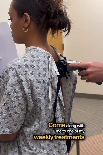
One of the hardest parts of living with an invisible illness is knowing something is wrong in your body while the outside world may not be able to see it.

For people living with Postural Orthostatic Tachycardia Syndrome, commonly known as POTS, or other forms of dysautonomia, this struggle can feel especially frustrating. The symptoms can be intense. The fatigue can be crushing. The heart palpitations, dizziness, brain fog, panic-like sensations, temperature changes, and exhaustion can affect nearly every part of daily life.

And yet, for many people, the search for answers is not simple.

Former Bachelorette Michelle Young has recently brought more public awareness to this experience by sharing her personal journey with dysautonomia and POTS. Her story resonates with so many people because it reflects something patients with invisible symptoms often know well: it can take a long time to understand what is happening inside the body.

Michelle has shared that a number of tests came back normal. At first, she thought her symptoms could have been related to stress or anxiety around her wedding. But the symptoms continued, and over time, she discovered that something much deeper was going on.

That experience is familiar to many patients.

Something feels wrong. The body does not feel like itself. Energy disappears. The nervous system feels overwhelmed. The heart races. The body feels reactive, unstable, or exhausted. But when testing does not clearly explain the pattern, patients can feel stuck between what they are experiencing and what can be objectively shown.

That is where the conversation around POTS, dysautonomia, and invisible illness becomes so important.

When symptoms are real but answers are hard to find

POTS is commonly associated with dysfunction in the autonomic nervous system. The autonomic nervous system controls many of the body’s automatic functions, including heart rate, blood pressure, temperature regulation, digestion, and the body’s response to stress.

When those automatic functions are not regulating properly, patients may experience symptoms that are difficult to predict and hard to explain. One day may look different from the next. A person may look fine on the outside while feeling completely depleted on the inside.

That is one of the most difficult parts of invisible illness.

Patients often know their body is struggling, but they may not have the words, the visuals, or the objective data to explain what is happening. They may hear that their symptoms could be stress, anxiety, hormones, lack of sleep, or simply “normal” test results.

For some patients, that creates a painful cycle.

They feel symptoms. They seek answers. They are told everything looks normal. They begin to question themselves. Then the symptoms continue.

Michelle’s story has struck a chord because she openly describes that kind of search. She has talked about debilitating symptoms, frustration, skepticism, and the need to understand what was happening in her nervous system.

That is why so many people are paying attention.

The emotional weight of invisible illness

Invisible illness carries a unique emotional burden.

When symptoms are not obvious to others, patients may feel like they have to prove that what they are experiencing is real. They may feel misunderstood by friends, family members, coworkers, or even healthcare providers. They may start to explain less because explaining becomes exhausting.

For many people, the hardest part is not only the symptoms themselves. It is the uncertainty.

Why am I so tired?

Why does my heart race?

Why do I feel stuck in fight or flight?

Why do normal activities feel impossible?

Why does my body feel like it cannot adapt?

These are deeply personal questions. They affect confidence, relationships, work, daily routines, and emotional well-being.

This is why Michelle’s comment about validation matters so much. In one of her reels, she shares that seeing how her body is doing in real time is validating when it comes to invisible illness.

That word, validation, is powerful.

Patients with chronic symptoms often do not need someone to exaggerate their experience or make promises. They need to be seen. They need clarity. They need a way to better understand what their body is expressing.

Objective scan visuals can help shift the conversation from, “I feel this,” to, “We can see patterns worth paying attention to.”

Why the autonomic nervous system matters in POTS and dysautonomia conversations

Michelle repeatedly points back to the autonomic nervous system in her story. She talks about dysautonomia, POTS, fight-or-flight, temperature regulation, heart rate, blood pressure, adaptability, and nervous system regulation.

That is important because these are not separate conversations. They are connected through the way the nervous system helps the body respond, adapt, and regulate.

The autonomic nervous system has two primary branches that are often discussed in this context: the sympathetic and parasympathetic systems.

The sympathetic system is commonly associated with fight-or-flight responses. It helps the body activate, respond, and mobilize energy.

The parasympathetic system is commonly associated with rest, recovery, regulation, and repair.

A well-regulated nervous system needs both systems to communicate and adapt appropriately. The goal is not simply to turn one system off and the other on. The goal is adaptability. The body needs the ability to respond when necessary and recover when appropriate.

When people describe feeling stuck in fight or flight, exhausted, reactive, or unable to recover, they are often describing the lived experience of poor adaptability.

That is why objective nervous system data can be such an important part of the conversation.

Why objective data can change the conversation

Michelle has shared that she was skeptical when she first began care because she had already tried many things that were not working. That skepticism is understandable.

Many patients with chronic symptoms have already tried multiple approaches. They have researched. They have changed routines. They have followed recommendations. They have tracked symptoms. They have hoped something would finally create clarity.

When a patient has tried many things without clear answers, words alone may not be enough.

Objective data creates a different kind of conversation.

Instead of beginning with assumptions, the doctor can begin with a baseline. A baseline gives the patient and provider a clearer starting point. It shows measurable patterns that can be reviewed, interpreted, and compared over time.

This is where INSiGHT scanning technology becomes meaningful for nervous system-based chiropractors.

INSiGHT scans do not diagnose POTS or dysautonomia. They provide objective neurological data that chiropractors can interpret in the context of a patient’s history, exam, and clinical picture.

That distinction matters.

The scans are not a replacement for clinical expertise. They support the doctor by creating objective examination data that can help communicate patterns in nervous system performance more clearly.

How INSiGHT scans help visualize nervous system patterns

INSiGHT scanning technology includes three core technologies: neuroPULSE, neuroTHERMAL, and neuroCORE. Each one helps measure a different aspect of nervous system performance.

neuroPULSE

neuroPULSE measures Heart Rate Variability, commonly known as HRV, in a clinical-grade scan. HRV helps provide insight into autonomic balance, autonomic activity, adaptability, and stress response.

For patients who feel stuck in fight or flight, neuroPULSE can help create a clearer conversation around how the body is adapting to stress and how the autonomic nervous system is responding.

This is especially important because HRV is often discussed casually through consumer wearables. neuroPULSE gives chiropractors a clinical-grade way to examine HRV patterns in the context of nervous system performance.

neuroTHERMAL

neuroTHERMAL helps identify temperature regulation and segmental stress patterns along the spine.

Temperature regulation is connected to autonomic nervous system activity, which makes this scan valuable for helping chiropractors visualize patterns that may not be obvious from symptoms alone.

For a patient who feels dysregulated, reactive, overheated, chilled, or inconsistent from day to day, the ability to visualize temperature regulation patterns can support a more objective conversation.

neuroCORE

neuroCORE uses surface electromyography, or sEMG, to measure energy and motor tone reactions. It helps show how muscle activity patterns are expressing through the spine and nervous system.

Many patients describe feeling tense, exhausted, weak, or physically drained. neuroCORE helps doctors communicate how stress and nervous system patterns may be showing up through muscle activity and motor tone reactions.

Together, these three scans help create a more complete picture of nervous system performance.

Why baseline and progress scans matter

One of the most valuable parts of Michelle’s second reel is that she shows the idea of a baseline scan compared to a more recent scan.

That matters because patients with invisible symptoms often want proof of progress.

They want to know whether anything is changing. They want to understand whether their body is adapting differently over time. They want to see more than a vague sense of improvement or decline.

A baseline scan gives the patient and doctor a starting point. A re-scan gives them a way to compare.

That comparison can make the care conversation more meaningful.

Instead of asking only, “How do you feel today?” the doctor can also ask, “What patterns are changing?”

Instead of relying only on memory, the doctor can review objective scan data.

Instead of leaving progress as a guess, the doctor can use re-scan comparisons to communicate change over time.

This is one of the core values of INSiGHT scanning technology. It helps chiropractors measure the baseline, re-scan over time, and communicate patterns of change in a way patients can see and understand.

Finding the right kind of provider

One of the clearest responses to Michelle’s reels is that people want to know where to find doctors who offer INSiGHT scans.

They are asking what kind of doctor does this. They are asking how to find a nervous system-based chiropractor. They are asking whether these scans are available near them.

That response reveals something important.

Patients are not only interested in the story. They are looking for access.

They want a provider who understands the nervous system. They want someone who can help them interpret what is happening beneath the surface. They want objective data that helps make invisible symptoms easier to discuss.

For nervous system-based chiropractors, this is a meaningful opportunity.

When a practice uses INSiGHT scanning technology, it is equipped to have a different kind of conversation. The doctor can show patients objective scan data. The doctor can explain nervous system patterns more clearly. The doctor can track change over time and help patients understand what their scans reveal.

That type of clarity matters deeply to patients who have spent months or years trying to make sense of what they feel.

Patients deserve clarity, not guesswork

POTS and dysautonomia can be deeply challenging conditions to live with. Invisible illness can be lonely, confusing, and exhausting. Patients may feel like they are constantly trying to explain something that others cannot see.

Michelle Young’s story has brought more attention to that reality.

It has also opened a larger conversation about the importance of objective nervous system data.

When patients can see patterns in their nervous system, the conversation changes. They can better understand what their body is expressing. Doctors can communicate with greater clarity. Re-scans can help show how patterns change over time.

For chiropractors, INSiGHT scanning technology provides a way to measure, communicate, and track nervous system performance using objective examination data.

For patients, that can mean something very simple and very powerful:

They can finally see patterns worth paying attention to.

Learn more about INSiGHT scanning technology

INSiGHT scanning technology helps chiropractors gather objective neurological data through neuroPULSE, neuroTHERMAL, and neuroCORE.

These scans help doctors create clearer conversations around stress response, adaptability, temperature regulation, motor tone reactions, and progress over time.

If you are a chiropractor who wants to bring objective nervous system scanning into your practice, contact us to learn more about INSiGHT scanning technology.

If you’re looking for chiropractors in your area that use INSiGHT technology, visit our directory.

Michelle Young, known from The Bachelor and The Bachelorette, is bringing new attention to INSiGHT scanning technology through her personal journey with dysautonomia and Postural Orthostatic Tachycardia Syndrome, commonly known as POTS.

Michelle shares on social media what it has been like to live with debilitating symptoms, searching for answers, and working with a nervous system-based chiropractor who uses INSiGHT scans to measure and track patterns in her nervous system.

Her story is resonating because it touches something many people understand deeply: the frustration of feeling that something invisible is happening inside the body, while searching for clear answers and objective validation.

Why Michelle Young’s Story Resonates With So Many People

Michelle’s story is powerful because it is personal, honest, and highly relatable to people living with chronic symptoms or invisible illness.

She explains that although she was diagnosed with dysautonomia about six months ago, she can trace symptoms back four or five years. Over time, those symptoms became more intense and eventually debilitating. She experienced extreme fatigue, brain fog, panic attacks, heart palpitations, hypoglycemia, and other symptoms tied to POTS.

After her wedding to Jack Leius in 2025, Michelle continued to experience health challenges and later shared that she had been severely exposed to mold and mycotoxins. She has since explained that she developed POTS after her autonomic nervous system “completely crashed.”

Her story has brought the autonomic nervous system and POTS conversation into the national spotlight. She is helping people understand that the nervous system plays a role in many automatic functions of the body, including heart rate, blood pressure, and temperature regulation.

“Dysautonomia is basically an overarching or umbrella term for a range of disorders where there’s a malfunction in the autonomic nervous system.”

In her first reel, she explains that when she began seeing a nervous system-based chiropractor, POTS had “completely taken over” her life. She shares that she was struggling to get out of bed, could hardly work, and was experiencing panic attacks and other symptoms at an all-time high.

She describes her first experience with INSiGHT scans:

“At my very first appointment, I went through a bunch of INSiGHT Scanning, which accurately tests your own central nervous system.”

She shares that her results showed that her body was “very much stuck in fight or flight” and that both her sympathetic and parasympathetic systems were exhausted.

In her second reel, Michelle goes deeper into the three INSiGHT scanning technologies used by her chiropractor (the neuroPULSE, neuroCORE, and neuroTHERMAL). Adding that “Neurological scans that give you a very detailed look on how your nervous system is functioning.”

To Michelle, the real validation comes from seeing what is happening in the body in real time:

“I think it’s pretty cool that they’re able to see how your body is doing in real time, which truthfully is validating at times when it comes to invisible illness.”

For people who feel symptoms deeply but struggle to explain them, objective data can create a bridge between experience and understanding. It gives the doctor and the patient something visual to discuss. It helps create a clearer conversation around patterns, baselines, and progress.

Michelle also highlights the importance of re-scanning over time. She shows an initial baseline scan that indicates her body is in a fight-or-flight pattern, and then compares it to a more recent scan showing that her body is becoming more stable.

That is a central strength of INSiGHT scanning technology. It gives chiropractors objective examination data they can interpret, communicate, and track over time.

What INSiGHT scans measure

INSiGHT scanning technology includes three core technologies: the neuroPULSE, neuroTHERMAL, and neuroCORE. Together, they help chiropractors gather objective neurological data related to nervous system performance.

Each technology measures a different aspect of nervous system function, giving the doctor a broader view of how the body is adapting, responding, and expressing stress patterns.

neuroPULSE

The neuroPULSE is designed to measure Heart Rate Variability, or HRV, in a clinical setting. HRV helps provide insight into autonomic balance, autonomic activity, adaptability, and stress response.

In simple terms, neuroPULSE gives the chiropractor objective data that helps show how the body is adapting to stress and whether patterns may be leaning toward fight-or-flight or recovery states.

neuroTHERMAL

The neuroTHERMAL helps chiropractors assess segmental stress patterns through temperature regulation. Because temperature regulation is influenced by the autonomic nervous system, this scan gives the doctor additional objective data to consider when evaluating nervous system patterns.

This can be especially meaningful for patients because it helps visualize patterns that are not always obvious from symptoms alone.

neuroCORE

The neuroCORE helps chiropractors measure energy and motor tone reactions. It provides objective data about muscle activity patterns and how the neuromuscular system is responding.

For the patient, this can make the conversation more visual. Instead of only talking about tension, fatigue, or stress patterns, the chiropractor has scan data that can help explain how those patterns are showing up through the body.

Why objective nervous system data matters

Patients often know what they feel. They may feel exhausted, overwhelmed, tense, foggy, anxious, dysregulated, or stuck in a stress pattern. But feelings alone can be hard to explain. They can also be hard to track over time.

That is where objective nervous system data becomes so valuable.

INSiGHT scans help chiropractors create a baseline. A baseline gives the chiropractor and patient a starting point. It allows the conversation to move from vague symptoms to visible patterns.

Then, with re-scans, the chiropractor can monitor how those patterns change over time. That makes progress easier to communicate because the doctor is not relying only on memory, symptom reporting, or guesswork. The scans provide objective examination data that the chiropractor can interpret in the context of the patient’s history, exam, and clinical picture.

When patients can see their scan results, the conversation becomes clearer. They can better understand what the doctor is observing. They can see why re-scans matter. They can recognize that nervous system performance is not static. It can shift over time, and those shifts can be measured and discussed.

That is the power of objective data. It brings clarity to the exam, meaning to the report of findings, and purpose to the re-scan conversation.

Learn more about INSiGHT scanning technology

INSiGHT scanning technology helps chiropractors measure, communicate, and track nervous system performance through objective examination data.

With neuroPULSE, neuroTHERMAL, and neuroCORE, doctors can create clearer conversations around autonomic patterns, stress response, adaptability, temperature regulation, motor tone reactions, and progress over time.

If you are a chiropractor who wants to bring objective nervous system scanning into your practice, contact our team to learn more about INSiGHT scanning technology. 

If you’re looking for chiropractors in your area that use INSiGHT technology, visit our directory.

The paraspinal region is one of the most important areas a chiropractor can evaluate, and one of the easiest to underestimate. Most patients walk in focused on low back pain, lower back stiffness, or that familiar feeling that they “pulled back” muscles after lifting, twisting, or simply doing too much for too long. But the paraspinal region tells a bigger story. It reveals how the spine is being stabilized, how the body is distributing load, and how the nervous system is adapting when movement becomes less efficient.

That is why the paraspinal region matters so much in chiropractic. This is not just a strip of tissue beside the spine. It is a highly active system of musculature, connective support, and neural relationships that helps the body stay upright, rotate, extend, and protect itself under stress. When the paraspinal muscle system loses coordination, the body often buys stability with stiffness. The result may look like muscle pain, severe pain, or chronic low back pain, but the deeper issue is often function.

For a Neurologically-Focused Chiropractor, the paraspinal region is not simply where symptoms show up. It is where strategy shows up. It is where you can begin to see whether the body is moving with options or moving with protection. And when that strategy becomes measurable through neurological scanning, the whole care conversation gets clearer.

What the paraspinal region is and why it matters in chiropractic

The paraspinal region refers to the tissues running vertically along both sides of the spine, from the cervical region through the thoracic spine and lumbar spine down toward the sacrum. This area sits location relative to each vertebra and spinous process and includes muscles around the spinal column that help guide motion and maintain stability. In chiropractic, that makes the paraspinal region one of the most clinically useful regions in the body.

The paraspinal muscles are a group, not a single structure. In fact, paraspinal muscles are a group of layered support systems that must work together if the spine is going to stay stable and still move well. These paraspinal muscle groups support posture, help control extension and rotation, and protect each intervertebral segment during movement. When this group of muscles works efficiently, the patient moves with confidence. When the system is overworked or poorly coordinated, patients become prone to pain, fatigue, and guarding.

That is why chiropractors pay close attention to the paraspinal region. A tight or tender paraspinal muscle is rarely just a local issue. It may be associated with paraspinal compensation, poor load sharing, or deeper neurological interference. Patients with similar symptoms can present with completely different motor strategies. One patient may hinge well through the lower back. Another may brace through the entire spine. Their symptoms may sound alike, but their nervous system strategy is not.

In day-to-day practice, this is one of the most valuable ideas to remember: the paraspinal region often shows how the body is protecting itself. It gives chiropractors insight into posture, movement tolerance, and the way the spine is being managed under load. That is what makes the assessment of paraspinal function so important.

Anatomy of the paraspinal region chiropractors need to know

The anatomy of the paraspinal is best understood as a layered system. The paraspinal region contains a broad muscle group, deeper segmental stabilizers, connective support, and neural relationships that all contribute to spinal control. The musculature in this area is not there simply to move the body. It helps coordinate the body’s response to gravity, load, and motion.

The best-known layer is the erector spinae. The erector spinae muscle system includes the iliocostalis, longissimus, and spinalis muscle groups. These extensor muscles help extend the spine, maintain upright posture, and manage longer-range trunk support. The erector spinae and lumbar erector spinae are especially important in the lumbar spine, where prolonged loading, poor endurance, or compensation patterns often show up first. In some back pain patients, you may even find a dominant left erector spinae pattern that reflects asymmetrical stabilization.

Below that larger system sits the transversospinalis layer, including the multifidus muscle. The lumbar multifidus and related segmental muscles are essential for fine control around each vertebra and spinous process. These muscles include deep stabilizers that help manage motion between levels rather than simply creating large movement. When the multifidus muscle or right multifidus underperforms, the body often shifts extra demand to the larger lumbar paraspinal muscle system. That can contribute to muscle fatigue, poor muscle strength, and inefficient movement in the lumbar spinal region.

The anatomy of the paraspinal also includes important relationships with nearby structures. The paravertebral and paravertebral muscles work beside the spine to support control and posture. The psoas muscle, abdominal muscles, and other back muscles influence how the paraspinal and psoas muscle systems share load. Muscle fibers in these regions must coordinate with the thoracic spine and upper back just as well as they do with the lower back. When one system loses efficiency, another muscles may try to compensate.

  • Erector spinae: The long extensor system that supports posture and extension.
  • Longissimus and iliocostalis: Key parts of the erector spinae that help manage trunk support and movement.
  • Multifidus muscle: A deep stabilizer that helps control motion at the segmental level.
  • Lumbar multifidus: Especially important in protecting the lumbar spine during load and transition.
  • Paravertebral muscles: Support structures that work with the broader paraspinal musculature.

That is the practical takeaway. The paraspinal muscle is not just a mover. The paraspinal musculature is a control system. It supports the spine, manages force, and helps determine whether the body moves with freedom or with stiffness.

Common paraspinal problems chiropractors see in practice

In practice, the paraspinal region is involved in both acute and chronic presentations. An acute complaint may begin with a muscle strain, an awkward lift, or a sudden disc irritation. The patient may describe severe pain, muscle spasms, or the sense that the lower back “locked up.” In those moments, the paraspinal muscle system is often acting protectively. The body is trying to reduce motion and create safety around the spine.

Over time, those patterns can become more complicated. Acute and chronic presentations often blend together when the original protection strategy never fully resolves. A patient with chronic low back pain may no longer present with obvious spasm, but the changes in the paraspinal region can still be significant. The lumbar paraspinal may remain overactive, the lumbar muscle system may lose endurance, and the body may keep relying on compensation rather than recovery. That is one reason chronic low back pain is so often associated with low back pain patterns that persist even when symptoms fluctuate.

Research has paid more attention to this in recent years. Chiropractors will see discussions of paraspinal muscle morphology, paraspinal muscle atrophy, muscle mass loss, and atrophy in the lumbar paraspinal region. These changes in the paraspinal system may be associated with low back pain, altered control, and long-term movement limitations. In some studies, paraspinal abnormalities and abnormalities in the paraspinal system are described alongside degenerative findings, soft tissue conditions, or intervertebral disc changes. That does not mean every patient follows the same path, but it does mean the paraspinal story is not always a simple one.

You may also see patients with an imbalanced lumbar paraspinal muscle pattern, asymmetry through the thoracic spine, or protective stiffness extending into the cervical paraspinal region. Some presentations are clearly associated with paraspinal overload. Others are associated with low back pain even when imaging does not look dramatic. In both cases, the chiropractor’s job is to ask a more useful question: can this patient stabilize the spine well, or is the body using protection as its only strategy?

  • Acute presentation: Muscle strain, guarding, disc irritation, or a sudden lower back flare-up.
  • Chronic presentation: Endurance loss, compensation, and chronic low back pain patterns.
  • Structural change: Atrophy, paraspinal muscle atrophy, or changes in the paraspinal seen over time.
  • Functional change: Reduced control, altered movement, poor muscle strength, and ongoing protection.

That is why these findings matter in chiropractic. A sore paraspinal muscle may reflect much more than local irritation. It may reveal how the nervous system is managing load, and whether the body still has enough reserve to move well.

How chiropractors assess the paraspinal region with more precision

The best assessment of paraspinal function is layered. It starts with observation. A chiropractor watches how the patient stands, hinges, rotates, extends, and transfers load through the spine. That movement story matters because posture, asymmetry, and guarding often show up before the patient has the language to describe them. This early assessment of paraspinal function is one of the most honest parts of the exam.

Hands-on evaluation still plays an essential role. Palpation, tenderness, tissue tone, and motion findings around each spinous process help build the clinical picture. So does medical history. The history of prior trauma, recurrent flare-ups, training habits, sedentary behavior, and previous disc episodes all influence how the chiropractor interprets the spine and surrounding musculature. This is where clinical reasoning begins to separate local irritation from deeper adaptation.

Still, structure is not the same as function. That is a critical point. MRI can describe tissue. CT scan can describe architecture. Radiology can add detail about a vertebra, spinous alignment, or an intervertebral disc. But an MRI scan does not tell you in real time how the nervous system is organizing the paraspinal musculature under load. It does not tell you whether the body is using the lumbar spine efficiently or bracing through the whole spinal system to reduce pain.

That is why chiropractors must move beyond static description. The assessment of paraspinal function should include what the patient can do, what the tissues reveal, and how the spine behaves under movement demand. When combined with objective neurological scanning, that process becomes far more precise.

Why neurological scanning changes the paraspinal conversation

If chiropractors want the paraspinal region to be more than a conversation about tightness or tenderness, they need objective analysis. This is where INSiGHT scanning technology becomes so useful. It helps make the paraspinal region measurable rather than mysterious. Instead of relying only on what you feel or what the patient says, you can support the analysis of paraspinal function with objective exam data.

The most direct technology here is neuroCORE. neuroCORE uses surface electromyography to evaluate paraspinal muscle activity and reveal patterns of asymmetry, overuse, exhaustion, and inefficient recruitment. This matters because the paraspinal muscle system is one of the clearest expressions of how the motor nervous system is organizing itself. When the erector spinae muscle is carrying too much of the load, when lumbar multifidus support is poor, or when the lumbar paraspinal is compensating for deeper dysfunction, neuroCORE helps make those patterns visible. That improves the assessment of paraspinal activity in a way palpation alone cannot.

Then there is neuroTHERMAL, which adds a segmental stress picture across the spinal column. The paraspinal region is not only a motor story. It is also an autonomic story. neuroTHERMAL helps visualize stress patterns that may be associated with segmental dysfunction and broader neurological distress. neuroPULSE then broadens the picture again by evaluating adaptability and reserve through heart rate variability. This matters when back pain patients are not just dealing with local tension, but with a nervous system that is stuck in protection and has less capacity to recover.

All three technologies work together through INSiGHT neuroTECH and Synapse software. That combination turns complex findings into scan reports the chiropractor can interpret and communicate more clearly. It is important to say this accurately: INSiGHT does not create the care plan. It provides objective exam data and reports. The chiropractor interprets those findings and then designs the care plan. That keeps the clinical decision-making exactly where it belongs, while giving the doctor stronger information to work from.

This is where the paraspinal conversation becomes much more useful in practice. A chiropractor can compare baseline and progress scans, explain why certain treatment options are being used, and support the treatment of paraspinal dysfunction with greater precision. That may include adjustments, progressive exercise, and other treatment options for paraspinal muscle coordination. It may also help explain why over-the-counter pain or pain relievers do not address the deeper functional issue. The goal is not just to reduce pain. The goal is to improve how the spine is being stabilized and how the nervous system is adapting over time.

  • neuroCORE: Helps document paraspinal muscle activity, asymmetry, and motor output patterns.
  • neuroTHERMAL: Helps visualize segmental stress patterns across the spine.
  • neuroPULSE: Helps explain adaptability and resilience when protection patterns persist.
  • Synapse software: Helps translate complex neurological data into clear reports.

In the end, that is why INSiGHT scanning belongs in the conversation. It brings objective data to a region that chiropractors already know matters deeply. It helps connect the paraspinal region back to nervous system performance, and it gives both doctor and patient a clearer way to see progress.

From sore tissue to smarter strategy

The paraspinal region deserves more than a quick note about tightness in the chart. It is one of the clearest places a chiropractor can observe stability, movement strategy, adaptation, and nervous system performance all at once. When the paraspinal muscle system is coordinated, the spine can move with more options. When it is not, the body often turns to stiffness, guarding, and compensation.

That is why this subject matters so much in chiropractic. The paraspinal region sits at the center of posture, load management, and movement quality. It is influenced by the erector spinae, multifidus muscle function, the lumbar paraspinal, the thoracic spine, and even the relationship between the paraspinal and psoas muscle systems. When these systems work well together, the patient has more adaptability. When they do not, the body often substitutes protection for performance.

For the chiropractor, the opportunity is simple but powerful. Evaluate the region carefully. Interpret it functionally. Understand when symptoms are associated with paraspinal compensation rather than just local irritation. And when possible, use neurological scanning to make those patterns visible. The neuroCORE helps document motor tone. The neuroTHERMAL helps visualize stress patterns. The neuroPULSE helps explain reserve and resilience. Together, they bring more clarity to the exam and more certainty to the report.

That is how the paraspinal region becomes more than a site of discomfort. It becomes a measurable part of the bigger neurological story. And when patients can see that story clearly, they are far more likely to understand the value of care and stay engaged in the process.

The Vagus Nerve conversation is everywhere right now. Patients ask about it. Social media talks about how to stimulate the vagus nerve. Wellness influencers promise ways to activate the vagus nerve with deep breathing, humming, cold showers, and supplements. Some of that conversation points in a useful direction. Much of it does not. For chiropractors, the real value of the vagus nerve is not hype. It is what this nerve reveals about adaptation, recovery, and nervous system performance.

The vagus nerve is one of the most important communication pathways in the body. It helps regulate heart rate, digestion, respiration, and many of the automatic processes that let a person shift out of sympathetic overdrive and into a more restorative state. That is why the vagus nerve keeps showing up in conversations about stress and anxiety, gastrointestinal function, inflammation, and heart rate variability. The subject matters. It just needs to be framed correctly.

In chiropractic, the vagus nerve should never be reduced to a trendy reset trick. The better conversation is about the autonomic nervous system, the parasympathetic nervous system, the sympathetic nervous system, and how the body adapts under load. Once you view the vagus nerve through that lens, it stops being a buzzword and starts becoming clinically useful.

What the Vagus Nerve Is and Why It Matters

The vagus nerve is the 10th Cranial nerve, and the nerve is one of 12 Cranial nerves that emerge from the brain rather than the spinal cord. In fact, the vagus nerve is one of the most far-reaching cranial nerves in the body. The vagus nerve is the longest of the 12 Cranial nerves, which is why it is often called the” wandering nerve”. That name fits because the nerve runs from the brainstem, through the foramen magnum,  down through the neck and into the chest and abdomen.

When chiropractors talk about the anatomy of the vagus, we are talking about a long cranial pathway with broad influence. The vagus nerve originates in the brainstem and extends through the chest and abdomen to structures involved in heart rate, lungs and digestive activity, and the digestive tract. This is why the vagus nerve helps regulate so many automatic processes. The nerve is also involved in motor and sensory functions, carrying nerve signals between the brain and body.  Its different divisions are intimately linked to emotional awareness, making it a critical pathway to express our human nature.  

The vagus nerve is one of the central players in the autonomic nervous system. More specifically, it is a major pathway of the parasympathetic nervous system. That means the vagus nerve helps support rest and digest physiology after the body has been in a “fight or flight” state. If the body cannot shift efficiently into recovery, repair, and regulation, patients may show signs of poor adaptability even when their complaints look unrelated on the surface.

For a chiropractor, that matters because the vagus nerve helps explain why people struggle differently under similar stress. One person adapts and recovers. Another stays wound up, depleted, and slow to regulate. The vagus nerve also helps us understand why complaints involving digestion, stress recovery, respiration, and heart rate can belong to the same larger story.

Why chiropractors should care about vagus nerve function

Vagus nerve function matters because it reflects how well the body is shifting between demand and recovery. The vagus nerve helps calm the body, lower your heart rate, and support regulation after stress. When that process is strong, patients often show better reserve and better adaptability. When it is weak or inconsistent, the body may stay locked into protective physiology longer than it should.

That does not mean every complaint is controlled by the vagus nerve. It does mean the vagus nerve has a role in regulating several systems chiropractors care about every day. If your clinical lens is neuro-centric, the vagus nerve deserves attention because it gives context to how the body is performing, not just how the patient is feeling.

How the Vagus Nerve Connects to Adaptability, Digestion, and Heart Rate

The vagus nerve helps the body do more than relax. It helps control digestion, supports bowel and gastrointestinal regulation, influences respiration, and helps coordinate communication between the brain and visceral organs via the vagus nerve. In practical terms, the vagus nerve conversation is really a conversation about adaptability. Can the body calm down after stress? Can it recover after demand? Can it organize resources well enough to support digestion, energy, and repair?

That is why vagal tone has become such a common phrase. In everyday clinical language, vagal tone refers to the influence of the vagus nerve within the autonomic nervous system. Higher vagal tone is usually associated with better recovery and more resilience. Lower vagal tone is often associated with poor recovery, reduced adaptability, and a body that has trouble shifting out of sympathetic overdrive.

One of the most useful ways to understand this is through heart rate variability. Heart rate variability is not simply measuring heart rate. It reflects the beat to beat changes in timing between contractions and gives insight into how adaptable the system may be. A patient can have a normal heart rate and still have reduced reserve. That is why heart rate variability matters so much in conversations about autonomic function and vagus nerve performance.

The vagus nerve also has a strong relationship to digestion. It influences the digestive system, the digestive tract, and several processes taking place in the abdomen. Patients with poor adaptation often describe digestive frustration, inconsistent appetite, bloating, sluggish digestion, or a body that never seems fully settled. Those signs are not proof of a single vagus issue, but they fit the bigger autonomic story.

The vagus nerve and the recovery side of physiology

The parasympathetic nervous system is the recovery side of physiology. It supports repair, restoration, and regulation. It counterbalances the Sympathetic influence on inflammation. The vagus nerve is one of its most important pathways. That is why the vagus nerve is so often discussed in relation to resting heart rate, recovery, sleep quality, and the body’s ability to move from demand into repair.

When people live in long-term neurological distress, the body may show signs of altered adaptability and increased inflammation. Some have an increased heart rate response to mild demand. Some stay tense after the stressor is gone. Some feel tired yet cannot settle. Some have trouble with digestion or recovery after exercise. These patterns do not all come from one source, but the vagus nerve gives chiropractors a meaningful way to talk about regulation instead of isolated symptoms.

Inflammation, immune regulation, and whole-body performance

The vagus nerve also shows up in the research on inflammation in the body and neuroimmune regulation. There is ongoing interest in how the vagus nerve may influence inflammatory signaling and immune homeostasis. That is one reason inflammation, inflammatory balance, and even chronic inflammation are now part of the broader vagus conversation.

Chiropractors should communicate this carefully. We do not need to overpromise. But it is reasonable to say the vagus nerve has a role in regulating more than mood or relaxation. It appears connected to broader adaptive functions, including gastrointestinal and inflammatory processes. That is one reason the subject matters for overall health and well-being, even though our focus remains on objective analysis and nervous system performance.

  • Heart rate and resting heart rate regulation
  • Heart rate variability and adaptive reserve
  • Digestion, bowel function, and gastrointestinal performance
  • Respiration and diaphragm driven calming responses
  • Inflammation, inflammatory signaling, and overall health

Vagus Nerve Stimulation and the Difference Between Medical VNS and Chiropractic

This is where chiropractors need to be precise. Vagus nerve stimulation is a real medical intervention. VNS is not a catchy wellness phrase when used in medicine. It refers to devices that stimulate the vagus nerve with electrical impulses or electrical stimulation in a therapeutic setting. In many cases, that means an implant or device implanted near the upper left side or left side of the chest, with impulses to the vagus nerve delivered through a lead system.

The left vagus nerve is commonly used in implant-based systems, which is why left vagus nerve anatomy is frequently discussed in the literature. The right vagus nerve is important anatomically too, but these details matter because medical VNS is highly specific. This is not the same as telling a patient to do breathing drills. It is a defined therapy used to treat certain conditions, including epilepsy and treatment-resistant depression. Vagus nerve stimulation may help in selected cases of migraine and cluster headaches as well, and research is ongoing in Alzheimer’s and other areas.

That distinction matters. Chiropractic should never imply that an adjustment is the same as an implant delivering electrical impulses to the vagus. A branch of the vagus nerve may be targeted in some noninvasive devices that stimulate peripheral pathways, but that still belongs to a separate medical conversation. Our job is different. We are looking at adaptability, regulation, and what objective findings reveal about the patient’s current nervous system status.

There are also many non-medical conversations around how to stimulate the vagus nerve. Deep breathing, slow inhale and exhale patterns, humming, singing, gargling, and exposure to cold are commonly discussed. Some of these may activate the vagus nerve or support vagus nerve function in a limited way. They may help calm heart rate, influence the diaphragm, or create a brief shift toward parasympathetic activity. But popular content often jumps too quickly from “this may stimulate” to “this fixes your vagus nerve.”

What the Vagus Nerve Means in a Chiropractic Framework

For chiropractors, the vagus nerve is not a stand-alone analysis. It is a doorway into a smarter discussion about function. Patients often arrive describing stress and anxiety, digestive strain, tension, poor recovery, unstable energy, or a body that cannot settle. The vagus nerve helps frame those experiences within the language of the autonomic nervous system and the central nervous system rather than reducing everything to isolated complaints.

That is important because symptoms alone are not enough. A patient may have digestive frustration, but the deeper question is whether the system is adapting well. A patient may have fatigue, but the better question is whether their regulatory reserve is depleted. A patient may struggle with recovery, but the more useful question is whether the body can organize itself efficiently after demand. The vagus nerve helps us ask better questions.

It also helps chiropractors communicate more clearly. Patients have heard of the vagus nerve. They may not know the anatomy of the vagus or understand that the vagus nerve is one major cranial pathway arising from the brainstem and traveling into the chest and abdomen, but they know it has something to do with calming down. That gives us a bridge. We can start with what they know and lead them into a more complete understanding of nervous system performance.

That is the real opportunity in the chiropractic profession. The vagus nerve also gives us a way to move the conversation beyond symptoms and into adaptation. Instead of talking only about how the patient feels, we can talk about what the body is doing, how nerve fibers are coordinating signals, and whether the system is showing resilience or depletion. That makes the conversation more meaningful and more clinically responsible.

How INSiGHT scanning technology makes this practical

This is where INSiGHT scanning technology matters. The vagus nerve is important, but chiropractors need more than theory. They need objective analysis. INSiGHT scanning technology does not diagnose a vagus lesion. What it does is help the chiropractor analyze the bigger autonomic and neurological picture that vagus conversations are pointing toward.

The INSiGHT neuroPULSE HRV gives a practical window into global autonomic balance and function. Since heart rate variability reflects the interplay between sympathetic and parasympathetic activity, it belongs directly in this conversation. When you are discussing vagal influence, adaptability, and recovery, HRV data becomes incredibly useful. It helps doctors move beyond vague impressions and into measurable patterns.

The INSiGHT neuroTHERMAL helps analyze autonomic trends along the spine, while the INSiGHT neuroCORE helps analyze postural tension and muscular energy output. Together, these technologies create a neuroTECH suite of analytical technologies which help the doctor see how the nervous system is adapting under load. Then the INSiGHT Synapse software translates those findings into scan views and reports that patients can understand. That is the strength of INSiGHT neuroTECH and Synapse software. It takes a subject that sounds abstract and makes it visible.

Once that happens, the vagus nerve conversation feels more complete. It becomes less about trendy phrases and more about objective exam data, clearer communication, and a more informed care plan. That is where chiropractic brings real value. We are not here to mimic medical VNS. We are here to analyze nervous system performance, explain what we find, and show patients proof your care is making a difference over time.

When the Vagus Nerve Stops Being a Buzzword

The vagus nerve deserves attention, but it deserves better attention than it usually gets. This nerve is one of 12 major Cranial pathways, the nerve is one of 12 structures that help connect the brain to the body, and the vagus nerve is the longest of them all. It matters because it touches recovery, digestion, heart rate, respiration, immune signaling, and adaptation. The vagus nerve helps tell the story of how well the body is regulating under load.

For chiropractors, that is where the subject becomes powerful. We do not have to turn the vagus nerve into a gimmick. We can use it as a doorway into a deeper conversation about the nervous system, adaptability, and objective assessment. When you explain the vagus nerve in that context, patients stop chasing trendy advice and start understanding function.

That is the goal. The better the doctor understands the vagus nerve, the better the conversation becomes. And when that conversation is supported by INSiGHT scanning technology, the invisible becomes visible. Patients see more clearly. Doctors communicate more clearly. And the care plan becomes grounded in objective findings, not guesswork.

+