Let me tell you something I’ve learned over the years: most people want to stick with chiropractic. They just don’t know what they’re sticking with.

Patients come in because of symptoms, but if you don’t provide a clear plan for what comes next, they’ll often leave as soon as those symptoms improve.

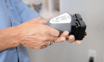
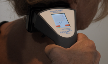
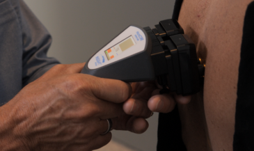
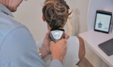
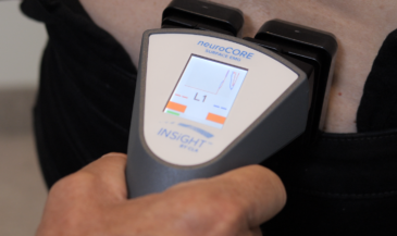
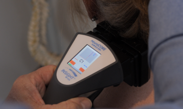
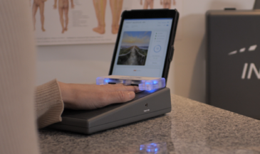
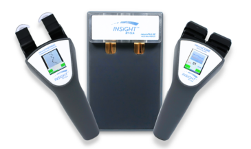
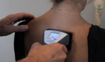
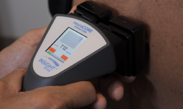
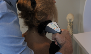
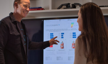
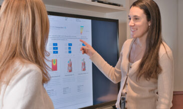
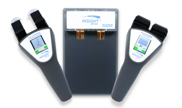
That’s why I developed a framework called “The Rule of 12.” It gives structure to your care plans and a practical, measurable way to keep patients engaged—not just for 12 visits, but for 12 weeks, 12 months, and beyond. When paired with INSiGHT scanning technology and the CORESCORE neurological report card, this framework turns early trust into long-term commitment.

When you give people a plan—and you show them that their nervous system is changing—you build the kind of trust that keeps families under care for years.

And you don’t need a complicated protocol to make it happen. You need a plan with rhythm. A system that aligns with the science of neurological change… and one that makes sense to real folks.

That’s what the Rule of 12 does.

Care Planning Needs Structure Patients Can Understand

Retention isn’t just a business metric—it’s the heartbeat of a thriving chiropractic practice. When patients commit to ongoing care, it means they’ve moved beyond reacting to symptoms and into a mindset of sustained neurological optimization. But commitment doesn’t happen by accident. It happens when your patients are guided—not just adjusted.

Most patient drop-offs don’t occur because care isn’t working. They happen because patients aren’t clear on what they’re committing to—or why it matters.

The Rule of 12 solves that.

By establishing a predictable sequence—12 initial visits with progress and comparative re-exams built into the first 12 weeks, expanding into 12 months of ongoing restorative   care—you give patients a simple, digestible roadmap for their healing journey. It’s the classic answer to the question: How do you eat an elephant? Answer: One bite at a time.

Rather than overwhelming new patients with an open-ended commitment, the Rule of 12 gives them a first milestone they can wrap their minds around. That initial 12-visit segment becomes the critical foundation to observe how well their nervous system is adapting under care.

This structure removes confusion. It shows patients what they’re committing to—and why it matters.

Click here to watch our Rule Of 12 webinar.

The First 12: Building the Foundation

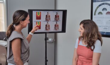
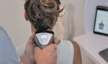
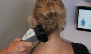
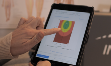
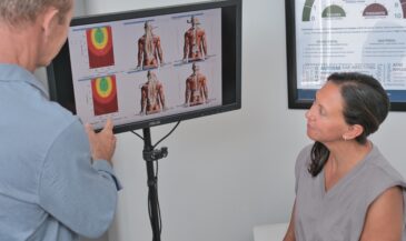
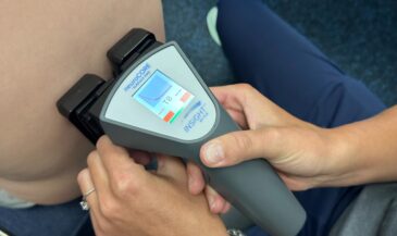
The first 12 visits serve a dual purpose: they initiate neurological changes and set the rhythm for future care and they typically address the issues that brought the patient to your office. By the end of this initial sequence, you’re ready to rescan and re-evaluate. In the Rule of 12, re-exams are the critical foundation for shifting the outcome conversation away from symptoms towards function and performance. These re-exams become the road signs on a map of their future neuro-spinal health. This first progress scan isn’t just for you—it’s a visual and objective confirmation for the patient that care is making a measurable difference.

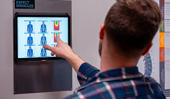
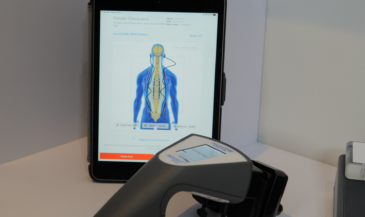
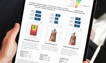
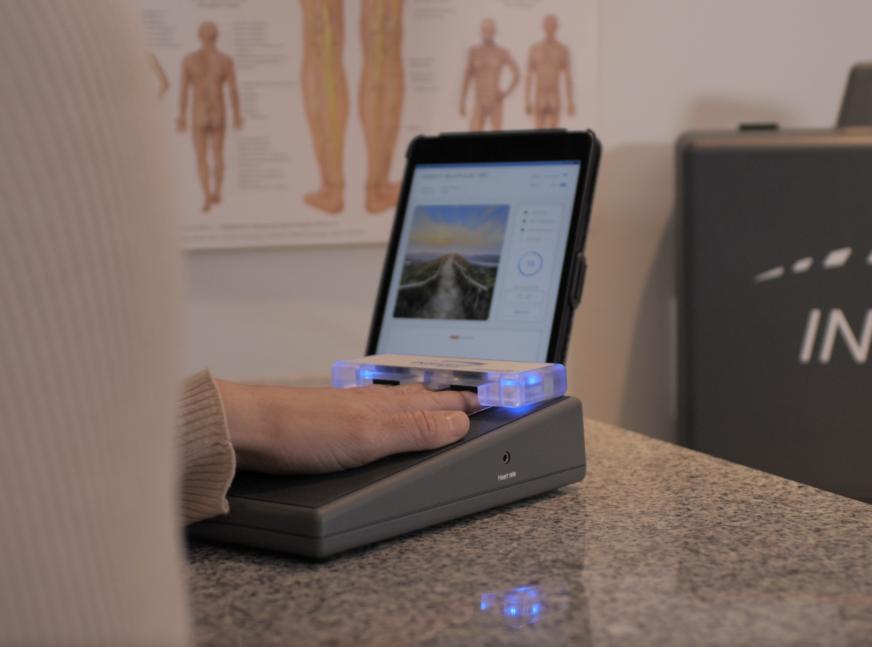
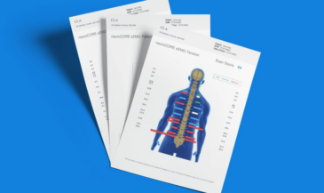
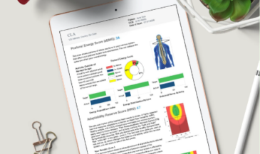
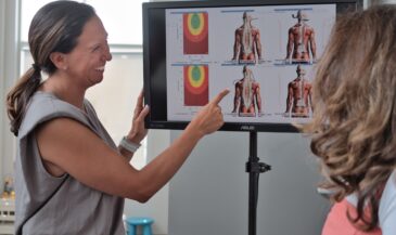
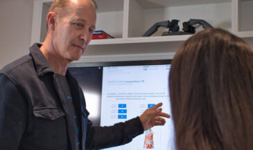
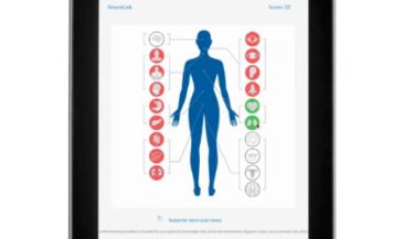
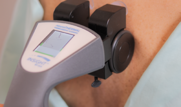
At this stage, you’ll deliver an updated CORESCORE report, which combines data from the neuroTHERMAL, neuroCORE, and neuroPULSE instruments to provide a snapshot of nervous system performance. This Progress Report describes the improvements and changes that you can have analyze=d.  In most cases, the deeper embedded habits within the nervous system have to be unwound before new and healthier patterns can replace them. This is where the CORESCORE shines—it helps the patient see the change, and that visibility confirms their path and creates momentum.

The Next 12: Establishing New Patterns

After the first 12 visits, you expand the lens toward a 12-week outcome. Why 12 weeks?

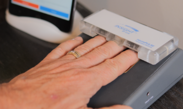
Because neuroplasticity takes time. The nervous system doesn’t just respond to one adjustment—it reprograms through consistency and repetition. In these first 12 weeks, you start to see the trends. You’ll be scanning every 12 visits, building a portfolio of change for each and every patient. From Progress Exams to Comparative exams, you’ll begin to track patterns deep within the nervous system. You’ll be watching neuroplasticity in living color!. And with each scan, you have another opportunity to connect the dots between what the patient is feeling—and what their nervous system is actually doing.

Whether you’re adjusting 3x/week or 4x/month, the key is this: every 12 visits includes a scan. Every scan includes a report. Every report is a chance to reinforce the value of care and deepen the patient’s trust.

The Long Game: 12 Months and Beyond

Once neurological adaptability starts trending upward and the patient has experienced care across 12 visits and 12 weeks, you now have the context to map out their performance-based future.

Twelve months becomes the new anchor. This isn’t just about maintenance—it’s about performance and potential elevation. You’re helping the patient stabilize their system, optimize their adaptability, and expand their capacity for life.

And you’re doing it with data—not guesswork. Each 12-visit checkpoint provides new insights, new conversations, and new opportunities to guide the patient into a lifestyle built around a well-adjusted nervous system.

Some may go on for 12 years. But it all starts with the first 12.

Scans That Motivate. Reports That Retain.

What makes this framework so effective is how it integrates scanning into care. You’re not just making clinical decisions—you’re showing patients their transformation.

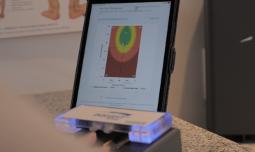
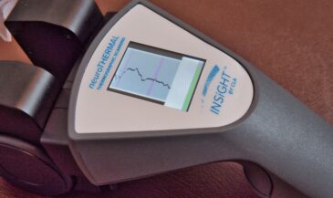
You’ll see HRV rise into the Green Zone, signaling recovery and reorganizational capacity. . You’ll watch sEMG go from high-tension patterns to calm postural balance. You’ll see thermal scans release older patterns and continue tostabilize and normalize. And every time, you’ll deliver a new CORESCORE report that shows how far they’ve come—and what’s possible next.

This is what builds retention: visible change, explained clearly, anchored to a care plan with structure.

Why the Rule of 12 Works

Let’s recap what makes this care planning method so powerful:

  • It’s simple: Patients can grasp the idea of 12 visits. It’s not abstract. It’s not indefinite.


  • It’s strategic: Each cycle corresponds to neurological adaptation timelines and meaningful scan intervals. Neuroplastic change requires time, consistency, and checkpoints.


  • It’s measurable: You’re integrating INSiGHT scans at every checkpoint to track real changes in adaptability, posture, and autonomic regulation.


  • It gives structure to your care plans: Each phase—12 visits, 12 weeks, 12 months—builds logically on the one before it.


  • It builds retention by creating rhythm: Every 12 visits comes with a scan, a conversation, and a renewed plan.


  • It keeps patients motivated with data: Each CORESCORE update becomes a progress milestone.


  • It’s simple to communicate: Patients can easily understand and commit to this approach.

The Rule of 12 Creates Lifelong Patients

The more neurological scans I’ve seen—the more I’ve realized just how powerful an organized care plan, basd on objective data little structure can be. The Rule of 12 gives you something simple, natural, and neurologically sound to build your care plans around. It helps your patients feel like they’re on a journey with clear roadsignscheckpoints, not just showing up for another visit. And when you combinepair that rhythm with INSiGHT scans and the CORESCORE, well… now you’ve got something people can see and understand. believe in.

When patients understand their care plan—and see their progress along the way—they don’t quit. They keep showing up. They bring their family. They refer their friends. And they begin to see chiropractic not as a quick fix, but as a long-term strategy for nervous system performance.

That’s the power of the Rule of 12.

It helps you deliver care with clarity. It gives patients a roadmap they can follow. And it turns every INSiGHT scan into an opportunity to re-engage, re-educate, and re-commit.

So if you’re looking to boost retention, simplify your care planning, and create more “aha!” moments with your patients, I can’t recommend the Rule of 12 enough. It’s not just good for business—it’s good for the nervous system. And when the nervous system’s happy, everything else starts working better too. Keep scanning, keep leading, and keep showing your community what true, measurable health looks like—one 12-visit step at a time.

Want even more examples of how you can implement the Rule of 12 in your practice? Click here to watch our Rule Of 12 webinar.

If now is the right time for you to explore the advantages of INSiGHT scanning while applying the Rule of 12, get in touch with an INSiGHT advisor. They can walk you through the steps of integrating INSiGHT scanning into your practice, so you can begin to share the power of chiropractic care using objective data and compelling reports.  Click here to book a no-obligation call.  

Get Started with INSiGHT Scanning

Take our Free Practice Strategy Assessment. A Personalized Guide and Expert Strategy Call to Help Determine How Scanning will Help you Grow
ABOUT THE AUTHOR

Dr. David Fletcher is actively involved in all aspects of innovation teaching and research connected to the INSiGHT™ scanning technologies. He is widely recognized for his ability to share his expertise in compelling and easy to understand ways.

Dr David is a renowned chiropractor who practiced for many years with his associates in a scan-centric thriving principled family-based practice in Toronto. He is a sought-after teacher mentor and keynote speaker who takes every opportunity to share the wisdom and the power of chiropractic as it is meant to be.

https://insightcla.com/wp-content/uploads/2022/07/david-1.png
Dr. David Fletcher
DC FRCCSS(C) – Founder & CEO CLA Inc.
LIKE THIS ARTICLE? HELP US SPREAD THE WORD

Be Seen. Be Heard. Be Certain

In today’s saturated healthcare market, chiropractors face an ongoing challenge: being visible. Discover key marketing tactics to ensure you can BE SEEN, BE HEARD, and BE CERTAIN in your practice!

Read these Next…

Get Informed

Join 23,121 other Chiropractors and receive topics covering day to day challenges of running your practice.

As a chiropractor, you’ve seen it countless times—patients walking in with debilitating headaches that have plagued them for months or even years. They’ve tried everything. Multiple medications. Lifestyle changes. Countless doctor visits. Yet the pain persists, radiating from the base of the skull upward, sometimes behind the eyes, creating a life of constant discomfort and frustration.

Here’s what most practitioners miss: About 25% of patients with a headache complaint have occipital neuralgia, not the migraines they’ve been diagnosed with. That’s one in four people receiving the wrong diagnosis and ineffective treatment.

Let’s be real—traditional treatment options for these headaches often focus exclusively on symptom management rather than addressing the underlying neurological dysfunction. Patients get trapped in an endless cycle of pain medications that mask symptoms temporarily while the root cause continues to worsen.

Occipital neuralgia—a condition characterized by inflammation or injury to the occipital nerves that run from the top of the spinal cord through the scalp—requires a fundamentally different approach than migraine treatment. This is where a neurologically focused chiropractic assessment transforms the game.

Understanding the Neurological Basis of Occipital Migraines

Occipital neuralgia is fundamentally a nervous system disorder, not just a pain syndrome. The greater and lesser occipital nerves emerge from between the C2 and C3 spinal nerves in the upper neck, weaving through muscles at the back of the head before branching throughout the scalp—sometimes reaching as far forward as the forehead.

These nerves serve as critical communication pathways, carrying sensory and motor information to the skin on your scalp. When functioning properly, this system works seamlessly. But when these nerves become irritated, compressed, or inflamed anywhere along their path, the result is the characteristic shooting, electric-like pain that defines occipital neuralgia.

Many practitioners confuse occipital neuralgia with migraines due to overlapping symptoms, but the distinctions are crucial for effective care:

  • Pain pattern: Occipital neuralgia typically begins at the base of the skull and radiates upward, while migraines often start frontally or behind the eyes
  • Duration: Occipital neuralgia pain is often brief and piercing—lasting seconds to minutes—compared to migraines that can persist for hours or days
  • Triggers: Simple movements like turning the head or brushing hair can trigger occipital neuralgia
  • Autonomic symptoms: Migraines commonly feature nausea and sensitivity to light, which are typically absent in pure occipital neuralgia

What makes occipital neuralgia particularly challenging is its connection to the broader neurological system. Subluxation in the upper cervical spine disrupts proper nerve function, which can create an effect that extends far beyond the immediate area of pain.

Causes and Symptoms of Occipital Neuralgia

Recognizing occipital neuralgia requires understanding its characteristic presentation. Unlike many headache disorders that develop gradually, occipital neuralgia often announces itself with sudden, intense pain that patients describe as shocking or electric.

The symptoms of occipital neuralgia patterns typically include:

  • Sharp, shooting pain that feels like an electric shock in the back of the head and neck
  • Piercing, throbbing, or burning sensations that start at the base of the skull and radiate upward
  • Unilateral or bilateral presentation affecting one or both sides of the head
  • Pain behind the eye on the affected side
  • Extreme scalp sensitivity, making even light touch, hair brushing, or lying on a pillow unbearable
  • Pain triggered by specific movements like turning the neck
  • Brief duration of intense episodes typically lasting seconds to minutes

The primary causes of occipital neuralgia center around irritation of the occipital nerves or muscle tension. Upper cervical subluxation is a critical factor that’s frequently overlooked. When vertebral misalignment occurs in the upper neck, it creates abnormal tension and fixation that can impact the occipital nerves as they exit the spine.

Beyond subluxation, several other factors can trigger or worsen occipital neuralgia:

  • Tight muscles in the posterior neck entrapping the nerves
  • Physical trauma to the head or neck
  • Degenerative disk disease in the cervical spine
  • Osteoarthritis affecting the upper spine
  • Poor posture creating chronic muscle tension

Certain systemic conditions can also contribute to occipital neuralgia, including diabetes, gout, blood vessel inflammation (vasculitis), and chronic infections.

Traditional Diagnosis vs. Neurological Assessment

Diagnosing occipital neuralgia has traditionally been a challenging process fraught with uncertainty. The conventional approach relies heavily on clinical examination, patient history, and the process of elimination. Most practitioners simply press on the occipital region to reproduce pain and then make a determination based on subjective reporting.

This traditional approach misses the underlying neurological dysfunction that’s actually causing the condition. When a patient describes head pain, the default diagnosis often becomes “migraine” without proper investigation of the occipital nerves and their function.

This is where nervous system-focused assessment transforms the diagnostic landscape. Rather than relying solely on subjective reporting, we can now objectively measure the function of the nervous system and identify specific patterns of dysfunction. This approach provides concrete, measurable data that guides precision care.

The INSiGHT scanning technology suite brings three critical assessment tools:

  • neuroCORE (Surface EMG) measures paraspinal muscle activity, detecting the dysponesis (wasted energy) and abnormal tension patterns that often accompany occipital neuralgia.
  • neuroPULSE (HRV) evaluates autonomic nervous system function through Heart Rate Variability assessment. This tells us how well your patient’s body is adapting to stress and reveals whether they’re stuck in a sympathetic dominant state—a common finding in chronic pain conditions.
  • neuroTHERMAL measures skin temperature patterns along the spine, detecting autonomic imbalances that affect blood vessel regulation. Temperature asymmetries can reveal areas of subluxation and nerve interference that directly impact the occipital nerves.

When combined, these three technologies create a comprehensive neural profile that reveals far more than what’s possible through traditional assessment alone. The resulting CORESCORE provides a single numerical representation of neural efficiency, allowing you to track progress objectively.

The Power of a Nervous System-Focused Approach

When you shift from symptom management to addressing the root neurological dysfunction in occipital neuralgia, everything changes. The focus moves from masking pain to restoring proper nervous system function—creating lasting results that transform your patients’ lives.

The nervous system-focused approach delivers several significant advantages:

First, it addresses the actual source of the problem, unlike pain medications or nerve blocks that temporarily deaden sensation, correcting subluxation and neurological dysfunction treats the underlying cause of occipital neuralgia.

Second, this approach provides objective documentation of both the condition and improvement. The INSiGHT scans create baseline measurements that allow you to track progress with precision.

For your patients, the benefits are profound:

  • Reduced reliance on medication as nervous system function improves
  • More accurate diagnosis that distinguishes occipital neuralgia from other headache types
  • Targeted care plans specific to their unique pattern of dysfunction
  • Visible progress tracking that reinforces commitment to care

For a practitioner, the neurologically-focused approach transforms how you practice:

  • Enhanced clinical certainty through objective measurements
  • Improved patient communication using visual scan results
  • Better care planning based on specific neurological findings
  • Increased patient retention as patients see measurable improvement

Implementing Neurologically-Focused Care for Occipital Migraines

Transforming your approach to occipital headaches begins with the implementation of a comprehensive assessment and care protocol:

Initial Assessment Protocol

Start with a complete neural profile using the INSiGHT scanning suite, followed by a targeted examination of the occipital and cervical regions, including palpation for subluxation, range of motion assessment, and specific orthopedic tests.

Precision Care Approach

Based on your assessment findings, develop a care plan that addresses the specific patterns of subluxation and neurological dysfunction identified:

  • Specific adjustments targeting the upper cervical spine where the occipital nerves emerge
  • Focused attention to transitional areas that often contribute to referred pain
  • Addressing any compensatory patterns revealed by the neuroCORE scan
  • Soft tissue techniques for hypertonic muscles that may be compressing the occipital nerves
  • Patient education regarding posture, ergonomics, and stress management

The key is individualization—each patient’s pattern of dysfunction is unique, and your care approach should reflect their specific neurological findings.

Progress Monitoring Framework

Regular re-assessment is critical for tracking progress:

  • Perform progress scans after 12 visits (following the “Rule of 12” approach)
  • Compare objective measurements from initial to progress scans to quantify the improvement
  • Track symptom changes, including frequency, intensity, and duration of occipital neuralgia episodes
  • Use the CORESCORE as a metric to demonstrate overall neurological improvement. Consider it to be their “neurological report card.”

This systematic monitoring creates accountability and provides the objective evidence needed to demonstrate that your care is creating real neurological change.

Your Pathway to Neurological Excellence

Occipital neuralgia represents an opportunity to demonstrate the profound impact of neurologically-focused chiropractic care. By addressing the root dysfunction rather than just managing symptoms, you can transform your approach to these challenging cases.

The journey begins with upgrading your assessment protocols to include objective neurological measurements. INSiGHT scanning technology provides the data you need to identify specific patterns of dysfunction, create targeted care plans, and track progress with precision.

Remember that every patient with occipital neuralgia represents not just a clinical challenge, but an opportunity to demonstrate the power of addressing the nervous system first. When you restore proper neurological function, the body’s innate healing capacity can express itself fully, often resolving conditions that have persisted despite years of symptomatic treatments.

At CLA, we’re committed to supporting your journey toward neurological excellence. The INSiGHT neuroTECH suite makes the invisible visible, transforming how you assess and care for these complex cases. Together, we can transform not just how occipital neuralgia is cared for, but how patients understand the central role of neurological function in their overall health.

Now that’s something to get excited about!

SOURCES

Harvard Health. (2024). Occipital neuralgia: Symptoms and treatments. https://www.health.harvard.edu/pain/occipital-neuralgia-symptoms-and-treatments

PubMed Central. (2021). Prevalence of Occipital Neuralgia at a Community Hospital-based Headache Clinic. https://pmc.ncbi.nlm.nih.gov/articles/PMC8101323/

National Center for Biotechnology Information. (2022). Anatomy, Head and Neck, Occipital Nerves. https://www.ncbi.nlm.nih.gov/books/NBK542213/

Kenhub. (2023). Occipital nerves. https://www.kenhub.com/en/library/anatomy/occipital-nerves

American Association of Neurological Surgeons (AANS). (2024). Occipital neuralgia. https://www.aans.org/patients/conditions-treatments/occipital-neuralgia/

WebMD. (2024). Occipital Neuralgia: What Is It?. https://www.webmd.com/migraines-headaches/occipital-neuralgia-symptoms-causes-treatments

Get Started with INSiGHT Scanning

Take our Free Practice Strategy Assessment. A Personalized Guide and Expert Strategy Call to Help Determine How Scanning will Help you Grow
ABOUT THE AUTHOR

Dr. David Fletcher is actively involved in all aspects of innovation teaching and research connected to the INSiGHT™ scanning technologies. He is widely recognized for his ability to share his expertise in compelling and easy to understand ways.

Dr David is a renowned chiropractor who practiced for many years with his associates in a scan-centric thriving principled family-based practice in Toronto. He is a sought-after teacher mentor and keynote speaker who takes every opportunity to share the wisdom and the power of chiropractic as it is meant to be.

https://insightcla.com/wp-content/uploads/2022/07/david-1.png
Dr. David Fletcher
DC FRCCSS(C) – Founder & CEO CLA Inc.
LIKE THIS ARTICLE? HELP US SPREAD THE WORD

Be Seen. Be Heard. Be Certain

In today’s saturated healthcare market, chiropractors face an ongoing challenge: being visible. Discover key marketing tactics to ensure you can BE SEEN, BE HEARD, and BE CERTAIN in your practice!

Read these Next…

Get Informed

Join 23,121 other Chiropractors and receive topics covering day to day challenges of running your practice

You know those moments when a patient describes feeling lightheaded, maybe getting a bit sweaty, and then suddenly needing to sit down? These experiences offer us an incredible opportunity to demonstrate the power of neurologically-focused assessment and care. 

When a patient experiences a vasovagal episode, it opens the door to a deeper conversation about their nervous system function and adaptive capacity.

The reality is striking—research has found that approximately 1 million Americans experience a syncope episode every year, with more than 80% of episodes of syncope being vasovagal. Yet most conventional approaches focus solely on the cardiovascular aspects, overlooking the critical neurological component that’s at the heart of these episodes.

Let’s be real—most practitioners are taught to view vasovagal episodes as simply “the common faint” without appreciating how they reflect information about a patient’s autonomic nervous system function and adaptive capacity. When we reduce these episodes to merely a cardiovascular event, we miss the opportunity to address the underlying neurological dysregulation.

Vasovagal episodes represent a window into autonomic nervous system function—specifically, how well the sympathetic and parasympathetic systems are balanced. When we understand these episodes through the lens of neurological function, we can move beyond simply managing episodes to improving the patient’s overall nervous system regulation and resilience.

The Neurophysiology of Vasovagal Episodes

Vasovagal syncope is fundamentally a neurological event masquerading as a cardiovascular problem. It represents the most common form of reflex syncope, accounting for over 85% of syncopal events in people younger than 40 years. At its core, it results from a temporary failure in the autonomic nervous system’s regulation, a drop in blood pressure, and heart rate, leading to reduced cerebral perfusion and loss of consciousness.

The key to understanding these episodes lies in recognizing them as a reflex arc with both afferent and efferent limbs. When triggered, the afferent limb sends signals to the central nervous system that initiate neurological events. The central nervous system then responds through the efferent limb with two critical actions: increased parasympathetic activity and heightened vagal tone, combined with decreased sympathetic activity that reduces vascular tone. This neurological double-hit dramatically reduces cardiac output and blood pressure, causing insufficient blood flow to the brain.

In patients prone to vasovagal episodes, we often see evidence of autonomic dysregulation that creates vulnerability. This dysregulation can manifest as:

  • Exaggerated parasympathetic responses to certain triggers
  • Inadequate sympathetic tone to maintain blood pressure during challenges
  • Poor coordination between the two branches of the autonomic system
  • Reduced overall adaptive capacity or “autonomic reserve”

Technical Breakdown of Vasovagal Episodes

To fully appreciate vasovagal episodes from a neurological perspective, we need to examine the distinct phases and components that make up these events.

The Three Phases of a Vasovagal Episode

  • Trigger Phase: The cascade begins with a trigger that activates the afferent limb of the vasovagal reflex. This trigger, combined with central hypovolemia, activates mechanoreceptors in the ventricle that signal through vagal afferents to the central nervous system.
  • Physiological Response Phase: Once triggered, the efferent limb produces a two-pronged neurological response: increased vagal firing causes a significant decrease in heart rate, while decreased sympathetic activity leads to reduced vascular tone. This drops mean arterial pressure below the threshold where cerebral autoregulation can maintain adequate blood flow.
  • Recovery Phase: When the patient falls or is placed supine, the increased circulating blood volume from the lower extremities combined with the reduced gravitational challenge allows rapid restoration of blood flow to the brain and recovery of consciousness.

Common Triggers Categorized by Neurological Impact

Triggers for vasovagal episodes vary widely but can be categorized based on how they impact the nervous system:

Emotional/Psychological Triggers:

  • Fear, anxiety, or intense emotional stress
  • The sight of blood, needles, or injury

Physical/Postural Triggers:

  • Prolonged standing, especially in warm environments
  • Sudden positional changes
  • Dehydration or reduced blood volume

Situational Triggers:

  • Intense pain
  • Coughing, sneezing, or straining
  • Urination (particularly in men)

Prodromal Symptoms and Autonomic Signaling

Most patients experience warning signs before losing consciousness—a critical window for intervention:

  • Lightheadedness and dizziness
  • Nausea
  • Pallor and cold, clammy skin 
  • Visual changes like tunnel vision
  • Weakness or fatigue

These reflex syncope symptoms aren’t random—they’re direct manifestations of specific autonomic nervous system changes that precede the full vasovagal episode.

Comparative Analysis: Beyond the Traditional Approach

When it comes to vasovagal attacks, the conventional medical model and a nervous system-focused approach represent fundamentally different paradigms in assessment and management.

Conventional Medical Approach:

  • Focuses primarily on the fainting event itself
  • Relies heavily on patient history and symptom description
  • Uses tests like orthostatic blood pressure and Electrocardiogram (ECG)
  • Aims to exclude dangerous causes rather than understand the underlying function

Nervous System-Focused Approach:

  • Examines the broader context of autonomic nervous system function
  • Utilizes objective measurements of neurological and autonomic balance
  • Employs advanced technologies to assess Heart Rate Variability
  • Aims to understand the patient’s unique autonomic signature and adaptive capacity

The diagnostic tools employed in these two approaches reveal another important contrast:

Conventional Diagnostic Methods:

  • Tilt-table testing primarily induces symptoms rather than explaining them
  • ECG captures only cardiac electrical activity, not autonomic regulation
  • Blood pressure measurements provide limited snapshots rather than regulatory patterns

INSiGHT Assessment Tools:

  • Heart Rate Variability (HRV) analysis via neuroPULSE technology quantifies autonomic balance and activity
  • Surface EMG through neuroCORE assessment reveals patterns of neuromuscular tension affecting posture and movements 
  • Thermographic scanning with neuroTHERMAL identifies dysautonomia patterns
  • Combined metrics create a CORESCORE that provides objective measurement of overall neurological efficiency

Perhaps the most significant difference lies in the management philosophy:

Reactive Symptom Management:

  • Wait for episodes to occur, then respond
  • Focus on avoiding known triggers
  • Medication only for severe cases

Proactive Nervous System Optimization:

  • Regular assessment of autonomic function with INSiGHT scanning technology
  • Targeted interventions to improve autonomic balance before episodes occur
  • Specific care to enhance nervous system resilience and adaptive capacity

Clinical Benefits of Neurological Assessment

Heart Rate Variability (HRV) assessment stands as perhaps the most valuable tool in understanding vasovagal tendencies. The neuroPULSE technology specifically measures what we call the Autonomic Balance Index (ABI) and Autonomic Activity Index (AAI)—key metrics that reveal a patient’s overall adaptive capacity and resilience to stress.

Patients prone to vasovagal episodes often display distinctive patterns in their HRV readings:

  • Low overall HRV (reduced adaptability and resilience)
  • Exaggerated parasympathetic responses to mild stressors
  • Imbalanced sympathetic/parasympathetic ratios, even between episodes

These patterns often appear in what we call the “distressed state” (Zone 3) of the HRV Rainbow Graph—indicating sympathetic dominance with depleted reserves. This state represents a nervous system that lacks the flexibility to appropriately respond to challenges.

The true clinical value of this assessment is its preventative power. By identifying these patterns before an episode occurs, you can initiate care that improves autonomic regulation and builds resilience.

Integrating Neurological Assessment

Start with a comprehensive baseline assessment that includes complete INSiGHT Scanning technology.

The “Rule of 12” approach is particularly effective for patients with vasovagal tendencies:

  • 12 initial visits within a condensed timeframe to engage nervous system retraining
  • Structured re-assessment at regular intervals (every 12 visits) to track progress

Remember that measurable improvements in autonomic function typically occur within a 12-week window of consistent care. Patients may report fewer prodromal symptoms or an increased ability to abort impending episodes before experiencing objective measurement changes.

Transforming Care Through Neurological Insight

The INSiGHT neuroTECH suite provides the missing piece that conventional approaches lack—objective measurement of nervous system function that can identify susceptibility before episodes occur and track improvements that symptom reporting alone might miss.

The most powerful shift occurs when patients move from seeing themselves as “prone to fainting” to understanding themselves as individuals with measurable autonomic patterns that can be optimized. This change in perspective transforms anxiety about unpredictable episodes into confidence in their improving nervous system function.

By bringing the power of neurological assessment to patients with vasovagal tendencies, you’re not just helping them avoid fainting—you’re offering a pathway to enhanced nervous system regulation that benefits their overall health and wellbeing. That’s the true promise of neurologically-focused care: transforming challenges into opportunities for comprehensive health improvement through better nervous system function.


SOURCES


ScienceDirect. (2021). The pathophysiology of vasovagal syncope: Novel insights. https://www.sciencedirect.com/science/article/pii/S1566070221001296

Barrow Neurological Institute. (2020). Syncope. https://www.barrowneuro.org/condition/syncope/

National Center for Biotechnology Information. (2023). Vasovagal Episode. https://www.ncbi.nlm.nih.gov/books/NBK470277/

PubMed Central. (2022). Syncopal reactions in blood donors: Pathophysiology, clinical course, and features. https://pmc.ncbi.nlm.nih.gov/articles/PMC11734782/

Get Started with INSiGHT Scanning

Take our Free Practice Strategy Assessment. A Personalized Guide and Expert Strategy Call to Help Determine How Scanning will Help you Grow
ABOUT THE AUTHOR

Dr. David Fletcher is actively involved in all aspects of innovation teaching and research connected to the INSiGHT™ scanning technologies. He is widely recognized for his ability to share his expertise in compelling and easy to understand ways.

Dr David is a renowned chiropractor who practiced for many years with his associates in a scan-centric thriving principled family-based practice in Toronto. He is a sought-after teacher mentor and keynote speaker who takes every opportunity to share the wisdom and the power of chiropractic as it is meant to be.

https://insightcla.com/wp-content/uploads/2022/07/david-1.png
Dr. David Fletcher
DC FRCCSS(C) – Founder & CEO CLA Inc.
LIKE THIS ARTICLE? HELP US SPREAD THE WORD

Be Seen. Be Heard. Be Certain

In today’s saturated healthcare market, chiropractors face an ongoing challenge: being visible. Discover key marketing tactics to ensure you can BE SEEN, BE HEARD, and BE CERTAIN in your practice!

Read these Next…

Get Informed

Join 23,121 other Chiropractors and receive topics covering day to day challenges of running your practice.

Have you ever had that patient who keeps coming back with the same persistent headaches—the one who’s seen multiple specialists, tried every medication, and still can’t find lasting relief? You’re not alone. 

Many of these chronic headache cases may actually be rooted in overlooked conditions like occipital neuralgia, which affects approximately 3.2% of patients, or cervicogenic headaches involving the greater occipital nerve, found in up to 4.1–20% of cases.

The frustration is real—both for you and your patients.

Most conventional approaches focus exclusively on symptom management, leaving patients caught in an endless cycle of temporary relief followed by recurring pain. Medications mask the problem while completely missing the underlying neurological dysfunction that drives these persistent symptoms.

Here’s the critical insight that’s often overlooked: the greater occipital nerve isn’t just another structure in the neck—it’s a primary communication pathway that reveals the true state of your patient’s neurological function.

This is where neurologically-focused care changes everything.

The Neuroanatomy of the Greater Occipital Nerve

The Greater Occipital Nerve (GON) originates as the medial branch of the dorsal ramus of the C2 spinal nerve, with some contribution from C3. Upon emerging, the nerve follows a complex journey through multiple tissue layers, creating several potential points of compression or irritation.

From its origin, the GON travels between the first and second cervical vertebrae, curving around the inferior oblique muscle of the head. It then pierces the semispinalis capitis muscle—a key point of potential entrapment—before continuing upward to penetrate the trapezius muscle. 

The final portion of its journey involves piercing the aponeurotic attachment where the trapezius and sternocleidomastoid muscles meet near the superior nuchal line, approximately 3 cm below and 1.5 cm lateral to the external occipital protuberance.

This complex path creates multiple potential compression sites:

  • Between the atlas (C1) and axis (C2) vertebrae—where a vertebral subluxation can directly impact nerve function
  • At the inferior oblique muscle—where muscle tension can compress the nerve
  • Through the semispinalis capitis muscle—a potential entrapment point
  • At the trapezius aponeurotic attachment—which has been found to be a major entrapment site
  • Along its superficial course—where it travels with the occipital artery in an intimate relationship

Once past these structures, the GON provides sensory innervation to the posterior scalp up to the vertex, the area above the ears, and the region over the parotid glands. What makes the GON particularly significant is its connection to the trigeminocervical complex—a group of neurons in the C2 dorsal horn that receive convergent input from both cervical and trigeminal nerves. 

Understanding the Function and Vulnerability of the Greater Occipital Nerve

Subluxation plays a pivotal role in GON dysfunction. Remember that subluxation isn’t just about misalignment—it’s a complex neurological dysfunction characterized by altered positioning within the neurospinal system, abnormal tension or fixation, and neurological interference. 

When subluxation affects the upper cervical spine, particularly at the C1-C2 level, it directly impacts the GON and can lead to occipital headaches.

Neurological interference disrupts the balance between nociception (the signaling of pain) and proprioception (the sense of body position). The primary factor that triggers neurological dysfunction is joint fixation and restricted movement, which affects proprioceptive input. Patients suffering from chronic musculoskeletal pain frequently demonstrate deficits in proprioception.

The GON’s intimate connection with the autonomic nervous system explains why patients with GON irritation often experience symptoms beyond simple pain—including dizziness, nausea, and visual disturbances. This could cause a shift toward sympathetic dominance—a state of “fight or flight” that affects everything from blood vessel tone to stress hormones.

Recognizing Greater Occipital Nerve Involvement in Patient Presentations

The classic presentation of occipital neuralgia includes a distinctive pattern of paroxysmal, shooting, or stabbing pain that originates at the base of the skull and radiates upward over the scalp. Patients often describe this pain as “electric,” “zapping,” or “shooting” in nature.

The hallmark signs and symptoms that suggest GON involvement include:

  • Unilateral pain in the posterior scalp that may radiate toward the vertex or behind the eye
  • Hypersensitivity of the scalp, sometimes so severe that patients report being unable to lay on a pillow or wash their hair
  • Tenderness to palpation over the nerve pathway, particularly at the superior nuchal line
  • Pain exacerbated by neck movement, especially extension and rotation
  • Associated symptoms like dizziness, nausea, or visual disturbances due to autonomic involvement
  • Numbness or paresthesia in the affected region
  • Trigger points at specific compression sites along the nerve’s course

What conventional assessment misses is the underlying neurological dysfunction that INSiGHT scanning can reveal.

The Neurological Cascade

When the GON becomes dysfunctional, it initiates a neurological cascade through the trigeminocervical complex, which can lead to central sensitization. This fundamentally alters how the brain processes sensory information, lowering the threshold for pain perception and expanding the receptive field.

This explains why patients with occipital neuralgia often experience:

  • Pain that radiates far beyond the GON’s anatomical distribution
  • Hypersensitivity to stimuli that wouldn’t normally be painful (allodynia)
  • Intensified pain response to normal painful stimuli (hyperalgesia)
  • Expansion of the affected area over time
  • Persistence of symptoms even after the initial irritation has been addressed

The autonomic component is equally significant. GON dysfunction typically shifts patients toward sympathetic dominance—a state of “fight or flight” that affects everything from blood vessel tone to stress hormones. This autonomic imbalance can be objectively measured using the neuroPULSE technology, which often shows decreased Heart Rate Variability in patients with chronic GON irritation.

Left untreated, this creates long-term changes through neuroplasticity that reinforce dysfunctional patterns, making the condition increasingly resistant to conventional treatments.

Comprehensive Assessment with INSiGHT Technology

The INSiGHT neuroTECH suite provides a comprehensive neural profile through three complementary technologies:

  • neuroCORE (sEMG) assesses paraspinal muscle activity, revealing patterns of tension, asymmetry, and dysponesis throughout the spine.
  • neuroPULSE (HRV) measures Heart Rate Variability to assess autonomic balance and function and calculate adaptability and neurological control. 
  • neuroTHERMAL uses infrared scanning to detect temperature asymmetries along the spine, indicating spinal nerve distress. 

The integration of these technologies creates a CORESCORE—a comprehensive metric that quantifies neural efficiency and provides an objective baseline for measuring progress.

The visual nature of INSiGHT scans transforms patient education. When patients can see the patterns of muscle tension, autonomic imbalance, and thermal asymmetry, they gain a deeper understanding of their condition beyond “just a headache.”

Nervous System Focused Care Approach

A nervous system focused approach requires a shift from symptom management to addressing underlying neural patterns. The cornerstone is addressing upper cervical subluxation, guided by objective findings from INSiGHT scans.

Beyond structural correction, comprehensive care includes:

  • Muscular component addressing: Reducing hypertonicity in the suboccipital muscles, semispinalis capitis, and trapezius.
  • Neural inflammation reduction: Supporting the body’s natural anti-inflammatory processes through specific adjustments and lifestyle modifications.
  • Autonomic balance restoration: Using Heart Rate Variability feedback to shift patients from sympathetic dominance toward balanced autonomic function. NeuroPULSE technology provides real-time feedback on this process.
  • Proprioceptive retraining: Improving position sense and movement patterns in the upper cervical region.

We typically recommend scanning every 12 visits to document changes, following the “Four-scan sequence”—Initial, Progress, Comparative, and Continuation assessments. As we see improvements in the CORESCORE, patients can benefit far beyond headache relief.

Elevating Your Approach to Headache Care

Understanding the greater occipital nerve provides a powerful window into nervous system function. INSiGHT technology serves as the bridge between clinical science and practical application.

To implement this approach in your practice, start with a thorough neural assessment using INSiGHT technology. This will help you establish baseline measurements and create care plans that tackle all aspects of GON dysfunction, including structural, muscular, neural, and autonomic factors. Regularly reassess to track progress objectively, and use scan visualizations to educate patients about their neural function.

With our understanding of the greater occipital nerve and INSiGHT technology, we can now address the true source of these conditions—restoring neural function, balance, and adaptability in ways that transform not just headaches but overall health.

Your patients are searching for this level of care—care that looks deeper, measures objectively, and addresses causes rather than symptoms. By mastering the assessment and care of greater occipital nerve dysfunction, you position yourself as the expert they’ve been looking for.


Sources

PubMed Central. (2023). Neuropathic Pain in the Emergency Setting: Diagnosis and Management. https://pmc.ncbi.nlm.nih.gov/articles/PMC10531819/

PubMed Central. (2024). Cervicogenic headache and occipital neuralgia. https://pmc.ncbi.nlm.nih.gov/articles/PMC11680101/

ScienceDirect. (n.d.). Greater occipital nerve. https://www.sciencedirect.com/topics/neuroscience/greater-occipital-nerve

PLOS ONE. (2018). Topographical study of the trapezius muscle, greater occipital nerve, and occipital artery for facilitating blockade of the greater occipital nerve. https://journals.plos.org/plosone/article?id=10.1371/journal.pone.0202448

PubMed. (2021). Greater occipital nerve block modulates nociceptive signals within the trigeminocervical complex. https://pubmed.ncbi.nlm.nih.gov/34312221/

Merck Manuals. (2024). Atlantoaxial Subluxation. https://www.merckmanuals.com/professional/musculoskeletal-and-connective-tissue-disorders/neck-and-back-pain/atlantoaxial-subluxation

PubMed Central. (2023). Role of proprioceptors in chronic musculoskeletal pain. https://pmc.ncbi.nlm.nih.gov/articles/PMC10988698/

Johns Hopkins Medicine. (n.d.). Occipital neuralgia. https://www.hopkinsmedicine.org/health/conditions-and-diseases/occipital-neuralgia

ScienceDirect. (n.d.). Occipital neuralgia. https://www.sciencedirect.com/topics/neuroscience/occipital-neuralgia

PubMed. (2003). The trigeminocervical complex and migraine: current concepts and synthesis. https://pubmed.ncbi.nlm.nih.gov/12946290/

American Migraine Foundation. (2019). What to know about allodynia. https://americanmigrainefoundation.org/resource-library/what-to-know-about-allodynia/

Get Started with INSiGHT Scanning

Take our Free Practice Strategy Assessment. A Personalized Guide and Expert Strategy Call to Help Determine How Scanning will Help you Grow
ABOUT THE AUTHOR

Dr. David Fletcher is actively involved in all aspects of innovation teaching and research connected to the INSiGHT™ scanning technologies. He is widely recognized for his ability to share his expertise in compelling and easy to understand ways.

Dr David is a renowned chiropractor who practiced for many years with his associates in a scan-centric thriving principled family-based practice in Toronto. He is a sought-after teacher mentor and keynote speaker who takes every opportunity to share the wisdom and the power of chiropractic as it is meant to be.

https://insightcla.com/wp-content/uploads/2022/07/david-1.png
Dr. David Fletcher
DC FRCCSS(C) – Founder & CEO CLA Inc.
LIKE THIS ARTICLE? HELP US SPREAD THE WORD

Be Seen. Be Heard. Be Certain

In today’s saturated healthcare market, chiropractors face an ongoing challenge: being visible. Discover key marketing tactics to ensure you can BE SEEN, BE HEARD, and BE CERTAIN in your practice!

Read these Next…

Get Informed

Join 23,121 other Chiropractors and receive topics covering day to day challenges of running your practice.

If you’ve been in practice for any time at all, you know the scene: a patient rolls in wearing their smartwatch, eager to talk about heart rate zones, “fat-burning workouts,” or what they heard on the latest health podcast. It’s the world we live in, isn’t it? And while I always love to see people taking an interest in their health, as a Neurologically-Focused Chiropractor, I know there’s a much bigger—and more important—story behind those heartbeats.

Most folks think that tracking heart rate is giving them the full picture. But all a heart rate monitor really tells you is how fast the heart is beating in a moment of time. It doesn’t tell you how well the system is managing effort, shifting gears, or bouncing back. That’s where Heart Rate Variability (HRV) comes in—and this is where our work as chiropractors truly shines.

HRV looks beyond the number. It analyzes the rhythm—those tiny, moment-to-moment fluctuations between heartbeats—and gives us insight into how adaptable the autonomic nervous system really is. It’s not about how hard someone is pushing; it’s about how well they’re recovering, restoring, and handling life’s demands.

And while wearables try to estimate HRV, they’re built for convenience—not clinical decision-making. With INSiGHT’s neuroPULSE, we’re using research-grade scanning technology that captures millisecond-level data, offering a clear, reproducible view of nervous system adaptability.

So in this article, we’re not just talking about heart rate zones—we’re going deeper. We’ll connect those zones to real neurological performance, walk through what they mean from a chiropractic standpoint, and show why HRV scanning gives you the objective data you need to truly guide care. When you stop chasing heart rate and start scanning for adaptability, everything changes—your care plans, your patient conversations, and the outcomes you can deliver.

Heart Rate Zones Explained: More Than Just Exercise

Heart rate zones have become a popular tool in fitness, wearable tech, and personal training—and for good reason. These zones offer a helpful way to gauge cardiovascular intensity and track how hard the heart is working during exercise. Each zone corresponds to a percentage of a person’s estimated maximum heart rate and is typically used to target specific training goals, from light recovery to peak performance.

Here’s a quick overview of the five traditional heart rate zones:

Zone

% Max Heart Rate

Intensity Level

Primary Energy Source

1

50–60%

Very Light / Recovery

Fat

2

60–70%

Light / Endurance

Fat

3

70–80%

Moderate / Aerobic

Fat / Carbohydrate Mix

4

80–90%

Hard / Threshold

Carbohydrates

5

90–100%

Maximum Effort

Carbohydrates


Zone 1: Recovery (50–60%)
Used for warm-ups, cool-downs, or active recovery. It’s low-effort and easy on the body—ideal for beginners or those easing back into movement.

Zone 2: Endurance (60–70%)
A steady, manageable effort level. Popular for longer sessions focused on fat-burning and building cardiovascular efficiency over time.

Zone 3: Aerobic Fitness (70–80%)
Often called the “tempo zone,” this range builds stamina and pushes aerobic capacity. Breathing gets heavier, but it’s still sustainable for moderate-length workouts.

Zone 4: Threshold Training (80–90%)
This is a challenging range that improves speed and performance. It’s not easy to maintain for long, and most people feel real effort here.

Zone 5: Peak Output (90–100%)
This is full capacity—sprinting, HIIT, or all-out effort. It’s only sustainable in short bursts and typically reserved for advanced training.

Traditional Fitness vs. Neurologically-Focused Heart Rate Training

Here’s what most health blogs get right: tracking heart rate and time in different zones can help people move more and get fitter. But here’s what they miss—true health isn’t about how high you can push. It’s about how well your system can bounce back.

Most training programs zero in on effort—spending more time in Zone 3 or Zone 4 to chase fat loss or performance. But what’s happening beneath that push? Is the nervous system adapting, or is it stuck in sympathetic overdrive?

That’s where Heart Rate Variability (HRV) comes in. And let’s be clear—HRV isn’t just another way of looking at heart rate. It’s an entirely different analysis.

Simply tracking heart rate with a wearable gives you a single number—how fast the heart is beating right now. For decades this metric was used to estimate the target heart rate and training zone. HRV, on the other hand, looks at the timing between each heartbeat. It tells us whether the nervous system is staying flexible, responsive, and resilient. High HRV signals adaptability. Low HRV flags neurological distress and diminished reserve.

And that’s exactly what INSiGHT’s neuroPULSE is designed to measure. Unlike fitness trackers, which focus on heart rate output, neuroPULSE delivers a clinical-grade analysis of autonomic activity. It assesses the balance between sympathetic and parasympathetic function with millisecond precision—giving you objective data on how well the system is adapting under pressure.

Here’s the real distinction:

Traditional Training:

  • Focuses on output—speed, reps, calorie burn
  • Ignores nervous system function
  • Can lead to overtraining and burnout if HRV isn’t considered

Neurologically-Focused Training:

  • Prioritizes adaptability and recovery
  • Uses HRV from the neuroPULSE to guide care plans
  • Tracks how well the system handles effort and restores balance
  • Moves the conversation from “How hard did you push?” to “How well are you adapting?”

That’s the difference between short-term gains and long-term health. And that’s where Neurologically-Focused Chiropractic Care thrives.

Why Heart Rate Alone Isn’t the Whole Story

Most people track heart rate to see how hard they’re working. But as Neurologically-Focused Chiropractors, we know the real question isn’t “How fast is the heart beating?”—it’s “How well is the nervous system adapting?” That’s where heart rate variability (HRV) comes in.

While heart rate gives you a single number in the moment, HRV reveals the deeper story—how flexibly and efficiently the autonomic nervous system is managing daily demands. It reflects the nervous system’s tone, balance, and reserve. If heart rate is a speedometer, HRV is the nervous system’s adaptability gauge.

That’s why the best care plans don’t just react to fitness trends—they align with how the nervous system is performing. With tools like the neuroPULSE, we can analyze these adaptability patterns with clinical precision. We move from guessing to knowing. And from explaining to showing.

Why Neurological Scanning Outperforms Tracking Alone

Wearables are great at tracking activity, but they miss the most important system of all: the nervous system. That’s where INSiGHT’s scanning technology changes the game.

With a full suite of tools—neuroPULSE for HRV, neuroTHERMAL for autonomic patterning, and neuroCORE for motor system tone—you’re not just looking at recovery or readiness. You’re visualizing how the whole system is operating.

Your care plans become clearer. Progress becomes measurable. And patients start to understand that what really matters isn’t how many steps they took or how fast their heart was beating—it’s how well their system is responding to life.

Here’s the bottom line: heart rate zones are helpful, but they’re only part of the picture. When you combine them with neurological scans, you give your patients something they’ve never had before—objective proof that their care is making a huge difference throughout their entire nervous system.. And that’s how you move from performance to potential.

Future-Proofing Your Practice: The Adaptability Advantage

If you’re looking for the next step in modern chiropractic, here it is: objective neurological scanning, paired with smart heart rate zone training, is changing the conversation for practitioners and patients alike..

No more guessing, no more hoping—now you can show, measure, and celebrate real transformation. With tools like neuroPULSE and its complimentary neurological instruments; neuroCORE EMG and the neuroTHERMAL you’re not just a chiropractor—you’re a guide to lifelong adaptability and resilience (adaptability as a marker of health).

Imagine explaining to a parent how their child’s adaptability is improving—or showing an athlete their progress not just in numbers, but in full-color scan views that reflect real nervous system change. That’s the INSiGHT difference. That’s the future of care grounded in adaptability.

So here’s my invitation: Lean in. Go beyond basic heart rate tracking. Bring heart rate zone education and HRV analysis into your care plans using tools like neuroPULSE. Let your scans guide the conversation—so adaptability becomes your clinical compass and your patients’ measure of progress.

Because when you make the nervous system visible—when you show how the body is truly responding, not just reacting—you don’t just shift a perspective. You change the direction of someone’s health trajectory. And that’s what this work is really all about.

Get Started with INSiGHT Scanning

Take our Free Practice Strategy Assessment. A Personalized Guide and Expert Strategy Call to Help Determine How Scanning will Help you Grow
ABOUT THE AUTHOR

Dr. David Fletcher is actively involved in all aspects of innovation teaching and research connected to the INSiGHT™ scanning technologies. He is widely recognized for his ability to share his expertise in compelling and easy to understand ways.

Dr David is a renowned chiropractor who practiced for many years with his associates in a scan-centric thriving principled family-based practice in Toronto. He is a sought-after teacher mentor and keynote speaker who takes every opportunity to share the wisdom and the power of chiropractic as it is meant to be.

https://insightcla.com/wp-content/uploads/2022/07/david-1.png
Dr. David Fletcher
DC FRCCSS(C) – Founder & CEO CLA Inc.
LIKE THIS ARTICLE? HELP US SPREAD THE WORD

Be Seen. Be Heard. Be Certain

In today’s saturated healthcare market, chiropractors face an ongoing challenge: being visible. Discover key marketing tactics to ensure you can BE SEEN, BE HEARD, and BE CERTAIN in your practice!

Read these Next…

Get Informed

Join 23,121 other Chiropractors and receive topics covering day to day challenges of running your practice.
+